This website uses cookies to ensure you get the best experience on our website.


- Table of Contents
Plan PBRM1 paraffin IHC around its expected nuclear location (UniProt). This guide highlights the observed nuclear and cytoplasmic tissue staining, a high-staining colon gland control, and uncertainty in the tissue profile (HPA tissue IHC).

Expected localisation, validated protocols, controls and antibodies — the at-a-glance facts below, then the full design guide.
| Expected localisation | Nuclear expected; nuclear and cytoplasmic staining observed (UniProt; HPA tissue IHC) | |
| Staining pattern | Nuclear/cytoplasmic staining in most tissues; high in colon glands (HPA tissue IHC) | |
| Antigen retrieval | EDTA pH 8.0 HIER, heat-mediated (datasheet M01130) | |
| Positive control | Cerebral cortex+4 more · see all | |
| Negative control | Liver+3 more · see all |
| Fixation | Keep fixation consistent across sections. (standard IHC practice; not target-specific) | |
| Caveat | Low staining/RNA concordance limits tissue expectations (HPA tissue IHC) | |
| Regulation | Widely expressed; low tissue specificity (UniProt; HPA tissue IHC) | |
| Isoform / epitope | 9 isoforms; isoform-specific staining cannot be assumed (UniProt) |
Use the catalog antibody’s IHC-P protocol (datasheet: M01130) alongside the published PBRM1 IHC workflows below (PMC5552448; PMC8870106; PMC6434066).
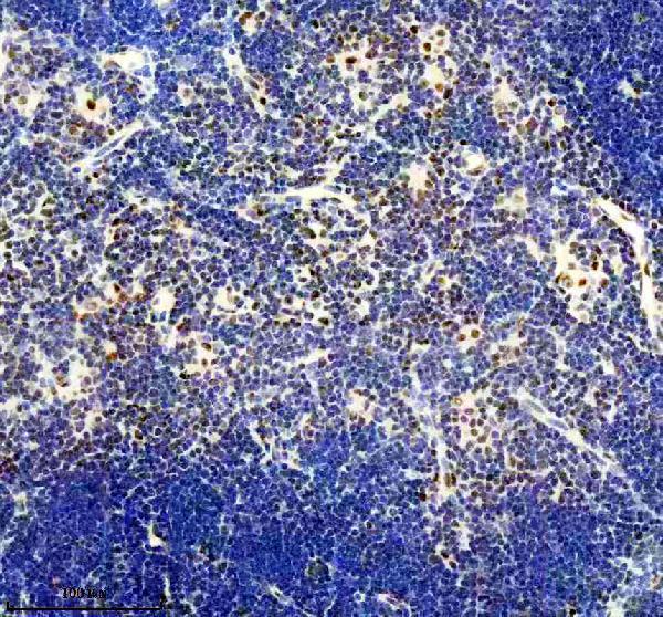
| Sample | Paraffin-embedded mouse thymus tissue; fixative not specified (datasheet M01130) |
| Fixation | Image fixative and duration unreported (datasheet M01130); verify before use. |
| Sectioning | 4–5 µm sections on charged slides (standard) |
| Deparaffinisation | Xylene, graded ethanol series to water (standard) |
| Antigen retrieval | Heat retrieval: EDTA pH 8.0 (datasheet M01130); 20 min, 95–100 °C (standard) |
| Peroxidase block | 3% H2O2, 10 min, room temperature (standard) |
| Blocking | 10% goat serum (datasheet M01130) |
| Primary antibody | Rabbit monoclonal (clone AFHG-16) anti-PBRM1, 1:50 (datasheet M01130) |
| Primary incubation | Overnight at 4 °C (datasheet M01130) |
| Detection | HRP-conjugated secondary, DAB chromogen (datasheet M01130) |
| Counterstain | Hematoxylin, blue, dehydrate and mount (standard) |
| Expected result | PBRM1-positive staining in neuronal cells of cerebral cortex (HPA tissue IHC: High). HPA tissue profile: Cytoplasmic and nuclear expression in most tissues. No signal in the no-primary control. |
PBRM1 is a nuclear chromatin regulator with no transmembrane segment (UniProt Q86U86: location, function, topology). Expect staining in nuclei across many cell types, including glandular cells in colon and duodenum and neurons in cerebral cortex (HPA tissue IHC: High). HPA also reports cytoplasmic staining in most tissues, but rates its tissue IHC profile Uncertain because antibody staining and RNA expression show low consistency (HPA tissue IHC: profile, reliability).
| Distinct nuclear staining in colon or duodenal glandular cells, with readable tissue structure and limited background. | This fits the expected compartment and two reported high staining cell populations (UniProt Q86U86: nucleus; HPA tissue IHC: High in colon and duodenal glandular cells). Interpret intensity relative to controls on the same run; HPA's tissue IHC reliability is Uncertain, so appearance alone does not establish antibody specificity (HPA tissue IHC: reliability; general IHC practice). |
| Strong cytoplasmic staining with little or no nuclear staining in an otherwise intact section. | An exclusively cytoplasmic result conflicts with UniProt's nuclear location and HPA's nucleoplasmic IF localization (UniProt Q86U86: nucleus; HPA subcellular ICC-IF: nucleoplasm). HPA does report cytoplasmic as well as nuclear tissue staining, so cytoplasmic signal alone is not proof of an artefact (HPA tissue IHC: profile). Check controls and detection background before assigning it to PBRM1 (general IHC practice). |
| Prominent staining in hepatocytes or prostate glandular cells. | These cell populations were reported as not detected in the supplied HPA tissue IHC examples (HPA tissue IHC: liver hepatocytes; prostate glandular cells). Unexpected signal may reflect cross-reactivity or endogenous detection activity; compare a no-primary control and the expected nuclear pattern (general IHC practice; UniProt Q86U86: nucleus). HPA's Uncertain reliability prevents treating either tissue as a definitive negative control (HPA tissue IHC: reliability). |
| Diffuse chromogen across nuclei, cytoplasm, and tissue spaces, obscuring cell boundaries. | A poorly resolved deposit cannot support a compartment-specific PBRM1 call (UniProt Q86U86: nucleus; general IHC practice). Consider nonspecific primary or secondary binding, endogenous detection activity, or excessive development as general IHC causes; use appropriate controls to identify the source (general IHC practice). HPA tissue staining levels do not diagnose the cause of background (HPA tissue IHC: profile). |
| No visible nuclear signal in colon or duodenal glandular cells. | These are reasonable positive-control candidates because HPA reports High staining there, although its tissue IHC reliability is Uncertain (HPA tissue IHC: colon and duodenum, reliability). First check that the section and detection controls worked, then review the catalog antibody's IHC-P instructions and the run conditions (general IHC practice). A blank section alone cannot distinguish technical failure from a true negative result (general IHC practice). |
| Tissue and cell selection | HPA reports High staining in colon and duodenal glandular cells and cerebral cortex neurons, but not detected in liver hepatocytes or prostate glandular cells (HPA tissue IHC). These are comparative examples, not guaranteed control outcomes: the overall tissue IHC reliability is Uncertain (HPA tissue IHC: reliability). |
| Antibody validation | HPA015629 is rated Uncertain for IHC and Enhanced for ICC; HPA059373 has no supplied IHC rating and is Enhanced for ICC (HPA antibodies). An ICC rating cannot establish performance in paraffin-section chromogenic IHC (HPA antibodies; general IHC practice). |
| Protein forms and epitope coverage | UniProt lists 9 PBRM1 isoforms and a single 1–1689 protein chain, with no signal peptide, propeptide, or transmembrane segment (UniProt Q86U86: isoforms, processing, topology). The supplied evidence gives no antibody epitope, so coverage of individual isoforms cannot be predicted (UniProt Q86U86; HPA antibodies). |
| IF/ICC Q&A: where should signal appear? | In IF/ICC, expect nucleoplasmic localization according to HPA, consistent with UniProt's nuclear assignment (HPA subcellular ICC-IF: nucleoplasm; UniProt Q86U86: nucleus). HPA lists RT-4, U-251MG, and U2OS as image-bearing cell lines; this localization reference does not supply an IF protocol or validate the IHC result (HPA subcellular ICC-IF; HPA antibodies). |
| Situation | Likely cause | Next action |
|---|---|---|
| No signal in a chosen high staining tissue. | The cause is unresolved from appearance alone; reagent, detection, section, or run conditions may have failed (general IHC practice). HPA's High rating is an observation with Uncertain overall tissue IHC reliability (HPA tissue IHC: reliability). | Check the run's positive and no-primary controls, then follow the catalog antibody's IHC-P instructions for retrieval, dilution, and detection (general IHC practice). Do not infer a PBRM1-specific fixation defect from this result. |
| Only cytoplasmic signal is visible. | This lacks the nuclear component expected from UniProt and HPA ICC-IF, although HPA tissue IHC reports mixed nuclear and cytoplasmic expression (UniProt Q86U86: nucleus; HPA subcellular ICC-IF: nucleoplasm; HPA tissue IHC: profile). | Inspect counterstained nuclei, compare expected positive cells, and review no-primary background (general IHC practice). Record the discordance rather than scoring cytoplasmic signal alone as confirmed PBRM1. |
| Chromogen appears in reported negative cell populations. | Hepatocytes and prostate glandular cells are listed as not detected by HPA; cross-reactivity or endogenous detection activity is possible (HPA tissue IHC: liver, prostate; general IHC practice). HPA's Uncertain reliability limits the strength of this comparison (HPA tissue IHC: reliability). | Compare a no-primary control and an expected positive tissue in the same run; investigate endogenous detection activity if the no-primary control stains (general IHC practice). |
| Diffuse background obscures nuclear boundaries. | Nonspecific binding, endogenous detection activity, or excessive chromogen development can produce background in chromogenic IHC (general IHC practice). The supplied PBRM1 sources do not identify which cause applies. | Review blocking, washes, antibody dilution, and development time against the validated IHC-P workflow; compare no-primary and positive controls (general IHC practice). |
| IF/ICC looks nucleoplasmic, but IHC is weak or atypical. | HPA's ICC ratings are Enhanced, while HPA015629's IHC rating is Uncertain and HPA059373 has no supplied IHC rating (HPA antibodies). The assays have different validation evidence (HPA antibodies). | Judge the paraffin-section result with IHC controls and the catalog antibody's IHC-P instructions (general IHC practice). Use the IF/ICC finding only as a localization comparison (HPA subcellular ICC-IF: nucleoplasm). |
| Staining varies between tissues or cell populations. | HPA reports High, Low, and not-detected examples despite low tissue RNA specificity and widely expressed PBRM1 (HPA tissue IHC: staining levels, RNA specificity; UniProt Q86U86: tissue specificity). HPA tissue IHC reliability is Uncertain (HPA tissue IHC: reliability). | Score the relevant cell population and nuclear compartment separately, alongside same-run controls; avoid treating whole-section intensity as a single result (UniProt Q86U86: nucleus; general IHC practice). |
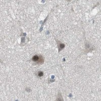


Comprehensive Human Protein Atlas IHC scoring per tissue (reliability: Uncertain — Low consistency between antibody staining and RNA expression data.). Rows are taken directly from the HPA tissue chart — click any row's HPA link to view the source.
| Tissue | Cell type | Level | Evidence | Source |
|---|---|---|---|---|
| Cerebral cortex | Neuronal cells | High | Protein (IHC) | HPA → |
| Cervix | Glandular cells | High | Protein (IHC) | HPA → |
| Colon | Glandular cells | High | Protein (IHC) | HPA → |
| Duodenum | Glandular cells | High | Protein (IHC) | HPA → |
| Endometrium | Glandular cells | High | Protein (IHC) | HPA → |
| Tissue | Cell type | Level | Evidence | Source |
|---|---|---|---|---|
| Liver | Hepatocytes | Not detected | Protein (IHC) | HPA → |
| Ovary | Ovarian stroma cells | Not detected | Protein (IHC) | HPA → |
| Prostate | Glandular cells | Not detected | Protein (IHC) | HPA → |
| Spleen | Cells in red pulp | Not detected | Protein (IHC) | HPA → |
These questions focus on nuclear PBRM1 staining in paraffin sections, with one entry on exploratory IF (UniProt Q86U86; datasheet M01130).
The catalog lists human, mouse and rat reactivity for two anti-PBRM1 antibodies (catalog applications/reactivity); M01130 has a mouse thymus paraffin-section IHC image (M01130 image caption).
M01130 renders with a chromogenic IHC image of mouse thymus in a paraffin section; IHC and IF/ICC are listed applications, and human, mouse and rat are listed reactivities (M01130 image caption; catalog applications/reactivity). A01130-1 is listed for IF/ICC with human, mouse and rat reactivity, but has no IHC application or IHC/IF image in the supplied catalog (A01130-1 catalog applications/reactivity/images).
Which to pick: Choose M01130 for paraffin-section tissue IHC: its own image caption documents mouse thymus, EDTA retrieval at pH 8.0 and a 1:50 primary dilution; the fixative is unreported (M01130 image caption). For IF/ICC, A01130-1 has a listed IF concentration of 5 μg/ml for human samples, while M01130 lists IF/ICC and a 1:50 IF dilution (A01130-1 dilution data; M01130 catalog applications/dilution). For work across human, mouse and rat, both list those species, but M01130 is the rabbit monoclonal with an IHC application and a mouse tissue IHC image (catalog host/clone/applications/reactivity; M01130 image caption).