PDIA6 / Protein disulfide-isomerase A6 · IHC design guide

Design Immunohistochemistry for PDIA6

Plan PDIA6 paraffin IHC around granular cytoplasmic staining, with high signal in marrow hematopoietic cells and pancreatic exocrine cells (HPA tissue IHC). Use the catalog antibody’s documented chromogenic workflow and compare staining across cell types (datasheet M03813-1; HPA tissue IHC).

Evidence assembled Oct 2026 · For research use; verify linked source records and product datasheet before use
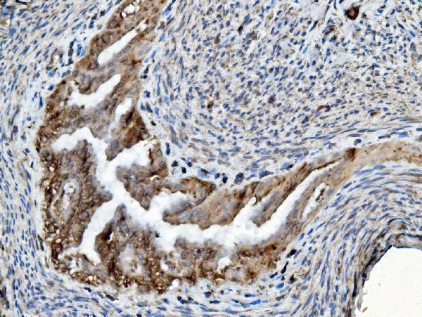
Immunohistochemistry protocol sheet for PDIA6 (IHC for PDIA6): expected localisation Granular cytoplasm observed; ER lumen expected (HPA tissue IHC; UniProt), antibody M03813-1, validated IHC image, and IHC protocol steps
Printable PDIA6 IHC protocol sheet — expected localisation Granular cytoplasm observed; ER lumen expected (HPA tissue IHC; UniProt), antibody M03813-1, controls and protocol steps. Open the full PDIA6 IHC guide →

PDIA6 Immunohistochemistry Experimental Design Guide

Expected localisation, validated protocols, controls and antibodies — the at-a-glance facts below, then the full design guide.

Must know before staining
Expected localisation Granular cytoplasm observed; ER lumen expected (HPA tissue IHC; UniProt)
Staining pattern Granular cytoplasmic staining across tissue cell types (HPA tissue IHC)
Antigen retrieval EDTA pH 8.0 HIER, heat-mediated (datasheet M03813-1)
Positive control ⓘ Bone marrow+4 more · see all
Negative control ⓘ None in HPA (detected in all 45 tissues); use no-primary + isotype controls
Important caveats
Reasons your staining may differ from the expected pattern.
Fixation Keep fixation consistent across paraffin sections. (standard IHC practice; not target-specific)
Caveat Marrow peroxidase can mimic DAB signal (HPA tissue IHC; standard IHC practice)
Regulation Low tissue specificity (HPA tissue RNA)
Isoform / epitope 5 isoforms; signal peptide cleaved; no fixed membrane side (UniProt)
Section 1

Recommended PDIA6 IHC & IF Protocols

Compare the catalog antibody’s IHC-P protocol with three published PDIA6 chromogenic IHC protocols (datasheet: M03813-1; PMC13444140; PMC6491656; PMC9449696).

Recommended immunohistochemistry (IHC-P) protocol parameters
SampleParaffin-embedded human bladder epithelial carcinoma tissue; fixative not specified (datasheet M03813-1)
FixationImage fixative and duration unreported (datasheet M03813-1); verify before use.
Sectioning4–5 µm sections on charged slides (standard)
DeparaffinisationXylene, graded ethanol series to water (standard)
Antigen retrievalHeat retrieval: EDTA pH 8.0 (datasheet M03813-1); 20 min, 95–100 °C (standard)
Peroxidase block3% H2O2, 10 min, room temperature (standard)
Blocking10% goat serum (datasheet M03813-1)
Primary antibodyMouse monoclonal (clone 3H5E7) anti-PDIA6, 2 μg/ml (datasheet M03813-1)
Primary incubationOvernight at 4 °C (datasheet M03813-1)
DetectionHRP-conjugated secondary, DAB chromogen (datasheet M03813-1)
CounterstainHematoxylin, blue, dehydrate and mount (standard)
Expected resultPDIA6-positive staining in hematopoietic cells of bone marrow (HPA tissue IHC: High). HPA tissue profile: Cytoplasmic expression with a granular pattern in all tissues. No signal in the no-primary control.
💡Decision noteFor the catalog antibody, start with heat-mediated EDTA retrieval at pH 8.0 (datasheet: M03813-1). Evaluate the published citrate conditions separately (PMC13444140; PMC6491656).
Section 2

What Is the Expected PDIA6 Staining Pattern?

PDIA6 is primarily an endoplasmic reticulum lumen protein with no transmembrane segment (UniProt Q15084: location and topology). In paraffin sections, expect granular cytoplasmic staining across tissues (HPA: tissue IHC profile), including High staining in pancreatic exocrine glandular cells, lung alveolar type II cells and bone marrow hematopoietic cells (HPA: tissue IHC). HPA rates tissue staining Enhanced, while reporting medium staining/RNA consistency and pending external verification (HPA: reliability).

What am I looking at on my slide?
Granular cytoplasmic staining is clear in pancreatic exocrine glandular cells.This matches an HPA High cell population and the reported tissue pattern (HPA: pancreas; tissue IHC profile). Assess cells and compartment together; HPA reports granular cytoplasmic expression across tissues, rather than a pancreas-exclusive pattern (HPA: tissue IHC profile).
Strong staining appears predominantly in nuclei.A nuclear-dominant result conflicts with HPA's granular cytoplasmic tissue pattern and supported ER location (HPA: tissue IHC; subcellular). Treat it as a possible staining artefact and review controls before scoring it as PDIA6.
Strong staining appears in urinary bladder urothelial cells.HPA records Low staining in these cells (HPA: urinary bladder). Markedly strong staining merits a cross-reactivity or endogenous-detection check; low is a comparator, not a claim that the cells must be unstained.
Chromogen forms a diffuse haze across cells and surrounding tissue.Haze that obscures cell boundaries cannot establish the granular cytoplasmic pattern (HPA: tissue IHC profile). In general IHC practice, background can arise from nonspecific detection; compare a control lacking primary antibody and review blocking and washes.
A known HPA-high tissue shows no discernible staining in its listed cell population.An absent signal in pancreatic exocrine glandular cells, for example, disagrees with the HPA High observation (HPA: pancreas). First establish whether the slide's staining run worked; one negative section does not establish absent PDIA6 expression.
💡Expected PDIA6 appearanceCall the result positive when distinct granular cytoplasmic chromogen appears in an HPA-high cell population, such as pancreatic exocrine glandular cells (HPA: tissue IHC); nuclear-dominant staining or diffuse haze does not match that pattern (HPA: tissue IHC profile).
How each factor affects the staining
Tissue and cell selectionUse an HPA High population, such as lung alveolar type II cells, as a positive-pattern reference (HPA: lung). HPA also lists low-staining populations, including cardiomyocytes; low means less staining, not a validated negative control (HPA: heart muscle).
Subcellular contextER lumen localization supports a cytoplasmic, organelle-associated interpretation (UniProt Q15084: location; HPA: supported ER location). UniProt also lists cell membrane and melanosome; these annotations do not turn a nuclear-dominant paraffin-section pattern into an expected result (UniProt Q15084: locations).
Antibody evidenceHPA lists Enhanced IHC validation for HPA034652, HPA034653 and CAB034347 (HPA: antibodies). Its tissue-level reliability still notes medium consistency with RNA and pending external verification (HPA: reliability); interpret a discordant slide with controls.
Isoforms and processingUniProt lists 5 isoforms and a cleaved signal peptide at residues 1–19 (UniProt Q15084: isoforms; processing). The payload gives no antibody epitope, so it cannot establish which isoforms or processed forms a particular stain detects.
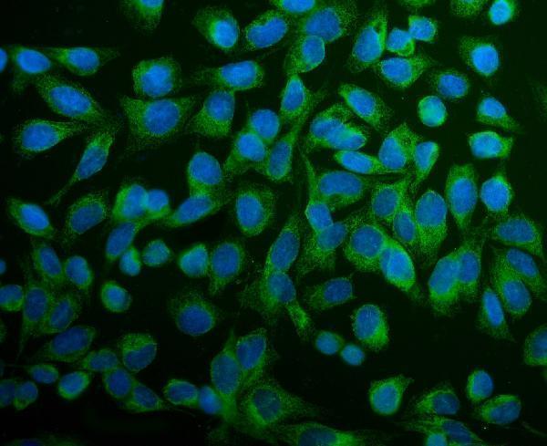
IF/ICC: where should signal appear?Mainly in the ER (HPA: supported ICC-IF location). HPA also reports primary cilium, centriolar satellite and basal body locations as uncertain; treat those as secondary observations, not the expected IHC scoring pattern (HPA: subcellular). IF/ICC has its own guide page.
Why is my staining missing, weak or wrong?
SituationLikely causeNext action
No chromogen in the expected cells of a positive reference sectionThe result conflicts with an HPA High population, such as pancreatic exocrine glandular cells (HPA: pancreas); the cause is undetermined.Check the staining run and antibody controls, then review the general IHC retrieval and detection workflow. No PDIA6-specific fixation sensitivity or retrieval requirement is supplied.
Diffuse brown background obscures the cytoplasmThe slide cannot resolve the granular cytoplasmic HPA pattern (HPA: tissue IHC profile); nonspecific detection is a possible general IHC cause.Compare a section processed without primary antibody. Review blocking, reagent concentration and washes as general IHC checks before assigning cell-level positivity.
Nuclei dominate the signalThis disagrees with the granular cytoplasmic tissue pattern and supported ER location (HPA: tissue IHC; subcellular).Review morphology and controls, then repeat or corroborate the stain if needed. Do not score nuclear-dominant signal as the expected PDIA6 pattern.
Unexpectedly strong urothelial stainingHPA lists urinary bladder urothelial cells as Low (HPA: urinary bladder); cross-reactivity or endogenous detection activity is possible.Compare an HPA-high tissue and a control lacking primary antibody. If the discrepancy persists, verify it with an independently validated antibody (HPA: antibodies).
Chromogen persists when primary antibody is omittedA primary-independent signal suggests endogenous detection activity or another background source (general IHC practice).Review detection-reagent controls and the blocking step appropriate to the chromogenic system. Reassess the test slide only after the control is clear.
Two antibody stains give different cell patternsHPA's Enhanced IHC listings support the listed antibodies, but tissue-level reliability still has medium staining/RNA consistency and awaits external verification (HPA: antibodies; reliability).Compare matched cell populations, compartment and controls under the same scoring criteria. Record the discordance instead of treating either isolated pattern as confirmed PDIA6 staining.

Sample controls for PDIA6 IHC & IF

🧪Run bone marrow first and expect hematopoietic cells to stain (HPA: High in hematopoietic cells). HPA detects PDIA6 in all 45 scored tissues, so use no-primary and isotype controls for the negative; unstained cells on the positive slide show local background but are not a validated PDIA6-negative population (HPA: no negative tissue rows).
Positive control tissue: Bone marrow (Hematopoietic cells, HPA High)
Negative control tissue: None in HPA: PDIA6 is detected in all 45 scored tissues. Use a no-primary (secondary-only) and an isotype control instead.
ICC-IF cell lines (HPA subcellular resource): HPA ICC-IF images show PDIA6 in A-431, U-251MG, U2OS, RPTEC/TERT1, hTERT-RPE1 (serum starved), with annotated localisation: Endoplasmic reticulum (supported) (HPA subcellular).
Technical controls: Include a no-primary, secondary-only section; a mouse IgG isotype control matched to the primary antibody’s subclass and concentration; and, if available, a PDIA6 knockout specimen or immunizing-peptide block (caption: mouse primary antibody; standard IHC controls). In bone marrow, quench endogenous peroxidase and check for pigment or blood-related DAB background (standard chromogenic IHC practice).
⚠️Feasibility: The selected M03813-1 paraffin-section caption does not state a fixative, and the supplied evidence reports no target-specific fixation window or fixation effect (caption: fixative unreported). Heat retrieval in EDTA at pH 8.0 is a documented starting condition, but retrieval dependence is unreported (caption: heat-mediated EDTA retrieval). Frozen sections or IF cannot be judged easier from the supplied evidence; bone marrow warrants care with endogenous peroxidase and blood-related background (HPA: bone marrow positive; standard IHC practice).

HPA tissue IHC evidence for PDIA6

Comprehensive Human Protein Atlas IHC scoring per tissue (reliability: Enhanced — Medium consistency between antibody staining and RNA expression data. Pending external verification.). Rows are taken directly from the HPA tissue chart — click any row's HPA link to view the source.

Positive expression · recommended positive controls

TissueCell typeLevelEvidenceSource
Bone marrow Hematopoietic cells High Protein (IHC) HPA →
Duodenum Endocrine cells High Protein (IHC) HPA →
Endometrium Glandular cells High Protein (IHC) HPA →
Epididymis Glandular cells High Protein (IHC) HPA →
Gallbladder Glandular cells High Protein (IHC) HPA →

Undetected expression · recommended negative controls

TissueCell typeLevelEvidenceSource
None in HPA: PDIA6 is detected in all 45 scored tissues. Use a no-primary (secondary-only) and an isotype control instead.
Section 3

Advanced PDIA6 IHC Tips

Troubleshoot PDIA6 staining in paraffin sections by checking retrieval, cellular pattern, controls, and scoring before interpreting DAB signal.

What retrieval should I start with for weak PDIA6 staining?
Start with heat-mediated antigen retrieval in EDTA at pH 8.0 for paraffin sections (datasheet M03813-1). The selected image used this retrieval before incubation with 2 μg/ml primary antibody overnight at 4°C, giving you a documented starting workflow (datasheet M03813-1). If staining is weak, check that sections received consistent heating and cooling, then compare retrieval duration on matched sections while keeping antibody concentration and detection constant (standard IHC practice). Include a known staining tissue and a no-primary control so a stronger DAB signal can be judged against background (HPA: high expression in pancreatic exocrine glandular cells; standard IHC practice).
How should I troubleshoot fixation-dependent loss of PDIA6 staining?
Target-specific sensitivity to fixation is unknown from the supplied evidence; the selected caption identifies a paraffin section but does not state its fixative (datasheet M03813-1). Record fixative, fixation duration, tissue thickness, and processing history for each specimen, then compare sections processed alike when investigating variable staining (standard IHC practice). Keep EDTA at pH 8.0 retrieval and the 2 μg/ml primary incubation constant during that comparison (datasheet M03813-1). Use a concurrently processed positive tissue and examine preservation of cell morphology, since damaged or unevenly processed regions can mislead chromogenic assessment (HPA: high expression in pancreatic exocrine glandular cells; standard IHC practice).
Which staining compartment should count as plausible PDIA6 signal?
Look first for granular cytoplasmic staining: that is the reported tissue IHC pattern, consistent with PDIA6's principal endoplasmic-reticulum localization (HPA: tissue IHC profile; HPA: supported endoplasmic-reticulum localization). PDIA6 is annotated in the endoplasmic-reticulum lumen and has no transmembrane segment, so a predominantly nuclear DAB pattern warrants a specificity check (UniProt Q15084 localization and topology; standard IHC practice). Cell-membrane and melanosome annotations exist, but they should not turn an isolated peripheral deposit into an assumed positive cell (UniProt Q15084 localization; standard IHC practice). Compare the pattern with cell morphology, a positive tissue, and a no-primary section before assigning an unusual compartment (standard IHC practice).
Could processing or isoforms explain inconsistent PDIA6 staining?
PDIA6 has 5 listed isoforms, and its precursor loses a 1–19 signal peptide to yield a 20–440 chain (UniProt Q15084 processing and isoforms). The record places thioredoxin domains at residues 20–133 and 154–287 and lists several phosphoserines, but the antibody epitope is not supplied (UniProt Q15084 domains and modified residues; datasheet M03813-1). Do not assign staining differences to one isoform or modification without epitope information and independent evidence (standard IHC practice). Compare matched sections under the documented EDTA pH 8.0 retrieval and 2 μg/ml incubation before considering an epitope-dependent explanation (datasheet M03813-1; standard IHC practice).
How can IF help resolve ambiguous cellular PDIA6 staining?
For a separate IF experiment, pair PDIA6 with a validated marker for the expected cell type, such as pancreatic exocrine glandular cells, to check cellular assignment (HPA: high expression in pancreatic exocrine glandular cells; standard IF practice). Choose a spectrally separated, preferably far-red fluorophore when tissue autofluorescence obscures shorter-wavelength signal, and inspect single-color controls before interpreting overlap (standard IF practice). Because PDIA6 is principally in the endoplasmic-reticulum lumen and has no transmembrane segment, test permeabilisation sufficient to expose an internal epitope while preserving morphology (UniProt Q15084 localization and topology; standard IF practice). The antibody epitope and an IF validation workflow are not supplied here, so verify the IF pattern independently (datasheet M03813-1; standard IF practice).
What should I check when PDIA6 DAB staining is diffuse?
The selected IHC procedure used 10% goat serum before primary antibody and an anti-mouse peroxidase detection system with DAB (datasheet M03813-1). Confirm an appropriate peroxidase block, washing, and a no-primary control when diffuse brown signal appears; these are general chromogenic IHC checks (standard IHC practice). Review whether antibody concentration or DAB development is driving background before changing retrieval, using 2 μg/ml primary overnight at 4°C as the documented reference (datasheet M03813-1; standard IHC practice). Score cellular granular cytoplasmic staining separately from pigment, extracellular deposits, and staining at section edges (HPA: tissue IHC profile; standard IHC practice).
How should I quantify PDIA6 across differently cellular sections? ⚠ ANSWER MARKED FOR VERIFICATION
Define the cell population and cytoplasmic compartment before scoring, since the reported tissue pattern is granular cytoplasmic and expression varies by cell type (HPA: tissue IHC profile). For intensity and prevalence, use an H-score from 0–300 or report the percentage of positive target cells with prespecified intensity thresholds (standard IHC practice). If counting positive cells per mm², also report target-cell density or tissue area so differences in cellularity are visible (standard IHC practice). Apply the same region-selection rules, retrieval, staining batch controls, and image thresholds across specimens, and exclude necrosis and damaged edges from the scored area (standard IHC practice).
How do I distinguish true PDIA6 positivity from artefact?
A plausible positive shows cellular granular cytoplasmic staining in an appropriate population, with the endoplasmic reticulum as the main supported location (HPA: tissue IHC profile; HPA: supported endoplasmic-reticulum localization). Check cell identity because HPA reports high staining in pancreatic exocrine glandular cells but low staining in urinary-bladder urothelial cells; the selected bladder carcinoma image does not establish a normal-urothelium pattern (HPA: tissue IHC cell levels; datasheet M03813-1). Predominantly nuclear signal, edge-only deposits, and staining in necrotic areas need control review before being called positive (UniProt Q15084 localization; standard IHC practice). Compare no-primary and peroxidase-block controls to identify detection background or endogenous enzyme activity (standard IHC practice).
Boster reagents

Best PDIA6 / Protein disulfide-isomerase A6 IHC Antibodies

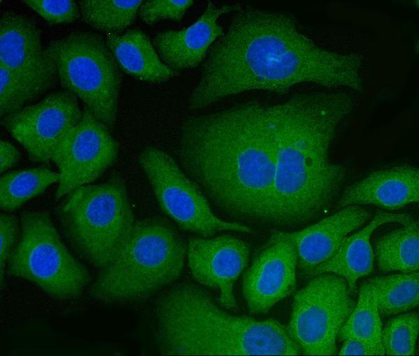
Anti-PDIA6 antibodies have IHC images from human cancer and mouse and rat ovary sections (catalog IHC captions), plus IF images from HepG2 and U20S cells (catalog IF captions).

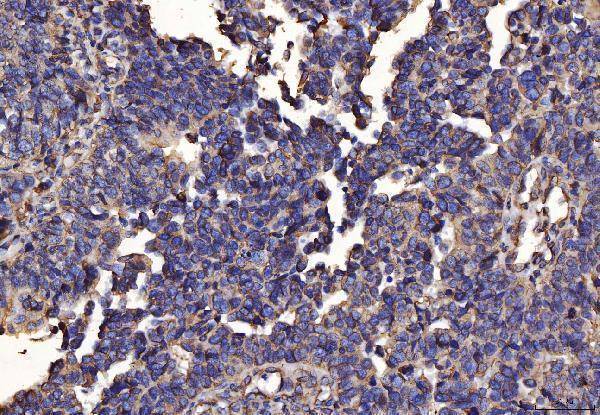
Real IHC data IHC analysis of PDIA6 using anti-PDIA6 antibody (M03813-1). PDIA6 was detected in a paraffin-embedded section of human bladder epithelial carcinoma tissue. Heat mediated antigen retrieval was performed in EDTA buffer (pH 8.0, epitope retrieval solution). The tissue section was blocked with 10% goat serum. The tissue section was then incubated with 2 μg/ml mouse anti-PDIA6 Antibody (M03813-1) overnight at 4°C. Peroxidase Conjugated Goat Anti-mouse IgG was used as secondary antibody and incubated for 30 minutes at 37°C. The tissue section was developed using HRP Conjugated Mouse IgG Super Vision Assay Kit (Catalog # SV0001) with DAB as the chromogen.
Anti-PDIA6 Antibody ® (monoclonal, 3H5E7)
Cat # M03813-1
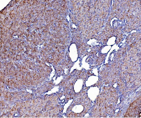
Real IHC data IHC analysis of PDIA6 using anti-PDIA6 antibody (A03813-2). PDIA6 was detected in paraffin-embedded section of mouse ovary tissue. Heat mediated antigen retrieval was performed in EDTA buffer (pH8.0, epitope retrieval solution). The tissue section was blocked with 10% goat serum. The tissue section was then incubated with 2μg/ml rabbit anti-PDIA6 Antibody (A03813-2) overnight at 4°C. Biotinylated goat anti-rabbit IgG was used as secondary antibody and incubated for 30 minutes at 37°C. The tissue section was developed using Strepavidin-Biotin-Complex (SABC) (Catalog # SA1022) with DAB as the chromogen.
Anti-PDIA6 Antibody ®
Cat # A03813-2

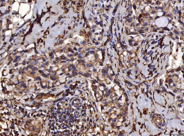
M03813-1 has paraffin-section IHC images from human bladder, breast, colorectal, and endometrial cancers, plus a HepG2 IF image (catalog IHC and IF captions). A03813-2 has paraffin-section IHC images from mouse and rat ovary and human breast and liver cancers, plus a U20S IF image (catalog IHC and IF captions).

Which to pick: Choose monoclonal M03813-1 for human paraffin-section IHC (catalog applications and M03813-1 IHC captions). Choose A03813-2 for human, mouse, or rat paraffin-section IHC; its own images cover all three species (catalog reactivity and A03813-2 IHC captions). Both support IF/ICC in human cells (catalog applications and IF captions); the fixative used for the paraffin sections is unreported (catalog IHC captions).

Each figure is that product's own IHC / IF validation image from its datasheet.

References

  1. UniProt Consortium. UniProt entry Q15084 (PDIA6_HUMAN, Protein disulfide-isomerase A6).
  2. Human Protein Atlas. PDIA6 tissue IHC expression (reliability: Enhanced).
  3. Human Protein Atlas. PDIA6 subcellular location (ICC-IF): Mainly localized to the endoplasmic reticulum. In addition localized to the centriolar satellites, primary cilium and basal body..
  4. Human Protein Atlas. PDIA6 antibody validation summary (3 antibodies).
  5. Endoplasmic reticulum stress-induced histone lactylation mediating immune escape of gastric cancer via PDIA6 overexpression in dendritic cells. Cell death & disease 2026 — PMC13444140.
  6. PDIA6 modulates apoptosis and autophagy of non-small cell lung cancer cells via the MAP4K1/JNK signaling pathway. EBioMedicine 2019 — PMC6491656.
  7. Protein disulfide isomerase A6 promotes the repair of injured nerve through interactions with spastin. Frontiers in molecular neuroscience 2022 — PMC9449696.
  8. PubMed PMID:7590364 — UniProt-cited evidence.
  9. PubMed PMID:14702039 — UniProt-cited evidence.
  10. PubMed PMID:15815621 — UniProt-cited evidence.