This website uses cookies to ensure you get the best experience on our website.


- Table of Contents
Plan PDK2 staining in paraffin sections using its observed cytoplasmic tissue pattern (HPA tissue IHC) and expected mitochondrial matrix location (UniProt). The guide covers fixation consistency, chromogenic detection and interpretation of variable staining intensity (HPA tissue IHC).

Expected localisation, validated protocols, controls and antibodies — the at-a-glance facts below, then the full design guide.
| Expected localisation | Observed cytoplasmic staining; mitochondrial matrix expected (HPA tissue IHC; UniProt) | |
| Staining pattern | Widespread cytoplasmic staining with variable intensity (HPA tissue IHC) | |
| Antigen retrieval | EDTA pH 8.0 HIER, heat-mediated (datasheet A03261) | |
| Positive control | Adrenal gland+4 more · see all | |
| Negative control | Soft tissue+1 more · see all |
| Fixation | Keep fixation conditions consistent across sections. (standard IHC practice; not target-specific) | |
| Caveat | Staining and RNA expression show medium consistency (HPA tissue IHC) | |
| Regulation | Expression varies by tissue (UniProt) | |
| Isoform / epitope | Two isoforms; epitope coverage is unspecified (UniProt) |
The catalog antibody protocol (datasheet A03261) is accompanied by one published IHC protocol using head and neck cancer tissue microarrays (PMC9727917).
| Sample | Paraffin-embedded human breast cancer tissue; fixative not specified (datasheet A03261) |
| Fixation | Image fixative and duration unreported (datasheet A03261); verify before use. |
| Sectioning | 4–5 µm sections on charged slides (standard) |
| Deparaffinisation | Xylene, graded ethanol series to water (standard) |
| Antigen retrieval | Heat retrieval: EDTA pH 8.0 (datasheet A03261); 20 min, 95–100 °C (standard) |
| Peroxidase block | 3% H2O2, 10 min, room temperature (standard) |
| Blocking | 10% goat serum (datasheet A03261) |
| Primary antibody | Rabbit anti-PDK2, 2-5μg/ml (datasheet A03261) |
| Primary incubation | Overnight at 4 °C (datasheet A03261) |
| Detection | HRP-conjugated secondary, DAB chromogen (datasheet A03261) |
| Counterstain | Hematoxylin, blue, dehydrate and mount (standard) |
| Expected result | PDK2-positive staining in glandular cells of adrenal gland (HPA tissue IHC: High). HPA tissue profile: Ubiquitous cytoplasmic expression. No signal in the no-primary control. |
Expect PDK2 staining mainly in the cytoplasm, consistent with its mitochondrial matrix location and lack of a transmembrane segment (UniProt Q15119). HPA describes ubiquitous cytoplasmic tissue staining, with high staining in selected glandular, epithelial, hematopoietic and germinal center cells (HPA tissue IHC). Tissue IHC reliability is Supported, with medium agreement between antibody staining and RNA expression (HPA tissue IHC).
| Cytoplasmic, granular staining in adrenal glandular cells, bronchial respiratory epithelial cells or bone marrow hematopoietic cells. | This fits the expected mitochondrial compartment (UniProt Q15119) and cell types scored High by HPA tissue IHC. Granularity is a plausible appearance for mitochondrial staining, but chromogenic IHC alone does not establish that every stained structure is a mitochondrion (general IHC practice). |
| Predominantly nuclear staining, with little or no cytoplasmic signal. | Recheck specificity and detection: the established location is mitochondrial (UniProt Q15119; HPA subcellular: supported), while HPA calls its IF nucleoplasmic assignment uncertain. Nuclear staining alone is therefore insufficient to call a correct PDK2 IHC pattern; it may reflect artefact or nonspecific staining. |
| Strong staining in soft tissue fibroblasts or vaginal squamous epithelial cells. | HPA reports PDK2 as Not detected in those cells (HPA tissue IHC). Consider cross-reactivity or endogenous detection activity, especially if staining persists in a no-primary control (general IHC practice). Check that tissue identity and the stained cell type match the reference before assigning a false positive. |
| Diffuse color across cells and extracellular areas, obscuring cytoplasmic detail. | This cannot be scored confidently against HPA’s cytoplasmic pattern (HPA tissue IHC). Excess detection reagent, incomplete blocking or inadequate washing can create background (general IHC practice). Assess a no-primary control and reduce background before interpreting the apparent tissue distribution. |
| No convincing signal in adrenal glandular cells or bone marrow hematopoietic cells. | Both are High reference cell populations (HPA tissue IHC), so their absence warrants a technical check. Verify that the cells are present and examine antibody dilution, antigen retrieval and detection controls (general IHC practice). The supplied sources do not establish a PDK2-specific fixation or retrieval failure. |
| Compartment | PDK2 is a mitochondrial matrix protein without a transmembrane segment (UniProt Q15119). Interpret tissue IHC against the HPA cytoplasmic pattern; HPA’s IF mitochondrial assignment is supported, while its nucleoplasmic assignment is uncertain (HPA tissue IHC; HPA subcellular). |
| Cell and tissue selection | HPA scores adrenal glandular cells and bronchial respiratory epithelial cells High, liver hepatocytes Low, and soft tissue fibroblasts Not detected (HPA tissue IHC). Compare matched cell populations within tissue; a whole-section impression can conceal these differences (general IHC practice). |
| Strength of reference evidence | HPA labels tissue IHC reliability Supported and describes medium agreement with RNA expression; the listed antibody HPA008287 is IHC Supported (HPA tissue IHC; HPA antibodies). Treat the reported distribution as a useful reference, with room for antibody and sample-level variation. |
| Isoforms and epitope coverage | UniProt lists two PDK2 isoforms (UniProt Q15119). Without an epitope map or isoform-specific validation in the supplied evidence, staining cannot identify which isoform contributes to a cell’s signal; review antibody documentation if isoform discrimination matters (general IHC practice). |
| Antigen retrieval | Retrieval is a standard paraffin-IHC workflow variable (general IHC practice). The supplied UniProt and HPA records provide no PDK2-specific retrieval condition or fixation-sensitivity claim, so optimize using assay controls rather than predicting an effect for a particular tissue. |
| Detection background | Endogenous enzyme activity and nonspecific reagent binding can produce chromogenic color independently of primary-antibody binding (general IHC practice). No-primary and detection controls help distinguish that background from the cytoplasmic cell pattern reported by HPA (HPA tissue IHC). |
| Situation | Likely cause | Next action |
|---|---|---|
| High reference cells have no staining. | Possible failed antibody or detection step, overly dilute reagent, or unsuitable retrieval (general IHC practice); no PDK2-specific fixation effect is established here. | Confirm the expected cells are present, run a positive tissue control, and check antibody dilution, retrieval and detection in sequence (general IHC practice; HPA tissue IHC: adrenal glandular cells High). |
| Only nuclei stain strongly. | A nuclear-only result does not match supported mitochondrial localization (UniProt Q15119; HPA subcellular). HPA’s nucleoplasmic IF assignment is uncertain (HPA subcellular). | Review morphology and a no-primary control, then reassess antibody specificity before scoring nuclei as PDK2 positive (general IHC practice; HPA subcellular). |
| Soft tissue fibroblasts show strong color. | That cell type is Not detected in the HPA tissue reference (HPA tissue IHC); cross-reactivity or endogenous detection activity is possible (general IHC practice). | Confirm the cell identity, inspect no-primary staining, and compare with an HPA High cell population processed in the same run (HPA tissue IHC; general IHC practice). |
| Color is diffuse across the section. | Background from incomplete blocking, excess reagent or insufficient washing may obscure the reported cytoplasmic pattern (general IHC practice; HPA tissue IHC). | Check no-primary and detection controls, then adjust blocking, reagent concentration and washes before scoring cellular intensity (general IHC practice). |
| Low reference cells look as dark as High reference cells. | Overdeveloped chromogen or saturated detection can compress apparent intensity differences (general IHC practice); HPA scores liver hepatocytes Low and adrenal glandular cells High (HPA tissue IHC). | Compare identifiable cell types under the same processing and shorten chromogen development or adjust detection if signal is saturated (general IHC practice; HPA tissue IHC). |
| IF shows mitochondria plus apparent nuclear signal. | HPA supports the mitochondrial IF location but marks nucleoplasmic localization uncertain (HPA subcellular). | Keep IF interpretation on its separate guide page; for this IHC assessment, prioritize the supported mitochondrial and tissue-cytoplasmic pattern and validate nuclear signal independently (HPA subcellular; HPA tissue IHC). |
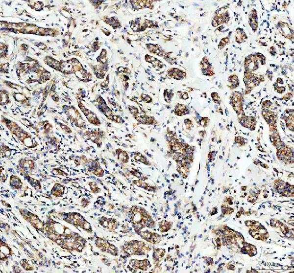
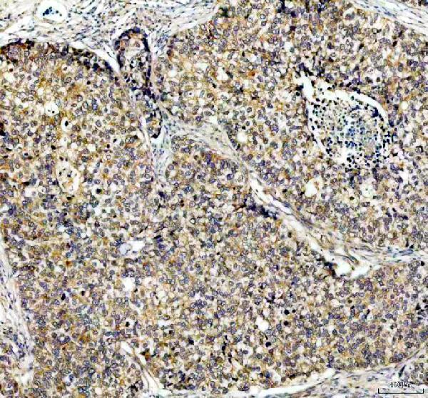
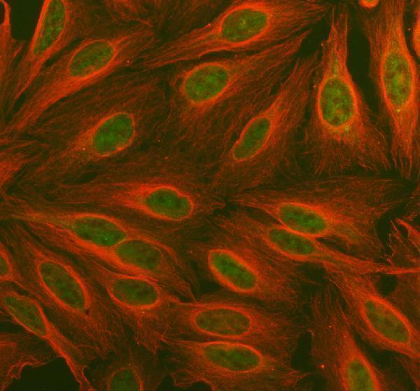
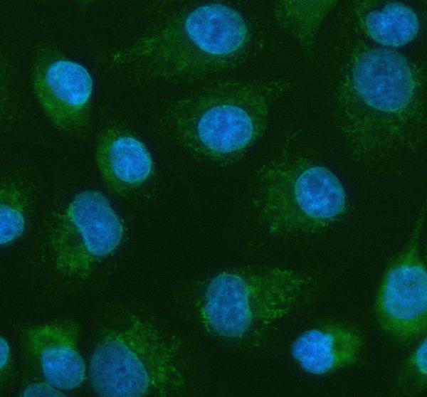
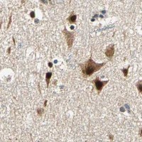


Comprehensive Human Protein Atlas IHC scoring per tissue (reliability: Supported — Medium consistency between antibody staining and RNA expression data.). Rows are taken directly from the HPA tissue chart — click any row's HPA link to view the source.
| Tissue | Cell type | Level | Evidence | Source |
|---|---|---|---|---|
| Adrenal gland | Glandular cells | High | Protein (IHC) | HPA → |
| Bone marrow | Hematopoietic cells | High | Protein (IHC) | HPA → |
| Bronchus | Respiratory epithelial cells | High | Protein (IHC) | HPA → |
| Cerebellum | Cells in molecular layer | High | Protein (IHC) | HPA → |
| Endometrium | Glandular cells | High | Protein (IHC) | HPA → |
Troubleshoot PDK2 chromogenic IHC by checking retrieval, staining controls, and whether the signal fits its mitochondrial localisation (UniProt Q15119; HPA subcellular).
A03261 has IHC images from human paraffin-embedded breast and lung cancer sections (A03261 IHC captions); PA1994 has an IF image from A549 cells (PA1994 IF caption).
A03261 will render with a human breast cancer section IHC figure; its catalog also documents IHC in human lung cancer sections and IF in U2OS cells (A03261 image captions). PA1994 will render with an A549 cell IF figure and is listed for IF/ICC (PA1994 IF caption and applications).
Which to pick: For tissue IHC, choose A03261: its rabbit antibody is listed for IHC and shown in paraffin-embedded human sections after EDTA pH 8 retrieval; the fixative is unreported (A03261 catalog applications and IHC captions). For IF/ICC, choose PA1994, a rabbit antibody shown in A549 cells at 5 μg/ml (PA1994 catalog applications and IF caption). For cross-species work, both list Human, Mouse and Rat reactivity, while the supplied tissue IHC images document human sections only (A03261 and PA1994 catalog reactivity; A03261 IHC captions).