Click image to see more details
Product Info Summary
| SKU: | PA1994 |
|---|---|
| Size: | 100 μg/vial |
| Reactive Species: | Human, Mouse, Rat |
| Host: | Rabbit |
| Application: | Flow Cytometry, IP, IF, ICC, WB |
Customers Who Bought This Also Bought

Product info
Product Name
Anti-PDK2 Antibody Picoband®
SKU/Catalog Number
PA1994
BA3775 is an alternative SKU for this antibody, used in previous lots.
Size
100 μg/vial
Form
Lyophilized
Description
Boster Bio Anti-PDK2 Antibody catalog # PA1994. Tested in Flow Cytometry, IP, IF, ICC, WB applications. This antibody reacts with Human, Mouse, Rat. The brand Picoband indicates this is a premium antibody that guarantees superior quality, high affinity, and strong signals with minimal background in Western blot applications. Only our best-performing antibodies are designated as Picoband, ensuring unmatched performance.
Storage & Handling
Store at -20˚C for one year from date of receipt. After reconstitution, at 4˚C for one month. It can also be aliquotted and stored frozen at -20˚C for six months. Avoid repeated freeze-thaw cycles.
Cite This Product
Anti-PDK2 Antibody Picoband® (Boster Biological Technology, Pleasanton CA, USA, Catalog # PA1994)
Host
Rabbit
Contents
Each vial contains 4 mg Trehalose, 0.9 mg NaCl and 0.2 mg Na2HPO4.
Clonality
Polyclonal
Isotype
Rabbit IgG
Immunogen
A synthetic peptide corresponding to a sequence at the C-terminus of human PDK2, identical to the related rat and mouse sequences.
Cross-reactivity
No cross-reactivity with other proteins
Reactive Species
PA1994 is reactive to PDK2 in Human, Mouse, Rat
Observed Molecular Weight
46 kDa
Calculated molecular weight
46.2 kDa
Background of PDK2
PDK2 (Pyruvate Dehydrogenase Kinase Isoenzyme 2), is an enzyme that in humans is encoded by the PDK2 gene. This gene encodes a member of the pyruvate dehydrogenase kinase family. The encoded protein phosphorylates pyruvate dehydrogenase, down-regulating the activity of the mitochondrial pyruvate dehydrogenase complex. Overexpression of this gene may play a role in both cancer and diabetes. Alternatively spliced transcript variants encoding multiple isoforms have been observed for this gene.
Antibody Validation
Boster validates all antibodies on WB, IHC, ICC, Immunofluorescence, and ELISA with known positive control and negative samples to ensure specificity and high affinity, including thorough antibody incubations.
Application & Images
Applications
PA1994 is guaranteed for Flow Cytometry, IP, IF, ICC, WB Boster Guarantee
Recommend Dilution
| Application | Dilution | Species |
|---|---|---|
| Western blot | 0.1-0.5μg/ml | Mouse, Rat |
| Immunocytochemistry/Immunofluorescence | 5 μg/ml | Human |
| Immunoprecipitation | 0.5-2 μg/ml | Human |
| Flow Cytometry(Fixed) | 1-3 μg/1x106 cells | Human |
Tested application
Suggested blocking solution with 5% non-fat milk or BSA; (*)Recommended protein loading: 20-40 µg per lane
Validation Images & Assay Conditions
Click image to see more details
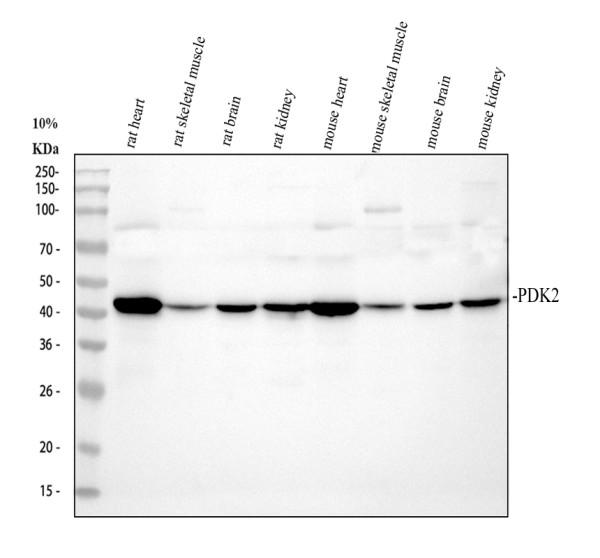
Western blot analysis of PDK2 using anti-PDK2 antibody (PA1994).
Electrophoresis was performed on a 10% SDS-PAGE gel at 80V (Stacking gel) / 120V (Resolving gel) for 2 hours. The sample well of each lane was loaded with 30 ug of sample under reducing conditions.
Lane 1: rat heart tissue lysates,
Lane 2: rat skeletal muscle tissue lysates,
Lane 3: rat brain tissue lysates,
Lane 4: rat kidney tissue lysates,
Lane 5: mouse heart tissue lysates,
Lane 6: mouse skeletal muscle tissue lysates,
Lane 7: mouse brain tissue lysates,
Lane 8: mouse kidney tissue lysates.
After electrophoresis, proteins were transferred to a nitrocellulose membrane at 150 mA for 50-90 minutes. Blocked the membrane with 5% non-fat milk/TBS for 1.5 hour at RT. The membrane was incubated with rabbit anti-PDK2 antigen affinity purified polyclonal antibody (PA1994) at 0.5 μg/mL overnight at 4°C, then washed with TBS-0.1%Tween 3 times with 5 minutes each and probed with a goat anti-rabbit IgG-HRP secondary antibody (Catalog # BA1054) at a dilution of 1:5000 for 1.5 hour at RT. The signal is developed using an ECL Plus Western Blotting Substrate (Catalog # AR1196-200) with Tanon 5200 system. A specific band was detected for PDK2 at approximately 46 kDa. The expected band size for PDK2 is at 46 kDa.

Click image to see more details
Immunoprecipitating (IP) PDK2 in Jurkat whole cell lysate.
Western blot analysis of PDK2 using anti-PDK2 antibody (PA1994);
Lane 1: Jurkat whole cell lysates (30ug);
Lane 2: Rabbit control IgG instead of anti-PDK2 antibody in Jurkat whole cell lysate;
Lane 3: anti-PDK2 antibody (2μg) + Jurkat whole cell lysate (500μg).
After electrophoresis, proteins were transferred to a membrane. Then the membrane was incubated with rabbit anti-PDK2 antigen affinity purified polyclonal antibody (PA1994) at a dilution of 0.5 μg/mL and probed with a goat anti-rabbit IgG-HRP secondary antibody (Catalog # BM2007). The signal is developed using ECL Plus Western Blotting Substrate (Catalog # AR1196-200). A specific band was detected for PDK2 at approximately 46 kDa. The expected band size for PDK2 is at 46 kDa.

Click image to see more details
IF analysis of PDK2 using anti-PDK2 antibody (PA1994).
PDK2 was detected in an immunocytochemical section of A549 cells. Enzyme antigen retrieval was performed using IHC enzyme antigen retrieval reagent (AR0022) for 15 mins. The cells were blocked with 10% goat serum. And then incubated with 5 μg/mL rabbit anti-PDK2 Antibody (PA1994) overnight at 4°C. DyLight®488 Conjugated Goat Anti-Rabbit IgG (BA1127) was used as secondary antibody at 1:500 dilution and incubated for 30 minutes at 37°C. The section was counterstained with DAPI. Visualize using a fluorescence microscope and filter sets appropriate for the label used.

Click image to see more details
Flow Cytometry analysis of PC-3 cells using anti-PDK2 antibody (PA1994).
Overlay histogram showing PC-3 cells stained with PA1994 (Blue line). To facilitate intracellular staining, cells were fixed with 4% paraformaldehyde and permeabilized with permeabilization buffer. The cells were blocked with 10% normal goat serum. And then incubated with rabbit anti-PDK2 Antibody (PA1994, 1 μg/1x106 cells) for 30 min at 20°C. DyLight®488 conjugated goat anti-rabbit IgG (BA1127, 5-10 μg/1x106 cells) was used as secondary antibody for 30 minutes at 20°C. Isotype control antibody (Green line) was rabbit IgG (1 μg/1x106) used under the same conditions. Unlabelled sample without incubation with primary antibody and secondary antibody (Red line) was used as a blank control.
Specific Publications For Anti-PDK2 Antibody Picoband® (PA1994)
Loading publications
Recommended Resources
Here are featured tools and databases that you might find useful.
- Boster's Pathways Library
- Protein Databases
- Bioscience Research Protocol Resources
- Data Processing & Analysis Software
- Photo Editing Software
- Scientific Literature Resources
- Research Paper Management Tools
- Molecular Biology Software
- Primer Design Tools
- Bioinformatics Tools
- Phylogenetic Tree Analysis
Customer Reviews
Have you used Anti-PDK2 Antibody Picoband®?
Share your experimental results or join a short interview to earn up to $1,000 in product credits or other rewards.
0 Reviews For Anti-PDK2 Antibody Picoband®
Customer Q&As
Have a question?
Find answers in Q&As, reviews.
Can't find your answer?
Submit your question
4 Customer Q&As for Anti-PDK2 Antibody Picoband®
Question
We appreciate helping with my inquiry over the phone. Here are the WB image, lot number and protocol we used for lung using anti-PDK2 antibody PA1994. Let me know if you need anything else.
Verified Customer
Verified customer
Asked: 2020-04-14
Answer
Thanks for the data. You have provided everything we needed. Our lab team are working to resolve your inquiry as quickly as possible, and we appreciate your patience and understanding! Please let me know if there is anything you need in the meantime.
Boster Scientific Support
Answered: 2020-04-14
Question
Do you have a BSA free version of anti-PDK2 antibody PA1994 available?
Verified Customer
Verified customer
Asked: 2019-07-31
Answer
I appreciate your recent telephone inquiry. I can confirm that some lots of this anti-PDK2 antibody PA1994 are BSA free. For now, these lots are available and we can make a BSA free formula for you free of charge. It will take 3 extra days to prepare. If you require this antibody BSA free again in future, please do not hesitate to contact me and I will be pleased to check which lots we have in stock that are BSA free.
Boster Scientific Support
Answered: 2019-07-31
Question
We are currently using anti-PDK2 antibody PA1994 for mouse tissue, and we are happy with the WB results. The species of reactivity given in the datasheet says human, mouse, rat. Is it likely that the antibody can work on dog tissues as well?
Verified Customer
Verified customer
Asked: 2017-07-19
Answer
The anti-PDK2 antibody (PA1994) has not been tested for cross reactivity specifically with dog tissues, though there is a good chance of cross reactivity. We have an innovator award program that if you test this antibody and show it works in dog you can get your next antibody for free. Please contact me if I can help you with anything.
Boster Scientific Support
Answered: 2017-07-19
Question
Does anti-PDK2 antibody PA1994 work for WB with lung?
R. Zhao
Verified customer
Asked: 2014-06-02
Answer
According to the expression profile of lung, PDK2 is highly expressed in lung. So, it is likely that anti-PDK2 antibody PA1994 will work for WB with lung.
Boster Scientific Support
Answered: 2014-06-02
