This website uses cookies to ensure you get the best experience on our website.


- Table of Contents
Plan paraffin-section PDPK1 IHC around generally cytoplasmic staining, with high signal in bone marrow hematopoietic cells and cerebellar Purkinje cells (HPA tissue IHC). Start the catalog antibody at 0.5–1 μg/mL (datasheet: PB9735), and score staining by cell type (standard IHC practice).

Expected localisation, validated protocols, controls and antibodies — the at-a-glance facts below, then the full design guide.
| Expected localisation | Generally cytoplasmic tissue staining (HPA tissue IHC) | |
| Staining pattern | Cytoplasmic staining in positive cells (HPA tissue IHC) | |
| Antigen retrieval | Citrate pH 6 HIER, heat-mediated (datasheet PB9735) | |
| Positive control | Bone marrow+4 more · see all | |
| Negative control | Adipose tissue |
| Fixation | Matched tissue-IHC evidence does not establish the fixation claim. Validate the specimen-specific method before use. (selected-SKU IHC image PB9735) | |
| Caveat | Block endogenous peroxidase in hematopoietic-rich controls (HPA tissue IHC; standard IHC practice) | |
| Regulation | Phospho-Tyr-9 rises in diseased tissue (UniProt) | |
| Isoform / epitope | 5 isoforms; epitope coverage needs checking (UniProt) |
The catalog antibody protocol is followed by 3 published PDPK1 IHC protocols for paraffin sections (PMC9195017; PMC3987083; PMC4270133).
| Sample | Paraffin-embedded Mouse Intestine tissue; fixative not specified (datasheet PB9735) |
| Fixation | Image fixative and duration unreported (datasheet PB9735); verify before use. |
| Sectioning | 4–5 µm sections on charged slides (standard) |
| Deparaffinisation | Xylene, graded ethanol series to water (standard) |
| Antigen retrieval | Heat retrieval: Citrate pH 6, 20 min (datasheet PB9735) |
| Peroxidase block | 3% H2O2, 10 min, room temperature (standard) |
| Blocking | 10% goat serum (datasheet PB9735) |
| Primary antibody | Rabbit anti-PDPK1, 0.5-1μg/ml (datasheet PB9735) |
| Primary incubation | Overnight at 4 °C (datasheet PB9735) |
| Detection | Streptavidin-biotin complex (SABC), DAB chromogen (datasheet PB9735) |
| Counterstain | Hematoxylin, blue, dehydrate and mount (standard) |
| Expected result | PDPK1-positive staining in hematopoietic cells of bone marrow (HPA tissue IHC: High). HPA tissue profile: General cytoplasmic expression. No signal in the no-primary control. |
PDPK1 is generally cytoplasmic in tissue IHC, with cell membrane, focal adhesion and context-dependent nuclear localization also reported (HPA: general cytoplasmic expression; UniProt O15530: subcellular location). Expect staining in hematopoietic cells, Purkinje cells, splenic red pulp cells and late spermatids (HPA: High in each). PDPK1 has no transmembrane segment, so membrane staining represents localization rather than an integral membrane protein (UniProt O15530: topology). HPA rates its tissue IHC evidence Supported, with medium agreement between staining and RNA expression (HPA: reliability).
| Cytoplasmic staining is strongest in bone marrow hematopoietic cells, cerebellar Purkinje cells, splenic red pulp cells or testicular late spermatids (HPA: High in each). | This fits the reported tissue IHC pattern (HPA: general cytoplasmic expression; High in the listed cells). Compare cells within the same section: HPA reports medium staining in bronchial respiratory epithelium and adrenal glandular cells, so uniform intensity across tissues is unnecessary (HPA: Medium in both). |
| Nuclear staining appears in neuronal cells, or focal adhesion or membrane staining appears in a relevant signaling context (UniProt O15530: subcellular location). | These compartments are biologically plausible: UniProt describes neuronal nuclear restriction and regulated membrane or focal adhesion localization (UniProt O15530: subcellular location). Judge the result against cell identity and context; HPA’s tissue IHC summary remains predominantly cytoplasmic (HPA: profile). |
| Signal outlines extracellular spaces, fills tissue-free areas or appears only as surface deposits (UniProt O15530: topology; HPA: general cytoplasmic expression). | Treat this as a likely staining artefact: PDPK1 lacks a transmembrane segment, and the supported tissue pattern is cellular (UniProt O15530: topology; HPA: profile). Genuine regulated membrane localization should track identifiable cells, not empty spaces (UniProt O15530: subcellular location). |
| Adipocytes stain strongly while expected positive cells do not (HPA: adipocytes Not detected; High in hematopoietic cells). | Suspect cross-reactivity or endogenous detection activity (standard IHC practice). HPA’s undetected adipocyte result is a reference pattern, not proof that every adipocyte must be negative; assess the antibody and detection controls before assigning specificity (HPA: adipocytes Not detected; reliability Supported). |
| The section has broad haze, or a known positive population has no visible signal (HPA: High in Purkinje cells and hematopoietic cells). | Haze limits compartment scoring; absent signal in a reported high-staining population makes a technical failure or unsuitable antibody conditions plausible (standard IHC practice; HPA: High in the listed cells). Review controls before interpreting either result as PDPK1 biology. |
| Tissue and cell selection (HPA: tissue IHC). | HPA reports High staining in hematopoietic cells, Purkinje cells, red pulp cells and late spermatids, but Not detected in adipocytes; choose comparison regions by cell type rather than whole-tissue averages (HPA: listed tissue levels). |
| Compartment and signaling state (UniProt O15530: subcellular location). | Insulin-associated membrane translocation and context-dependent nuclear or focal adhesion localization can alter the visible distribution; a membrane or nuclear component alone does not establish antibody failure (UniProt O15530: subcellular location). |
| Antibody validation (HPA: antibody records). | HPA lists IHC as Supported for HPA035199 and CAB004272; HPA008062 has no IHC status listed (HPA: antibody validation). Supported tissue evidence has medium staining–RNA agreement, so controls remain useful (HPA: reliability). |
| Isoforms and epitope coverage (UniProt O15530: isoforms). | UniProt lists 5 isoforms; the supplied record does not map the catalog antibody’s epitope to them (UniProt O15530: isoforms; supplied evidence). An unexpected cell pattern cannot be assigned to a specific isoform from these data. |
| IF/ICC Q&A: where should signal appear? (HPA: subcellular ICC-IF). | Mainly in cytosol, with additional primary cilium tip localization (HPA: cytosol enhanced; cilium tip approved). This is ICC-IF evidence, so apply its compartment expectation cautiously to paraffin tissue IHC (HPA: assay-specific records). |
| Situation | Likely cause | Next action |
|---|---|---|
| No staining in a high-reference cell population (HPA: High in bone marrow hematopoietic cells). | Primary antibody, detection or retrieval conditions may be inadequate (standard IHC practice); the HPA reference pattern alone cannot identify which step failed. | Check the positive control and omitted-primary control; then review the antibody’s IHC validation and optimize one general IHC variable at a time (HPA: antibody records; standard IHC practice). |
| Diffuse staining obscures cell boundaries (HPA: general cytoplasmic expression). | Nonspecific primary binding or detection background may be contributing (standard IHC practice). A diffuse field cannot reliably establish PDPK1 localization. | Inspect omitted-primary and detection controls, then adjust blocking, washing or antibody concentration according to the assay setup (standard IHC practice). |
| Strong adipocyte signal dominates the section (HPA: adipocytes Not detected). | Cross-reactivity or endogenous detection activity is possible (standard IHC practice); HPA’s negative reference is not an absolute biological exclusion. | Check omitted-primary and relevant endogenous-activity controls, and compare with a reported high-staining cell population on a suitable section (standard IHC practice; HPA: High populations). |
| Nuclear signal is scored as automatically wrong (UniProt O15530: neuronal nuclear localization). | The interpretation overlooks cell context: UniProt describes neuronal nuclear restriction, while HPA summarizes tissue IHC as generally cytoplasmic (UniProt O15530: subcellular location; HPA: profile). | Identify the stained cell population and compare cytoplasmic and nuclear signal within it; use antibody and detection controls before calling the compartment artefactual (standard IHC practice). |
| Membrane staining is either rejected or called universally positive (UniProt O15530: regulated membrane localization). | PDPK1 can translocate to the membrane, but its tissue IHC profile is generally cytoplasmic and it has no transmembrane segment (UniProt O15530: subcellular location and topology; HPA: profile). | Score membrane signal only in identifiable cells, record its distribution, and check whether cytoplasmic signal and controls support the interpretation (HPA: profile; standard IHC practice). |
| A weak tissue is used as the sole negative or positive check (HPA: Low in cerebral cortex neuronal cells). | HPA reports cell-specific variation despite low tissue RNA specificity; low or undetected staining in one population cannot validate the whole run (HPA: tissue IHC and RNA specificity). | Include a reported high-staining population and a reported undetected population when available, and interpret each by its named cell type (HPA: High populations; adipocytes Not detected). |


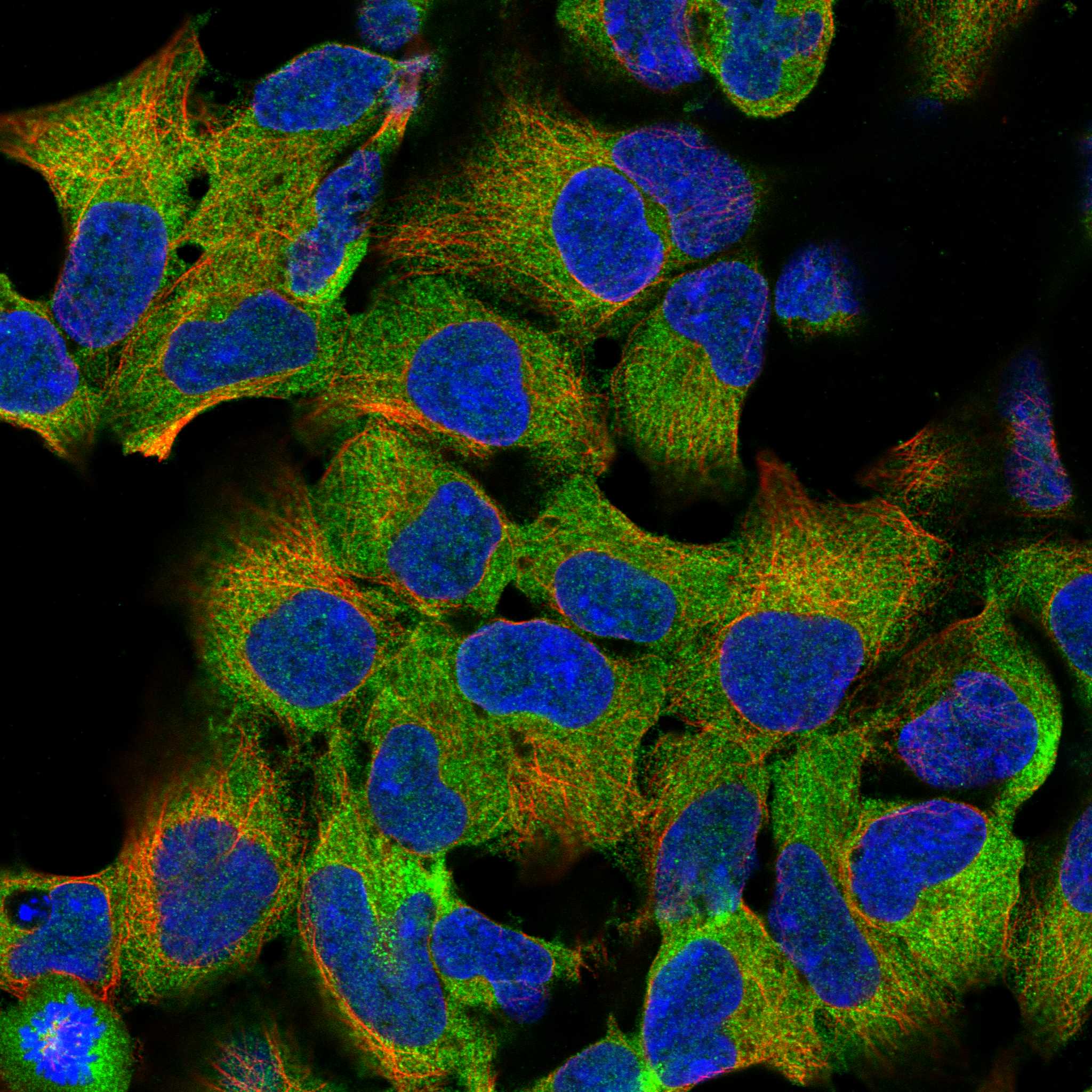
Comprehensive Human Protein Atlas IHC scoring per tissue (reliability: Supported — Medium consistency between antibody staining and RNA expression data.). Rows are taken directly from the HPA tissue chart — click any row's HPA link to view the source.
| Tissue | Cell type | Level | Evidence | Source |
|---|---|---|---|---|
| Bone marrow | Hematopoietic cells | High | Protein (IHC) | HPA → |
| Cerebellum | Purkinje cells | High | Protein (IHC) | HPA → |
| Spleen | Cells in red pulp | High | Protein (IHC) | HPA → |
| Testis | Elongated or late spermatids | High | Protein (IHC) | HPA → |
| Adrenal gland | Glandular cells | Medium | Protein (IHC) | HPA → |
| Tissue | Cell type | Level | Evidence | Source |
|---|---|---|---|---|
| Adipose tissue | Adipocytes | Not detected | Protein (IHC) | HPA → |
Use the catalog antibody’s paraffin-section result as the starting point, then assess PDPK1 staining by cell type and compartment.
Anti-PDPK1 IHC images cover human kidney, lung cancer and placenta, mouse intestine, and rat testis (catalog IHC image alts); a PC-12 IF image is available (M01159 IF image alt).
PB9735 is shown in paraffin sections of mouse intestine, rat testis, and human lung cancer, and in a frozen human placenta section (PB9735 IHC image alts). M01159 is shown in a paraffin section of human kidney and in PC-12 cells by IF (M01159 image alts).
Which to pick: For tissue IHC, PB9735 has paraffin-section images across mouse, rat, and human samples; its captions specify citrate retrieval, 1 μg/ml primary antibody, and DAB detection (PB9735 IHC image alts). For IF/ICC, choose M01159: it is a rabbit monoclonal, lists IF/ICC, and has a PC-12 IF image (M01159 catalog and IF image alt). For cross-species work, both list human, mouse, and rat reactivity (catalog); M01159 also has a human kidney paraffin-section IHC image (M01159 IHC image alt), and neither antibody’s paraffin-section caption reports the fixative (catalog IHC image alts).