PELP1 / Proline-, glutamic acid- and leucine-rich protein 1 · IHC design guide

Design Immunohistochemistry for PELP1

Use this guide to assess PELP1 nuclear staining in paraffin sections (HPA tissue IHC). Breast glandular cells show high staining (HPA tissue IHC); A02530 has an IHC dilution range of 1:50–1:100 (datasheet: A02530), and some tumors show cytoplasmic localization (UniProt).

Evidence assembled Oct 2026 · For research use; verify linked source records and product datasheet before use
Immunohistochemistry protocol sheet for PELP1 (IHC for PELP1): expected localisation Ubiquitous nuclear staining across tissues (HPA tissue IHC), antibody M02530, validated IHC image, and IHC protocol steps
Printable PELP1 IHC protocol sheet — expected localisation Ubiquitous nuclear staining across tissues (HPA tissue IHC), antibody M02530, controls and protocol steps. Open the full PELP1 IHC guide →

PELP1 Immunohistochemistry Experimental Design Guide

Expected localisation, validated protocols, controls and antibodies — the at-a-glance facts below, then the full design guide.

Must know before staining
Expected localisation Ubiquitous nuclear staining across tissues (HPA tissue IHC)
Staining pattern High nuclear staining in breast glandular cells (HPA tissue IHC)
Antigen retrieval Tris-EDTA pH 9.0 HIER, 95–98 °C, 20 min (rule: nuclear antigen)
Positive control ⓘ Adrenal gland+4 more · see all
Negative control ⓘ Adipose tissue
Important caveats
Reasons your staining may differ from the expected pattern.
Fixation Keep fixation consistent across compared paraffin sections. (selected-SKU IHC image M02530)
Caveat Some tumors show predominantly cytoplasmic PELP1 (UniProt)
Regulation Widely expressed; regulation unreported (UniProt)
Isoform / epitope No annotated isoforms, signal peptide or propeptide (UniProt)
Section 1

Recommended PELP1 IHC & IF Protocols

The catalog antibody’s IHC-P protocol accompanies four published PELP1 workflows for medulloblastoma, paraspinal muscle, glioma, and lung adenocarcinoma (PMC7021561; PMC4821488; PMC7147719; PMC8262236).

Recommended immunohistochemistry (IHC-P) protocol parameters
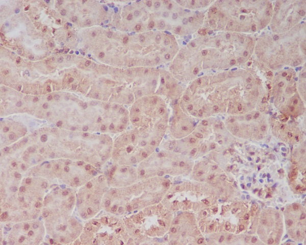
SampleParaffin-embedded mouse kidney tissue; fixative not specified (datasheet M02530)
FixationImage fixative and duration unreported (datasheet M02530); verify before use.
Sectioning4–5 µm sections on charged slides (standard)
DeparaffinisationXylene, graded ethanol series to water (standard)
Antigen retrievalHeat-induced epitope retrieval in Tris-EDTA buffer, pH 9.0, 20 min at 95–98 °C (standard rule: nuclear antigen)
Peroxidase block3% H2O2, 10 min, room temperature (standard)
Blocking10% normal serum of the secondary host, 30 min, room temperature (standard)
Primary antibodyRabbit monoclonal (clone COD-16) anti-PELP1, 1:50 (datasheet M02530)
Primary incubationOvernight at 4 °C (standard)
DetectionHRP-polymer secondary, DAB chromogen 5–10 min (standard)
CounterstainHematoxylin, blue, dehydrate and mount (standard)
Expected resultPELP1-positive staining in glandular cells of adrenal gland (HPA tissue IHC: High). HPA tissue profile: Ubiquitous nuclear expression. No signal in the no-primary control.
💡Decision noteStart with Tris-EDTA pH 9.0 HIER at 95–98 °C for 20 min (page retrieval rule); citrate pH 6.0 is a published muscle-section alternative (PMC4821488).
Section 2

What Is the Expected PELP1 Staining Pattern?

PELP1 is mainly nucleoplasmic, with nucleolar staining also expected; low cytoplasmic levels are reported (UniProt Q8IZL8; HPA subcellular). In paraffin tissue sections, expect predominantly nuclear staining across many cell types, including breast glandular cells and bone marrow hematopoietic cells reported as High (HPA tissue IHC). HPA describes the tissue pattern as ubiquitous nuclear expression with Supported reliability (HPA tissue IHC). PELP1 has no transmembrane segment (UniProt Q8IZL8 topology).

What am I looking at on my slide?
Distinct nuclear staining in breast glandular cells or bone marrow hematopoietic cells (HPA tissue IHC).This fits the reported High tissue staining and predominantly nuclear pattern (HPA tissue IHC). Assess staining within the identified cells, alongside section morphology, rather than judging the entire section by its overall darkness (standard IHC practice).
Strong, widespread cytoplasmic staining with little nuclear signal in an otherwise expected positive tissue (HPA tissue IHC).Check for an artefact before scoring this as the usual pattern: HPA reports ubiquitous nuclear expression (HPA tissue IHC). Cytoplasmic PELP1 can be biologically plausible in a subset of breast tumors and high-grade endometrial tumors (UniProt Q8IZL8); tissue context matters.
Staining chiefly in adipocytes while expected positive cells show little or none (HPA tissue IHC).Adipocytes are reported as Not detected, whereas several listed cell populations stain High (HPA tissue IHC). Consider antibody cross-reactivity or endogenous detection activity; this pattern alone cannot distinguish those causes (standard IHC practice).
Diffuse color over cells and surrounding tissue, with nuclear borders hard to resolve (standard IHC practice).Diffuse background prevents a reliable localisation call (standard IHC practice). Review detection and blocking controls, then compare staining in known positive cells with background in the same run (standard IHC practice). HPA's nuclear profile does not explain a particular run's background (HPA tissue IHC).
No visible nuclear staining in breast glandular cells or bone marrow hematopoietic cells (HPA tissue IHC).These are reported High and can serve as expected positive tissue comparators (HPA tissue IHC). A blank result warrants a run-level check of the antibody and detection steps before interpreting other tissues as negative (standard IHC practice).
💡Expected PELP1 appearanceCall an IHC result consistent with PELP1 when identifiable cells show predominantly nuclear staining, with clearly visible signal in a reported High population such as breast glandular cells (HPA tissue IHC); widespread staining of adipocytes without the expected nuclear signal is suspect (HPA tissue IHC; standard IHC practice).
How each factor affects the staining
Tissue and cell selection (HPA tissue IHC).Reported High populations include breast glandular, bronchial respiratory epithelial, cerebellar Purkinje, and bone marrow hematopoietic cells; hepatocytes are listed Low and adipocytes Not detected (HPA tissue IHC). Use these levels as tissue context, not a guarantee for every cell in a section (standard IHC practice).
Compartment and tumor context (UniProt Q8IZL8; HPA subcellular).HPA reports nucleoplasm and nucleoli in ICC-IF, while UniProt describes mainly nucleoplasmic PELP1 with low cytoplasmic levels (HPA subcellular; UniProt Q8IZL8). UniProt also reports predominantly cytoplasmic localisation in high-grade endometrial tumors; record compartment separately from intensity when scoring (UniProt Q8IZL8; standard IHC practice).
IHC antibody evidence (HPA antibodies; HPA tissue IHC).HPA060760 has Supported IHC status, and the tissue profile has Supported reliability based on staining and RNA consistency (HPA antibodies; HPA tissue IHC). Those summaries support the reported pattern but do not validate a different antibody or a particular experimental run (standard IHC practice).
Can IF/ICC show the same localisation? (HPA subcellular; HPA antibodies).Yes: HPA reports enhanced nucleoplasmic and nucleolar localisation in ICC-IF, with Enhanced ICC status for HPA053966 and HPA056028 (HPA subcellular; HPA antibodies). This is an IF/ICC interpretation cue, not an IHC protocol or proof that the IHC antibody has ICC validation (HPA antibodies).
Why is my staining missing, weak or wrong?
SituationLikely causeNext action
Expected positive cells have no staining (HPA tissue IHC).A run failure is possible when a reported High population is blank (HPA tissue IHC; standard IHC practice).Check a known positive section from the same run, then verify primary antibody application, detection reagents and counterstain visibility (standard IHC practice). Do not assign PELP1 absence from one failed control (standard IHC practice).
Signal is mainly cytoplasmic in normal tissue expected to show nuclear staining (HPA tissue IHC).Background or misplaced staining is possible; the usual tissue profile is nuclear (HPA tissue IHC; standard IHC practice).Check morphology and background controls, then repeat or compare with an independently validated antibody if available (standard IHC practice). Record tumor type before dismissing cytoplasmic signal, given UniProt's tumor-localisation reports (UniProt Q8IZL8).
Adipocytes stain strongly but nearby expected positive cells do not (HPA tissue IHC).Cross-reactivity or endogenous detection activity is plausible; adipocytes are reported Not detected (HPA tissue IHC; standard IHC practice).Use an appropriate negative detection control and compare staining with a reported High cell population on the same run (standard IHC practice; HPA tissue IHC). A control result is needed to narrow the cause (standard IHC practice).
Diffuse background obscures nuclei (standard IHC practice).Excess detection signal or inadequate blocking may reduce contrast (standard IHC practice).Target-specific fixation effects are not established by the supplied assay evidence. Verify with a matched IHC source before attributing a result to fixation.
Hepatocytes look weaker than breast glandular cells (HPA tissue IHC).That contrast agrees with reported Low hepatocyte and High breast glandular staining (HPA tissue IHC).Score each identified cell population against its reported level and compartment; do not use breast intensity as a universal threshold (HPA tissue IHC; standard IHC practice).
Nucleolar staining is hard to distinguish in chromogenic IHC (HPA subcellular).HPA's nucleolar localisation comes from ICC-IF; chromogenic section morphology may limit compartment resolution (HPA subcellular; standard IHC practice).First assess the dominant nuclear pattern in identifiable cells (HPA tissue IHC). Use the separate IF/ICC evidence when a nucleolar localisation call is essential (HPA subcellular; standard IHC practice).

Sample controls for PELP1 IHC & IF

🧪Run adrenal gland first: its glandular cells should stain for PELP1 (HPA: High in adrenal gland glandular cells). Run adipose tissue as the negative comparator (HPA: Not detected in adipocytes); on the adrenal slide, background and morphologically identifiable cells without specific staining should remain clear, without assuming that every nonglandular cell is PELP1-negative (UniProt: widely expressed).
Positive control tissue: Adrenal gland (Glandular cells, HPA High)
Negative control tissue: Adipose tissue (HPA Not detected)
ICC-IF cell lines (HPA subcellular resource): HPA ICC-IF images show PELP1 in CACO-2, MCF-7, U2OS, NIH 3T3, with annotated localisation: Nucleoplasm (enhanced), Nucleoli (enhanced) (HPA subcellular).
Technical controls: Include no-primary (secondary-only) and isotype controls matched to the primary antibody’s host species and immunoglobulin class, plus PELP1-knockout material as a biological negative where available (standard IHC practice). For chromogenic adrenal IHC, block endogenous peroxidase and check background with the no-primary control; assess endogenous biotin if using avidin–biotin detection (standard IHC practice).
⚠️Feasibility: A target-specific fixation window and retrieval dependency are unreported in the supplied evidence; optimize antigen retrieval with the catalog antibody’s IHC-P protocol (supplied application evidence). The M02530 mouse-kidney paraffin-section caption does not state a fixative, so its fixative is unreported (M02530 caption). The evidence does not establish whether frozen sections or IF are easier; adrenal background should be checked against the negative controls before scoring chromogenic staining (standard IHC practice).

HPA tissue IHC evidence for PELP1

Comprehensive Human Protein Atlas IHC scoring per tissue (reliability: Supported — High consistency between antibody staining and RNA expression data.). Rows are taken directly from the HPA tissue chart — click any row's HPA link to view the source.

Positive expression · recommended positive controls

TissueCell typeLevelEvidenceSource
Adrenal gland Glandular cells High Protein (IHC) HPA →
Appendix Glandular cells High Protein (IHC) HPA →
Bone marrow Hematopoietic cells High Protein (IHC) HPA →
Breast Glandular cells High Protein (IHC) HPA →
Bronchus Respiratory epithelial cells High Protein (IHC) HPA →

Undetected expression · recommended negative controls

TissueCell typeLevelEvidenceSource
Adipose tissue Adipocytes Not detected Protein (IHC) HPA →
Section 3

Advanced PELP1 IHC Tips

Troubleshoot PELP1 staining in paraffin sections by checking retrieval, nuclear localisation, background and compartment-specific scoring before interpreting cytoplasmic signal.

How should I retrieve PELP1 in paraffin sections when nuclear staining is weak?
Use Tris-EDTA at pH 9.0 for heat-induced retrieval at 95–98 °C for 20 min (page retrieval rule: nuclear antigen). After cooling, compare staining in a consistently processed positive section while keeping antibody and detection conditions fixed (standard IHC practice). Score nucleoplasmic and nucleolar signal separately, because both are expected locations (HPA subcellular: nucleoplasm and nucleoli), and inspect whether stronger retrieval also raises diffuse background (standard IHC practice). If staining remains weak, test an alternative retrieval buffer or duration on adjacent sections as a documented fallback, then select the condition that preserves morphology and improves specific nuclear contrast (standard IHC practice).
Could fixation explain weak or uneven PELP1 staining?
Target-specific sensitivity of PELP1 to fixation is unknown from the supplied evidence (fixation evidence: not supplied). The selected antibody image shows staining in paraffin-embedded mouse kidney, but its caption does not state a fixative (M02530 tissue-IHC caption). Record fixative, fixation duration and processing history for each specimen, and compare adjacent, similarly processed sections before changing retrieval (standard IHC practice). If weak staining tracks with processing batches, hold retrieval at Tris-EDTA pH 9.0, 95–98 °C for 20 min while checking morphology and nuclear contrast (page retrieval rule: nuclear antigen; standard IHC practice). Do not infer fixation sensitivity from the protein's localisation or modifications (UniProt Q8IZL8; fixation evidence: not supplied).
Should PELP1 staining be nuclear, nucleolar or cytoplasmic?
Expect prominent nucleoplasmic and nucleolar staining in many cells (HPA subcellular: enhanced nucleoplasm and nucleoli; HPA tissue IHC: ubiquitous nuclear expression). Low cytoplasmic signal is also described, and predominantly cytoplasmic localisation has been reported in subsets of breast tumors and high-grade endometrial tumors (UniProt Q8IZL8 localisation). Inspect nuclear and cytoplasmic compartments separately at the same exposure or chromogen development endpoint, using a counterstain to define nuclei (standard IHC practice). Before calling a cytoplasmic shift, confirm preserved morphology, a reproducible cellular distribution across the section and low signal in the detection-only control (standard IHC practice).
How can epitope uncertainty affect PELP1 staining across specimens?
The supplied record lists 0 isoforms and no annotated transmembrane segment, but it does not identify the catalog antibody's epitope (UniProt Q8IZL8; antibody evidence: epitope not supplied). PELP1 has recorded modified residues, including phosphoserines at positions 13, 477 and 481, and phosphothreonine at 488 (UniProt Q8IZL8 modified residues). Those annotations alone do not show that modification changes this antibody's binding (antibody evidence: epitope not supplied). If specimens differ, compare adjacent sections under identical retrieval and detection conditions, then check the antibody's documented epitope before proposing an epitope-specific explanation (standard IHC practice).
How should I investigate PELP1 localisation with multiplex IF?
Use the separate IF/ICC workflow to compare PELP1 with a marker identifying the cell population under study, while scoring nuclear and cytoplasmic signal within the same cells (standard IF practice). Nucleoplasm and nucleoli are expected locations, and HPA lists ICC/IF images from CACO-2, MCF-7, U2OS and NIH 3T3 cells (HPA subcellular). Choose fluorophores after checking tissue autofluorescence and single-label controls, favouring a detection channel with clear signal above that background (standard IF practice). Because PELP1 has no transmembrane segment and is mainly nuclear, use controlled permeabilisation for access to intracellular epitopes, then verify that nuclear morphology remains intact (UniProt Q8IZL8 topology and localisation; standard IF practice).
What should I check when PELP1 chromogenic staining looks diffuse?
Compare the stained section with a detection-only control and inspect chromogen outside intact cells, since those patterns can reveal nonspecific detection or deposits (standard IHC practice). Include a peroxidase block before chromogenic detection and assess residual endogenous enzyme signal in the control (standard IHC practice). Check whether Tris-EDTA retrieval at pH 9.0, 95–98 °C for 20 min preserves morphology and clear nuclear contrast; excessive background after processing warrants a controlled condition comparison (page retrieval rule: nuclear antigen; standard IHC practice). Treat widespread nuclear staining cautiously as background evidence by itself, because PELP1 has a ubiquitous nuclear tissue profile (HPA tissue IHC: ubiquitous nuclear expression).
How should I score PELP1 when both nuclear and cytoplasmic signals appear? ⚠ ANSWER MARKED FOR VERIFICATION
Define the analysed cell population and compartment before scoring, then apply the same definition to every section (standard IHC practice). Record the percentage of positive cells and, where intensity is reproducible, a compartment-specific H-score using intensity categories 0–3 (standard IHC practice). Normalise positive counts to the number of evaluable cells in the defined population, or report positive-cell density per mm² of viable analysed tissue (standard IHC practice). Report nuclear and cytoplasmic results separately, because predominantly nuclear expression is the broad reference pattern while cytoplasmic predominance is described in particular tumor settings (HPA tissue IHC; UniProt Q8IZL8 localisation).
When is apparent cytoplasmic PELP1 staining convincing rather than artefactual?
First establish that the signal lies within intact, correctly identified cells rather than at section edges, necrotic areas or chromogen deposits (standard IHC practice). Compare those cells with internal nuclear staining: PELP1 is broadly nuclear, although cytoplasmic predominance is reported in subsets of breast tumors and high-grade endometrial tumors (HPA tissue IHC; UniProt Q8IZL8 localisation). Check a detection-only control for endogenous enzyme or detection signal, and repeat the assessment away from damaged regions (standard IHC practice). A reproducible compartment shift in viable cells supports a localisation observation; staining alone does not establish the underlying signalling mechanism (standard IHC interpretation; UniProt Q8IZL8 function).
Boster reagents

Best PELP1 / Proline-, glutamic acid- and leucine-rich protein 1 IHC Antibodies

Two anti-PELP1 antibodies list IHC and IF applications (catalog applications). M02530 lists Human reactivity; A02530 lists Human, Mouse and Rat reactivity (catalog reactivity).

Real IHC data Immunohistochemical analysis of paraffin-embedded mouse kidney, using PELP1 Antibody.
Anti-PELP1 Rabbit Monoclonal Antibody
Cat # M02530
Real IF data Immunofluorescence analysis of L929 cells using PELP1 Rabbit pAb at dilution of 100 . Blue: DAPI for nuclear staining.
Anti-PELP1 Antibody
Cat # A02530

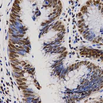
M02530 lists IHC and IF/ICC and shows IHC staining of paraffin-embedded mouse kidney (M02530 applications and image caption). A02530 lists IHC and IF/ICC and shows IF staining of L929 and U2OS cells (A02530 applications and IF image captions).

Which to pick: For tissue IHC, M02530 has the paraffin-section figure (M02530 image caption); its listed reactivity is Human (M02530 catalog reactivity), and the caption does not report a fixative (M02530 image caption). For IF/ICC, A02530 has cell IF figures and is a rabbit polyclonal (A02530 IF image captions and catalog description). For work across Human, Mouse and Rat, A02530 lists all three species, while the rabbit monoclonal M02530 lists Human reactivity (catalog reactivity, host and clone).

Each figure is that product's own IHC / IF validation image from its datasheet.

References

  1. UniProt Consortium. UniProt entry Q8IZL8 (PELP1_HUMAN, Proline-, glutamic acid- and leucine-rich protein 1).
  2. Human Protein Atlas. PELP1 tissue IHC expression (reliability: Supported).
  3. Human Protein Atlas. PELP1 subcellular location (ICC-IF): Localized to the nucleoplasm and nucleoli..
  4. Human Protein Atlas. PELP1 antibody validation summary (3 antibodies).
  5. PELP1 signaling contributes to medulloblastoma progression by regulating the NF-κB pathway. Molecular carcinogenesis 2020 — PMC7021561.
  6. Expression of Estrogen Receptor Coactivator Proline-, Glutamic Acid- and Leucine-Rich Protein 1 within Paraspinal Muscles in Adolescents with Idiopathic Scoliosis. PloS one 2016 — PMC4821488.
  7. PELP1 promotes glioblastoma progression by enhancing Wnt/β-catenin signaling. Neuro-oncology advances 2019 — PMC7147719.
  8. Overexpression of PELP1 in Lung Adenocarcinoma Promoted E(2) Induced Proliferation, Migration and Invasion of the Tumor Cells and Predicted a Worse Outcome of the Patients. Pathology oncology research : POR 2021 — PMC8262236.
  9. PubMed PMID:15489334 — UniProt-cited evidence.
  10. PubMed PMID:11481323 — UniProt-cited evidence.
  11. PubMed PMID:12415108 — UniProt-cited evidence.