This website uses cookies to ensure you get the best experience on our website.


- Table of Contents
Plan chromogenic PFKFB2 IHC in paraffin sections using cytoplasmic staining as the expected pattern (HPA tissue IHC). Compare test sections with tissue controls, and interpret results in light of the uncertain tissue staining record (HPA tissue IHC).

Expected localisation, validated protocols, controls and antibodies — the at-a-glance facts below, then the full design guide.
| Expected localisation | Cytoplasmic staining in most tissues (HPA tissue IHC) | |
| Staining pattern | Cytoplasmic staining in glandular and glial cells (HPA tissue IHC) | |
| Antigen retrieval | EDTA pH 8.0 HIER, heat-mediated (datasheet A05977) | |
| Positive control | Appendix+4 more · see all | |
| Negative control | Adipose tissue+4 more · see all |
| Fixation | Matched tissue-IHC evidence does not establish the fixation claim. Validate the specimen-specific method before use. (selected-SKU IHC image A05977) | |
| Caveat | Heart specificity contrasts with undetected cardiomyocytes (UniProt; HPA tissue IHC) | |
| Regulation | Staining intensity varies by tissue (HPA tissue IHC) | |
| Isoform / epitope | 2 isoforms; confirm epitope coverage (UniProt) |
The catalog antibody has an IHC-P protocol (datasheet A05977). One published PFKFB2 IHC protocol using paraffin-embedded GIST tissue is listed below (PMC10918489).
| Sample | Paraffin-embedded human cortical adenomas tissue; fixative not specified (datasheet A05977) |
| Fixation | Image fixative and duration unreported (datasheet A05977); verify before use. |
| Sectioning | 4–5 µm sections on charged slides (standard) |
| Deparaffinisation | Xylene, graded ethanol series to water (standard) |
| Antigen retrieval | Heat retrieval: EDTA pH 8.0 (datasheet A05977); 20 min, 95–100 °C (standard) |
| Peroxidase block | 3% H2O2, 10 min, room temperature (standard) |
| Blocking | 10% goat serum (datasheet A05977) |
| Primary antibody | Rabbit anti-PFKFB2, 2-5 μg/ml (datasheet A05977) |
| Primary incubation | Overnight at 4 °C (datasheet A05977) |
| Detection | Streptavidin-biotin complex (SABC), DAB chromogen (datasheet A05977) |
| Counterstain | Hematoxylin, blue, dehydrate and mount (standard) |
| Expected result | PFKFB2-positive staining in glandular cells of appendix (HPA tissue IHC: Medium). HPA tissue profile: Cytoplasmic expression in most tissues. No signal in the no-primary control. |
In paraffin-section IHC, expect mainly cytoplasmic staining in selected glandular and glial cells; HPA describes cytoplasmic expression across most tissues (HPA tissue IHC). Interpret this pattern cautiously: the tissue IHC reliability is Uncertain, pending external verification (HPA tissue IHC). UniProt assigns no subcellular location and reports no transmembrane segment (UniProt O60825 topology). ICC-IF reports an enhanced nucleoplasmic pattern, so assay context matters (HPA subcellular ICC-IF).
| Cytoplasmic staining in appendix or colon glandular cells, or cerebral cortex glial cells. | This fits the reported Medium cell-level IHC pattern (HPA tissue IHC). Judge the stained cells against adjacent architecture and controls; a matching pattern remains provisional because the tissue IHC assessment is Uncertain (HPA tissue IHC). |
| Predominantly nuclear staining on the chromogenic tissue slide, with little matching cytoplasmic signal. | Treat this as a compartment mismatch requiring an artefact check against controls and morphology (HPA tissue IHC; standard IHC practice). Nuclear signal is not automatically false: HPA reports enhanced nucleoplasmic staining by ICC-IF, a separate assay (HPA subcellular ICC-IF). |
| Strong signal in cardiomyocytes or adipocytes, or across cell types expected to be negative. | HPA reports PFKFB2 as Not detected in cardiomyocytes and adipocytes by tissue IHC (HPA tissue IHC). Check antibody cross-reactivity and endogenous detection activity before assigning such staining to PFKFB2 (standard IHC practice). |
| Uniform brown haze over cells, stroma, and the negative-control section. | A broadly shared background pattern cannot establish cell-specific PFKFB2 staining (standard IHC practice). Inspect blocking, washing, chromogen development, and the detection-only control; HPA's reported signal is assigned to particular cells (HPA tissue IHC). |
| No visible signal in appendix glandular cells or cerebral cortex glial cells. | These are reported Medium examples, so absence warrants a run check, including control performance and detection settings (HPA tissue IHC; standard IHC practice). They are reference examples rather than guaranteed positives because HPA rates tissue IHC Uncertain (HPA tissue IHC). |
| Tissue and cell selection | Appendix and colon glandular cells and cerebral cortex glial cells are Medium IHC examples; cardiomyocytes and adipocytes are Not detected examples (HPA tissue IHC). Compare like cell types. |
| Strength of IHC evidence | The tissue profile is marked Uncertain and pending external verification; antibody HPA049975 is also IHC Uncertain (HPA tissue IHC; HPA antibodies). Treat agreement as supportive, not definitive. |
| IHC versus ICC-IF localisation | Tissue IHC reports mainly cytoplasmic expression, whereas ICC-IF reports enhanced nucleoplasmic localisation (HPA tissue IHC; HPA subcellular ICC-IF). Interpret each assay with its own controls. |
| IF/ICC Q: What compartment should be checked? | A: Check the nucleoplasm in ICC-IF; HPA lists A-431, HeLa, U2OS, and SiHa images and rates the listed antibodies ICC Enhanced (HPA subcellular ICC-IF; HPA antibodies). This does not validate nuclear tissue IHC. |
| Molecular features | UniProt reports 2 isoforms, no transmembrane segment, no signal peptide, and no propeptide (UniProt O60825). Antibody epitope coverage is unspecified, so an isoform-specific staining prediction is unsupported. |
| Heart as a reference tissue | UniProt lists heart tissue specificity, while HPA reports cardiomyocytes Not detected by tissue IHC (UniProt O60825; HPA tissue IHC). Do not assume heart muscle will be an IHC positive control. |
| Situation | Likely cause | Next action |
|---|---|---|
| Expected glandular or glial cells are unstained. | A staining-run failure is possible; these cells show Medium signal in HPA examples, with Uncertain IHC reliability (HPA tissue IHC). | Review section integrity, detection-only and run controls, antibody dilution, and chromogen development (standard IHC practice). Avoid declaring biological absence from one run. |
| Nuclei dominate the tissue-section result. | That differs from the cytoplasmic tissue IHC profile, although nucleoplasm is reported in ICC-IF (HPA tissue IHC; HPA subcellular ICC-IF). | Check counterstain and detection-only controls, then assess whether cytoplasmic signal is independently present (standard IHC practice). Report the assay-specific discrepancy. |
| Cardiomyocytes stain strongly. | This conflicts with the HPA Not detected cardiomyocyte IHC result, despite UniProt's heart tissue-specificity entry (HPA tissue IHC; UniProt O60825). | Inspect no-primary and tissue controls for endogenous activity or nonspecific detection (standard IHC practice); do not use this signal alone to validate specificity. |
| Brown staining spreads across tissue and background. | Diffuse signal can arise from nonspecific detection or excess chromogen development (standard IHC practice); it obscures HPA's cell-level pattern (HPA tissue IHC). | Compare the no-primary control, review blocking and washes, and assess chromogen timing before scoring cells (standard IHC practice). |
| A reported negative cell type appears positive. | Cross-reactivity or endogenous detection activity is possible (standard IHC practice); HPA lists adipocytes and bone-marrow hematopoietic cells as Not detected (HPA tissue IHC). | Compare morphology, a detection-only control, and reported positive cell types on the same run before attributing the signal to PFKFB2 (standard IHC practice; HPA tissue IHC). |
| Replicate sections differ in staining intensity. | Variation in section handling or development can change apparent intensity (standard IHC practice); the HPA tissue IHC assessment remains Uncertain (HPA tissue IHC). | Run comparable sections with consistent retrieval, antibody dilution, and development, then score cell type and compartment alongside intensity (standard IHC practice). |


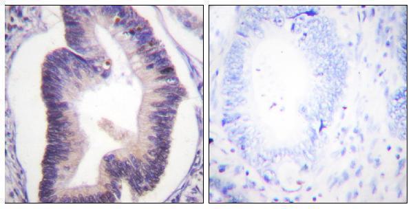
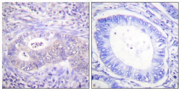


Comprehensive Human Protein Atlas IHC scoring per tissue (reliability: Uncertain — Pending external verification.). Rows are taken directly from the HPA tissue chart — click any row's HPA link to view the source.
| Tissue | Cell type | Level | Evidence | Source |
|---|---|---|---|---|
| Appendix | Glandular cells | Medium | Protein (IHC) | HPA → |
| Caudate | Glial cells | Medium | Protein (IHC) | HPA → |
| Cerebral cortex | Glial cells | Medium | Protein (IHC) | HPA → |
| Colon | Glandular cells | Medium | Protein (IHC) | HPA → |
| Duodenum | Glandular cells | Medium | Protein (IHC) | HPA → |
| Tissue | Cell type | Level | Evidence | Source |
|---|---|---|---|---|
| Adipose tissue | Adipocytes | Not detected | Protein (IHC) | HPA → |
| Bone marrow | Hematopoietic cells | Not detected | Protein (IHC) | HPA → |
| Bronchus | Respiratory epithelial cells | Not detected | Protein (IHC) | HPA → |
| Cerebellum | Cells in granular layer | Not detected | Protein (IHC) | HPA → |
| Cervix | Glandular cells | Not detected | Protein (IHC) | HPA → |
Troubleshoot PFKFB2 staining in paraffin sections by checking the documented retrieval condition, cellular pattern, controls, and scoring method.
Three antibodies have human paraffin-section IHC images (catalog IHC captions); A05977 also has HeLa-cell IF data (A05977 IF caption). All list Human, Mouse, and Rat reactivity (catalog).
A05977 has IHC images from human cortical adenoma, esophageal squamous carcinoma, liver cancer, and lung cancer paraffin sections, plus IF data in HeLa cells (A05977 image captions). A30440 has a human colon carcinoma paraffin-section IHC image; P30440 has one for phospho-Ser483 PFKFB2 in human colon carcinoma (A30440 and P30440 IHC captions).
Which to pick: For tissue IHC, choose A05977 when a documented retrieval and detection workflow is useful (A05977 IHC caption); A30440 covers total PFKFB2 and P30440 targets phospho-Ser483 in paraffin-embedded human colon carcinoma, with the fixative unreported for both (A30440 and P30440 IHC captions). For IF/ICC, A05977 has a HeLa-cell IF image, while A30440 and P30440 list IF/ICC applications without IF images in this payload (catalog applications and image captions). All three list Human, Mouse, and Rat reactivity, but their supplied IHC images use human tissue; A30440 and P30440 are described as polyclonal (catalog reactivity, dilution details, and IHC captions).