This website uses cookies to ensure you get the best experience on our website.


- Table of Contents
Plan PGM1 staining in paraffin sections using hepatocytes as a positive reference for cytoplasmic and nuclear signal (HPA tissue IHC). The guide covers fixation, staining interpretation, and the HPA warning that the antibody may detect protein from more than one gene (HPA tissue IHC).

Expected localisation, validated protocols, controls and antibodies — the at-a-glance facts below, then the full design guide.
| Expected localisation | Cytoplasm; hepatocytes also show nuclear staining (HPA tissue IHC) | |
| Staining pattern | Hepatocytes: cytoplasmic and nuclear staining (HPA tissue IHC) | |
| Antigen retrieval | Citrate pH 6 HIER, heat-mediated (datasheet M02318) | |
| Positive control | Liver+4 more · see all | |
| Negative control | Adipose tissue+4 more · see all |
| Fixation | Use formaldehyde-fixed paraffin sections (datasheet M02318 IHC-P) (selected-SKU IHC image M02318) | |
| Caveat | Staining may include protein from more than one gene (HPA tissue IHC) | |
| Regulation | RNA enriched: skeletal muscle, tongue (HPA tissue RNA) | |
| Isoform / epitope | 3 isoforms; epitope coverage is unspecified (UniProt) |
The catalog antibody’s IHC-P protocol is paired with published PGM1 staining protocols for melanoma and colorectal cancer tissue microarrays (PMC13483203; PMC9134613).
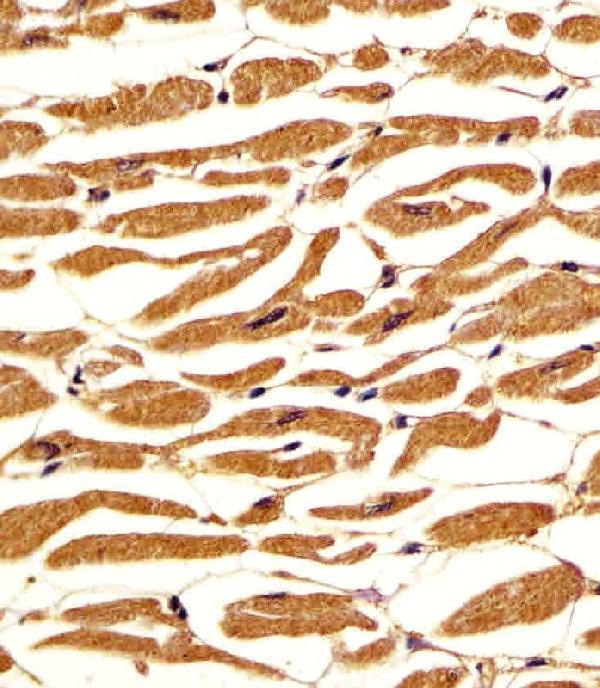
| Sample | Formaldehyde-fixed, paraffin-embedded human heart tissue (datasheet M02318) |
| Fixation | Image formalin-fixed; duration unreported (datasheet M02318); verify before use. |
| Sectioning | 4–5 µm sections on charged slides (standard) |
| Deparaffinisation | Xylene, graded ethanol series to water (standard) |
| Antigen retrieval | Heat retrieval: Citrate pH 6 (datasheet M02318); 20 min, 95–100 °C (standard) |
| Peroxidase block | 3% H2O2, 10 min, room temperature (standard) |
| Blocking | 10% normal serum of the secondary host, 30 min, room temperature (standard) |
| Primary antibody | Rabbit anti-PGM1, 1:25 (datasheet M02318) |
| Primary incubation | 1 hours at 37°C (datasheet M02318) |
| Detection | HRP-polymer secondary, DAB chromogen 5–10 min (standard) |
| Counterstain | Hematoxylin, blue, dehydrate and mount (standard) |
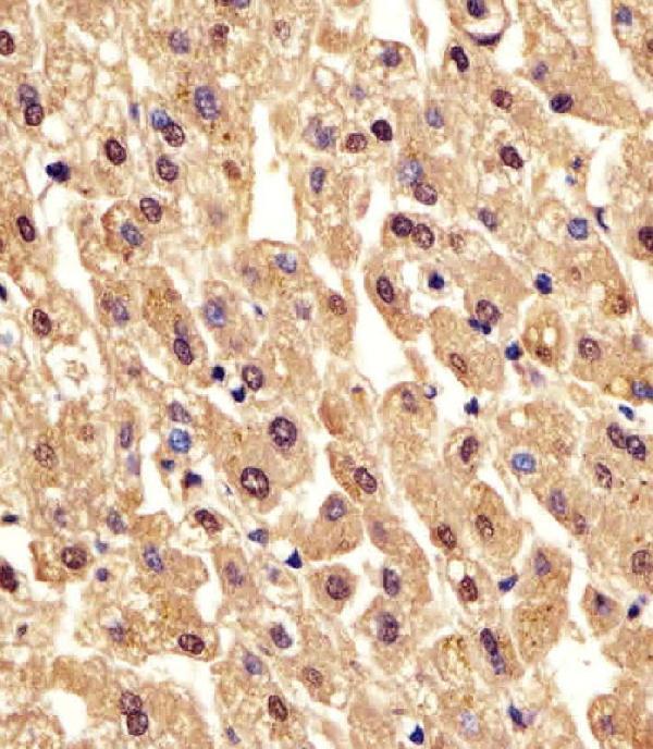
| Expected result | PGM1-positive staining in hepatocytes of liver (HPA tissue IHC: High). HPA tissue profile: Cytoplasmic expression in several different tissue types. Cytoplasmic and nuclear expression in hepatocytes. No signal in the no-primary control. |
PGM1 is a cytoplasmic protein with no transmembrane segment (UniProt P36871: cytoplasm; topology). In paraffin section IHC, expect cytoplasmic staining in hepatocytes and several other cell types, with cytoplasmic and nuclear staining possible in hepatocytes (HPA tissue IHC). HPA rates the tissue profile Enhanced but reports only medium consistency with RNA and cautions that its antibodies may target proteins from more than one gene (HPA tissue IHC).
| Strong hepatocyte staining, with cytoplasmic signal and some nuclear signal. | This fits the reported high hepatocyte expression and permitted nuclear component (HPA tissue IHC: liver, High; hepatocyte profile). Judge signal in identified hepatocytes, because an overall liver score can hide staining in other cells (standard IHC interpretation). |
| Predominantly cytoplasmic staining in adrenal or colon glandular cells, breast myoepithelial cells, kidney tubule cells, or hippocampal glia. | These named populations have Medium staining in the HPA tissue profile (HPA tissue IHC). A weaker result than liver can therefore be compatible with the reference pattern; compare the same cell type and compartment (HPA tissue IHC; standard IHC interpretation). |
| Predominantly nuclear staining outside hepatocytes, or staining confined to membranes. | These compartments differ from the reported cytoplasmic tissue pattern; HPA additionally permits nuclear staining in hepatocytes (HPA tissue IHC). Check morphology, counterstain, and controls before calling an unexpected compartment PGM1 (standard IHC interpretation). |
| Strong staining in HPA listed non-detected populations, or color over tissue edges and unrelated cells. | Adipocytes and lung alveolar cells, for example, are reported Not detected (HPA tissue IHC). Unexpected cell staining raises cross-reactivity; broad color may also reflect endogenous detection activity or nonspecific background (standard IHC interpretation). |
| No hepatocyte signal, including on a liver control section. | This conflicts with HPA High hepatocyte staining (HPA tissue IHC: liver). First assess control quality, retrieval, primary antibody use, and detection performance (standard IHC practice). One failed section cannot establish that PGM1 is absent from the specimen. |
| Cell type and tissue choice | Liver hepatocytes are a High reference; adrenal and colon glandular cells, breast myoepithelial cells, kidney tubule cells, and hippocampal glia are Medium (HPA tissue IHC). Match the cell population, not just the organ name. |
| Strength of the HPA reference | The tissue profile is Enhanced, yet RNA and staining show medium consistency and HPA cautions that antibodies may target proteins from more than one gene (HPA tissue IHC). Treat unexpected staining as provisional. |
| Antibody specific validation | HPA lists IHC Enhanced for HPA024190, HPA024637, and CAB004666, and IHC Approved for HPA046329 (HPA antibodies). Those ratings describe the listed antibodies; they do not establish validation for the catalog antibody. |
| Isoforms and processing | UniProt lists three isoforms, one chain spanning residues 1–562, and no signal peptide or propeptide (UniProt P36871). The supplied records do not identify the catalog antibody epitope or show isoform specific IHC staining. |
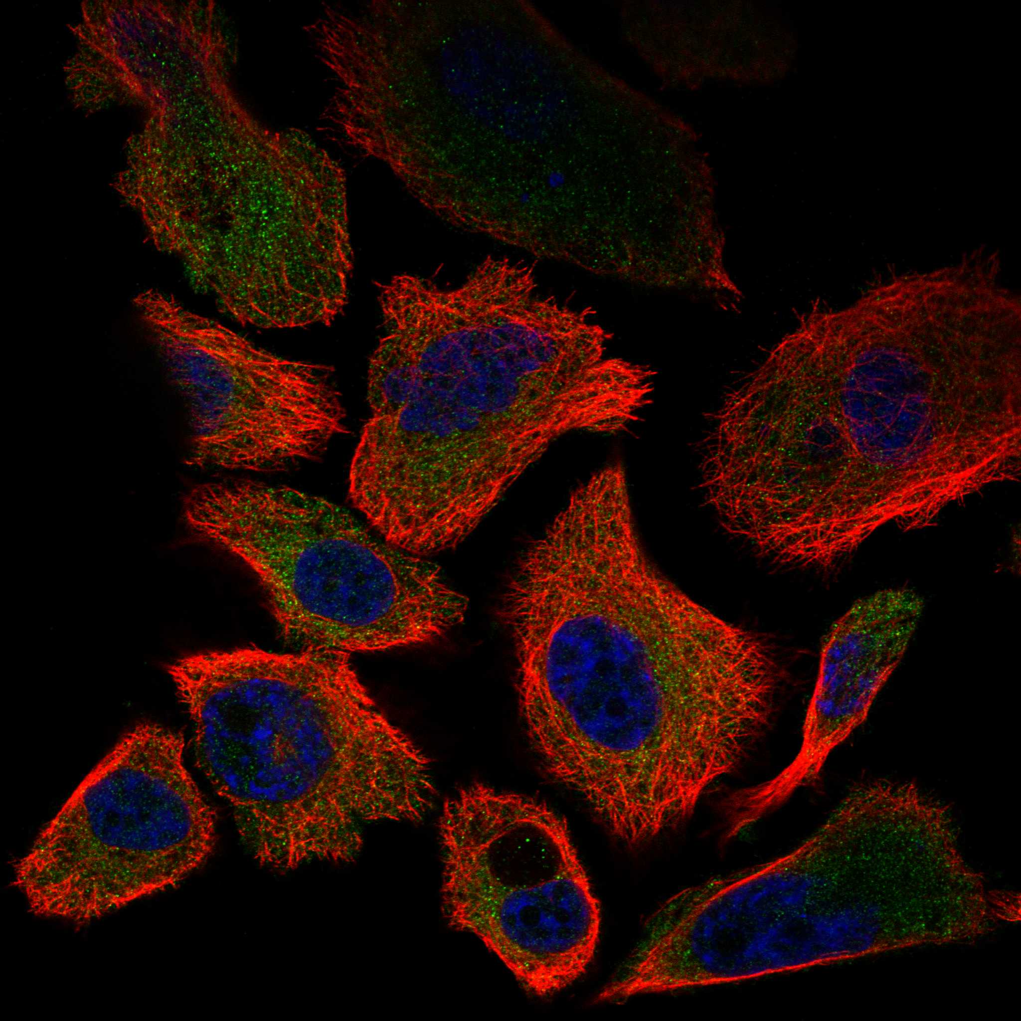
| IF/ICC: what pattern should appear? | Cytosolic staining is supported in HPA ICC-IF, with a caution that the antibodies may target proteins from multiple genes (HPA subcellular). This is an interpretation reference; IF/ICC belongs to its own guide page. |
| Situation | Likely cause | Next action |
|---|---|---|
| Liver control is blank. | A failed control or IHC workflow is possible when expected High hepatocyte staining is absent (HPA tissue IHC: liver; standard IHC practice). | Check tissue preservation, retrieval, primary antibody application, detection reagents, and counterstain against a concurrently processed positive control (standard IHC practice). Do not infer a PGM1 specific retrieval requirement from HPA. |
| All cells have pale, even chromogen. | Nonspecific background, residual endogenous enzyme activity, or excessive detection signal can obscure cell boundaries (standard chromogenic IHC practice). | Inspect a no primary control; review blocking, washes, detection chemistry, and development time (standard IHC practice). Interpret staining only after the control identifies the background source. |
| Adipocytes or lung alveolar cells stain strongly. | These cell populations are reported Not detected, so cross-reactivity or endogenous detection activity warrants investigation (HPA tissue IHC; standard IHC interpretation). | Compare neighboring positive cell types, examine a no primary control, and confirm cell identity on the counterstain (standard IHC practice). Do not use an organ wide negative label. |
| Signal appears confined to cell membranes. | PGM1 is cytoplasmic and lacks a transmembrane segment (UniProt P36871); the reported tissue pattern is cytoplasmic, with a hepatocyte nuclear component (HPA tissue IHC). | Recheck cellular boundaries and staining controls; score membrane only signal as discordant until independently verified (standard IHC interpretation). |
| Hepatocyte nuclei stain along with cytoplasm. | HPA explicitly reports cytoplasmic and nuclear hepatocyte expression (HPA tissue IHC). Nuclear color alone does not establish an artefact. | Confirm that the cytoplasmic hepatocyte pattern and controls are credible, then record nuclear staining separately (HPA tissue IHC; standard IHC scoring). |
| A Medium reference tissue looks weaker than liver. | HPA reports Medium staining for the named non-liver populations and High staining for hepatocytes (HPA tissue IHC). | Compare like cell populations and score intensity by compartment; avoid treating weaker staining alone as assay failure (HPA tissue IHC; standard IHC scoring). |



Comprehensive Human Protein Atlas IHC scoring per tissue (reliability: Enhanced — Medium consistency between antibody staining and RNA expression data. Caution, targets protein from more than one gene.). Rows are taken directly from the HPA tissue chart — click any row's HPA link to view the source.
| Tissue | Cell type | Level | Evidence | Source |
|---|---|---|---|---|
| Liver | Hepatocytes | High | Protein (IHC) | HPA → |
| Adrenal gland | Glandular cells | Medium | Protein (IHC) | HPA → |
| Breast | Myoepithelial cells | Medium | Protein (IHC) | HPA → |
| Colon | Glandular cells | Medium | Protein (IHC) | HPA → |
| Duodenum | Glandular cells | Medium | Protein (IHC) | HPA → |
| Tissue | Cell type | Level | Evidence | Source |
|---|---|---|---|---|
| Adipose tissue | Adipocytes | Not detected | Protein (IHC) | HPA → |
| Cerebellum | Cells in granular layer | Not detected | Protein (IHC) | HPA → |
| Cerebral cortex | Glial cells | Not detected | Protein (IHC) | HPA → |
| Endometrium | Cells in endometrial stroma | Not detected | Protein (IHC) | HPA → |
| Esophagus | Squamous epithelial cells | Not detected | Protein (IHC) | HPA → |
Troubleshoot PGM1 staining by checking retrieval, tissue handling, cellular localisation and controls before comparing chromogenic IHC results across sections.
Anti-PGM1 options include an IHC-P antibody with human heart and liver staining images and an IF/ICC antibody listed for human, mouse and rat (M02318 image captions; M02318-1 catalog: applications/reactivity).
The M02318 card shows staining in human heart paraffin sections; a second catalog caption describes human liver paraffin sections (M02318 image captions). M02318-1 is listed for IF/ICC in human, mouse and rat, but has no IF image in the payload (M02318-1 catalog: applications/reactivity; IF image list).
Which to pick: Choose M02318 for tissue IHC: this rabbit polyclonal is listed for IHC-P in human and mouse, with its own human heart and liver paraffin-section captions (M02318 catalog: applications/reactivity/clonality; M02318 image captions). Choose M02318-1 for IF/ICC or rat samples: this rabbit monoclonal is listed for IF/ICC in human, mouse and rat at 1:50–1:200, though no IF image is supplied (M02318-1 catalog: applications/reactivity/clonality/dilution; IF image list). For cross-species IHC, M02318 lists mouse reactivity, but its pictured tissue is human; its captions give conflicting fixative descriptions, paraformaldehyde and formaldehyde (M02318 catalog: reactivity; M02318 image captions).