This website uses cookies to ensure you get the best experience on our website.


- Table of Contents
Plan PHOX2A chromogenic IHC around high staining in adrenal medullary cells (HPA tissue IHC) and expected nuclear localisation (UniProt). Use the catalog antibody’s 2.5 μg/mL starting concentration (datasheet) and compare staining with HPA tissue controls (HPA tissue IHC).

Expected localisation, validated protocols, controls and antibodies — the at-a-glance facts below, then the full design guide.
| Expected localisation | Nuclear localisation is expected; tissue staining compartment is unconfirmed (UniProt) | |
| Staining pattern | High staining in adrenal medullary cells; compartment unreported (HPA tissue IHC) | |
| Antigen retrieval | Tris-EDTA pH 9.0 HIER, 95–98 °C, 20 min (rule: nuclear antigen) | |
| Positive control | Adrenal gland+1 more · see all | |
| Negative control | Adipose tissue+4 more · see all |
| Fixation | Keep fixation consistent across sections (standard IHC practice; not target-specific) | |
| Caveat | A section lacking adrenal medullary cells may miss the positive population (HPA tissue IHC) | |
| Regulation | Adrenal gland–enriched expression (HPA tissue RNA) | |
| Isoform / epitope | No annotated isoforms or processing; epitope effects unknown (UniProt) |
The catalog antibody provides the IHC-P protocol; the published PHOX2A immunohistochemistry protocol below uses mouse and human tissue with fluorescent detection.
| Sample | Tissue sections; selected-image fixative not specified (standard IHC workflow) |
| Fixation | Image fixative and duration unreported (datasheet A05780); verify before use. |
| Sectioning | 4–5 µm sections on charged slides (standard) |
| Deparaffinisation | Xylene, graded ethanol series to water (standard) |
| Antigen retrieval | Heat-induced epitope retrieval in Tris-EDTA buffer, pH 9.0, 20 min at 95–98 °C (standard rule: nuclear antigen) |
| Peroxidase block | 3% H2O2, 10 min, room temperature (standard) |
| Blocking | 10% normal serum of the secondary host, 30 min, room temperature (standard) |
| Primary antibody | Rabbit anti-PHOX2A, 2.5 μg/mL (datasheet A05780) |
| Primary incubation | Overnight at 4 °C (standard) |
| Detection | HRP-polymer secondary, DAB chromogen 5–10 min (standard) |
| Counterstain | Hematoxylin, blue, dehydrate and mount (standard) |
| Expected result | PHOX2A-positive staining in medullary cells of adrenal gland (HPA tissue IHC: High). HPA tissue profile: Expression in adrenal medulla. No signal in the no-primary control. |
PHOX2A is a nuclear protein with no transmembrane segment (UniProt O14813 localization and topology). In paraffin section IHC, expect staining in adrenal medullary cells, where HPA reports high expression, and potentially in preleptotene spermatocytes, where it reports medium expression (HPA tissue IHC). HPA rates the tissue pattern Enhanced because antibody staining agrees strongly with RNA expression (HPA tissue IHC reliability).
| Distinct nuclear staining in adrenal medullary cells; neighboring cells show little or no signal. | This matches the principal observed tissue pattern: high staining in medullary cells and adrenal enriched RNA expression (HPA tissue IHC). Judge the result by cell identity and nuclear placement together; staining somewhere in the adrenal gland alone is less informative (UniProt O14813 localization; HPA tissue IHC). |
| Nuclear staining in preleptotene spermatocytes is weaker than the adrenal medullary signal. | A medium testicular signal is compatible with the reported pattern (HPA tissue IHC: Medium in preleptotene spermatocytes; High in adrenal medullary cells). Do not require testis to match adrenal intensity, or treat all testicular cell types as established positives. |
| The strongest staining fills cytoplasm or outlines cell membranes while nuclei remain clear. | That distribution conflicts with the nuclear assignment and lack of a transmembrane segment (UniProt O14813 localization and topology). Consider nonspecific staining or a detection artefact, then reassess nuclear detail and controls before assigning PHOX2A positivity (general IHC practice). |
| Strong chromogen appears in cell types listed as undetected, or across many unrelated cells. | For example, HPA reports no detection in adipocytes of adipose tissue or hematopoietic cells of bone marrow (HPA tissue IHC). Broad staining there raises concern for cross-reactivity or endogenous detection activity; those HPA observations do not establish that every cell in either tissue must be negative. |
| No nuclear signal appears in identifiable adrenal medullary cells. | The result misses the strongest supplied positive reference (HPA tissue IHC: High in adrenal medullary cells). Check section identity, antibody and detection controls, and the validated staining conditions before interpreting the slide as PHOX2A negative (general IHC practice). |
| Cell identity and tissue context | Adrenal medullary cells are the strongest supplied tissue reference; preleptotene spermatocytes have medium staining (HPA tissue IHC). Use the named cells when comparing slides: the HPA levels describe observed cells, not a blanket score for every cell in those organs. |
| Antibody evidence for IHC | HPA rates the tissue IHC profile Enhanced and lists HPA078004 as IHC Enhanced (HPA tissue IHC; HPA antibodies). Those ratings support the reported pattern; they do not establish the performance of every other antibody or every staining condition. |
| Protein location and processing | PHOX2A is nuclear, with no transmembrane segment, signal peptide, propeptide, or annotated processed fragment (UniProt O14813). Read a discrete nuclear result as the expected compartment; the supplied record gives no basis for expecting a membrane or secreted staining pattern. |
| IF/ICC Q: where should fluorescence appear? | A: Mainly in the nucleoplasm (HPA subcellular ICC-IF: supported). HPA lists SH-SY5Y and U2OS images and marks HPA053810 and HPA065621 ICC Approved (HPA subcellular; HPA antibodies). This supports an IF localization comparison, not an IHC-P protocol. |
| Fixation and retrieval evidence | Target-specific fixation effects are not established by the supplied assay evidence. Verify with a matched IHC source before attributing a result to fixation. |
| Situation | Likely cause | Next action |
|---|---|---|
| Adrenal medulla has no visible nuclear staining. | A missed positive tissue pattern may reflect a staining or detection failure; HPA reports high medullary cell staining (HPA tissue IHC). | Confirm medullary cells are present, inspect a working positive control, and check primary antibody, retrieval, and detection steps against the IHC-validated antibody's instructions (general IHC practice). |
| Nuclei are clear but cytoplasm is strongly colored. | The compartment conflicts with PHOX2A's nuclear location (UniProt O14813). Nonspecific binding or detection background is possible (general IHC practice). | Compare with a primary-omission control; review blocking, antibody concentration, wash stringency, and counterstain visibility (general IHC practice). Require a cell-specific nuclear pattern for interpretation. |
| Many cell types show uniform chromogen, including cells expected to be undetected. | Widespread staining conflicts with the restricted HPA observations and may reflect cross-reactivity or endogenous detection activity (HPA tissue IHC; general IHC practice). | Inspect a primary-omission control and the detection-system blocking steps; compare the same run with adrenal medulla and an HPA-listed undetected cell type (general IHC practice; HPA tissue IHC). |
| All sections show diffuse haze that obscures nuclei. | Excess antibody or incomplete blocking or washing can raise background in chromogenic IHC (general IHC practice). The supplied sources do not define a PHOX2A-specific background mechanism. | Review the antibody's IHC-P dilution guidance, blocking and washes; adjust one workflow variable at a time while preserving the positive tissue control (general IHC practice). |
| Testis stains faintly while adrenal medulla stains strongly. | Different strengths can reflect the reported levels: medium in preleptotene spermatocytes and high in adrenal medullary cells (HPA tissue IHC). | Identify preleptotene spermatocytes before scoring. Compare nuclear signal with local background and the adrenal control; do not demand equal intensity across tissues (HPA tissue IHC; general IHC practice). |
| ICC-IF fluorescence looks nucleoplasmic, but IHC-P looks extranuclear. | The supported IF location is nucleoplasmic, while UniProt assigns PHOX2A to the nucleus (HPA subcellular ICC-IF; UniProt O14813). The assays and listed antibody validations differ (HPA antibodies). | Evaluate each assay with its own controls and antibody validation. For IHC-P, recheck cell identity, nuclear counterstain, and chromogenic background before accepting extranuclear signal (general IHC practice). |


Comprehensive Human Protein Atlas IHC scoring per tissue (reliability: Enhanced — High consistency between antibody staining and RNA expression data.). Rows are taken directly from the HPA tissue chart — click any row's HPA link to view the source.
| Tissue | Cell type | Level | Evidence | Source |
|---|---|---|---|---|
| Adrenal gland | Medullary cells | High | Protein (IHC) | HPA → |
| Testis | Preleptotene spermatocytes | Medium | Protein (IHC) | HPA → |
| Tissue | Cell type | Level | Evidence | Source |
|---|---|---|---|---|
| Adipose tissue | Adipocytes | Not detected | Protein (IHC) | HPA → |
| Appendix | Glandular cells | Not detected | Protein (IHC) | HPA → |
| Bone marrow | Hematopoietic cells | Not detected | Protein (IHC) | HPA → |
| Breast | Adipocytes | Not detected | Protein (IHC) | HPA → |
| Bronchus | Respiratory epithelial cells | Not detected | Protein (IHC) | HPA → |
Troubleshoot PHOX2A staining in chromogenic paraffin section IHC using nuclear localisation, documented tissue patterns, and matched controls.
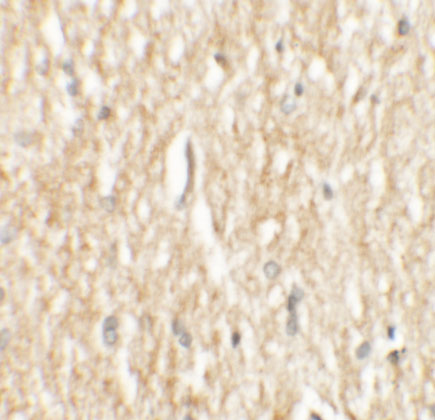
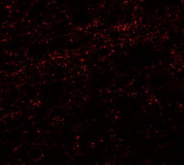
A05780 has human brain IHC and IF images (catalog: image captions) and lists Human, Mouse and Rat reactivity (catalog: reactivity).
A05780 is listed for IHC-P and IF (catalog: applications). Its images show IHC and IF in human brain tissue (catalog: IHC and IF image captions).
Which to pick: For paraffin-section IHC, choose A05780: IHC-P is listed (catalog: applications), and its human brain IHC image uses 2.5 μg/mL (catalog: IHC image caption); the fixative is unreported (catalog: IHC image caption). For IF, A05780 has a human brain image at 20 μg/mL (catalog: IF image caption); ICC validation and clonality are unreported (catalog: applications; catalog: clone). For cross-species planning, A05780 lists Human, Mouse and Rat reactivity (catalog: reactivity), while its supplied IHC and IF images show human brain tissue (catalog: IHC and IF image captions).