This website uses cookies to ensure you get the best experience on our website.


- Table of Contents
Plan PI4KB paraffin IHC around cytoplasmic staining in most tissues (HPA tissue IHC). The catalog antibody has an IHC dilution of 2–5 μg/ml (datasheet A04249-2); compare staining across relevant cell types and tissues (HPA tissue IHC).

Expected localisation, validated protocols, controls and antibodies — the at-a-glance facts below, then the full design guide.
| Expected localisation | Cytoplasmic in most tissues (HPA tissue IHC) | |
| Staining pattern | Adipocytes and glandular cells: cytoplasmic staining (HPA tissue IHC) | |
| Antigen retrieval | EDTA pH 8.0 HIER, heat-mediated (datasheet A04249-2) | |
| Positive control | Adipose tissue+4 more · see all | |
| Negative control | Cervix+4 more · see all |
| Fixation | Keep fixation consistent across paraffin sections. (standard IHC practice; not target-specific) | |
| Caveat | Ovarian follicle cells undetected despite high ovarian expression (HPA tissue IHC; UniProt) | |
| Regulation | No intensity regulator specified (UniProt) | |
| Isoform / epitope | 3 isoforms; epitope coverage unknown (UniProt) |
The catalog antibody protocol specifies EDTA pH 8.0 heat retrieval (datasheet A04249-2). One published PI4KB IHC protocol qualifies for a parameter table (PMC12134136).
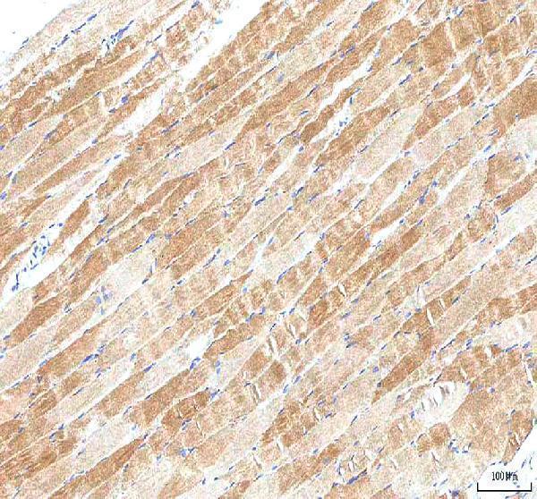
| Sample | Paraffin-embedded human skeletal muscle tissue; fixative not specified (datasheet A04249-2) |
| Fixation | Image fixative and duration unreported (datasheet A04249-2); verify before use. |
| Sectioning | 4–5 µm sections on charged slides (standard) |
| Deparaffinisation | Xylene, graded ethanol series to water (standard) |
| Antigen retrieval | Heat retrieval: EDTA pH 8.0 (datasheet A04249-2); 20 min, 95–100 °C (standard) |
| Peroxidase block | 3% H2O2, 10 min, room temperature (standard) |
| Blocking | 10% goat serum (datasheet A04249-2) |
| Primary antibody | Rabbit anti-PI4KB, 2-5 μg/ml (datasheet A04249-2) |
| Primary incubation | Overnight at 4 °C (datasheet A04249-2) |
| Detection | HRP-conjugated secondary, DAB chromogen (datasheet A04249-2) |
| Counterstain | Hematoxylin, blue, dehydrate and mount (standard) |
| Expected result | PI4KB-positive staining in adipocytes of adipose tissue (HPA tissue IHC: Medium). HPA tissue profile: Cytoplasmic expression in most tissues. No signal in the no-primary control. |
PI4KB should appear mainly cytoplasmic in tissue sections, with a perinuclear or Golgi emphasis possible because it associates with endomembranes despite having no transmembrane segment (HPA tissue IHC; UniProt Q9UBF8 topology and subcellular location). Look for staining in the specific cell populations documented by HPA, such as adipocytes, neurons and glandular cells. The tissue IHC profile is Approved, with medium consistency between staining and RNA data (HPA tissue IHC).
| Cytoplasmic staining in adipocytes, neuronal cells or glandular cells; sometimes concentrated near the nucleus. | This fits the reported tissue profile and PI4KB's Golgi and perinuclear localisation (HPA tissue IHC; UniProt Q9UBF8 subcellular location). HPA lists medium staining in adipose adipocytes, cortical neurons and duodenal glandular cells; compare the matching cell population, not the entire section (HPA tissue IHC). |
| Predominantly nuclear staining, or a crisp plasma membrane rim without appreciable cytoplasmic staining. | These compartments do not match the reported cytoplasmic tissue profile or supported Golgi localisation (HPA tissue IHC; HPA subcellular ICC-IF). Treat the pattern as suspect; review morphology and controls before scoring it as PI4KB. Membrane association alone does not predict a cell surface rim (UniProt Q9UBF8 subcellular location). |
| Strong staining in a cell population HPA lists as not detected, such as esophageal squamous cells or endometrial stromal cells. | Possible explanations include antibody cross-reactivity or endogenous detection activity; staining alone cannot distinguish them (general IHC practice). HPA's finding is specific to the named cells and should not be extended to every cell in that tissue (HPA tissue IHC). |
| Weak, widespread colour across cells and empty spaces, obscuring intracellular boundaries. | This is more consistent with diffuse assay background than the reported cytoplasmic cellular pattern (HPA tissue IHC; general IHC practice). Examine a no-primary control and the distribution of signal relative to cell outlines before interpreting intensity (general IHC practice). |
| No visible signal in a documented medium-staining cell population, such as adipose adipocytes or cerebellar Purkinje cells. | The result does not reproduce that HPA observation, but one negative section does not establish absent protein (HPA tissue IHC). Verify that the intended cells are present, then check assay controls and detection performance (general IHC practice). HPA rates tissue IHC consistency with RNA as medium (HPA tissue IHC). |
| Cell population and tissue choice | HPA reports medium staining in selected adipocytes, neurons and glandular cells, low staining in skeletal myocytes and kidney tubule cells, and no detection in specific other populations (HPA tissue IHC). Score the identified cells against their own reference level; a low-staining population is a weak positive comparator. |
| Compartment and topology | UniProt places PI4KB at the Golgi, rough ER and mitochondrial outer membrane and in the perinuclear cytoplasm, while reporting no transmembrane segment (UniProt Q9UBF8 topology and subcellular location). HPA describes tissue staining broadly as cytoplasmic and supports Golgi localisation by ICC-IF (HPA tissue IHC; HPA subcellular ICC-IF). |
| Differences between source summaries | UniProt describes broad expression with high levels in ovary, yet HPA reports no detection in ovarian follicle cells (UniProt Q9UBF8 tissue specificity; HPA tissue IHC). These statements cover different kinds of evidence and cellular scope; do not convert whole-tissue expression into an expected positive follicle-cell stain. |
| Antibody evidence | The three listed antibodies have Approved IHC status; only CAB022071 also has Supported ICC status (HPA antibody validation). Approved tissue IHC carries medium staining–RNA consistency in this record (HPA tissue IHC). Interpret unusual staining with controls rather than treating the status as proof that every stained cell is specific. |
| Isoforms and epitope coverage | UniProt lists three PI4KB isoforms and a processed chain spanning residues 2–816 (UniProt Q9UBF8 isoforms and processing). No antibody epitope or isoform coverage is supplied, so this record cannot predict whether the catalog antibody distinguishes isoforms or whether a particular tissue result reflects epitope selection. |
| Situation | Likely cause | Next action |
|---|---|---|
| Known HPA medium-staining cells remain blank. | The observed result conflicts with the selected HPA reference population; the source does not establish a PI4KB-specific cause (HPA tissue IHC). | Confirm the reference cells are in the section and review positive, no-primary and detection controls before changing general IHC conditions (general IHC practice). |
| Only nuclei or cell borders stain. | The pattern conflicts with cytoplasmic tissue staining and supported Golgi localisation (HPA tissue IHC; HPA subcellular ICC-IF). | Recheck cell boundaries and counterstain, then compare with a no-primary control and an independently validated antibody where available (general IHC practice). |
| A population listed as not detected stains strongly. | Cross-reactivity or endogenous detection activity is possible; the HPA call applies to the specified cells (HPA tissue IHC; general IHC practice). | Identify the stained cell type precisely and compare no-primary and detection controls before assigning PI4KB positivity (general IHC practice). |
| Colour is diffuse across tissue and blank spaces. | Noncellular or widespread background prevents a reliable match to HPA's cytoplasmic pattern (HPA tissue IHC; general IHC practice). | Inspect a no-primary control and reassess blocking, washes and chromogen development as general IHC workflow checks (general IHC practice). |
| Ovarian follicle cells are negative despite UniProt's high ovarian expression summary. | HPA specifically lists follicle cells as not detected; UniProt's tissue-wide statement does not specify those cells (HPA tissue IHC; UniProt Q9UBF8 tissue specificity). | Report the cell-specific negative result and use an HPA-listed medium-staining population for a positive comparator (HPA tissue IHC). |
| Q: Should IF/ICC show the same diffuse cytoplasmic appearance as IHC? | A: HPA supports Golgi localisation in ICC-IF, while its tissue IHC summary describes cytoplasmic staining in most tissues (HPA subcellular ICC-IF; HPA tissue IHC). | For IF/ICC, assess whether signal concentrates at the Golgi; use the separate IF/ICC guide for assay setup (HPA subcellular ICC-IF). |
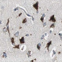
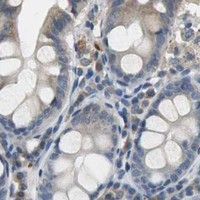
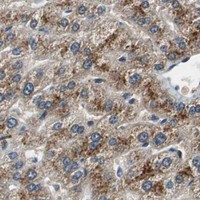
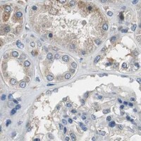
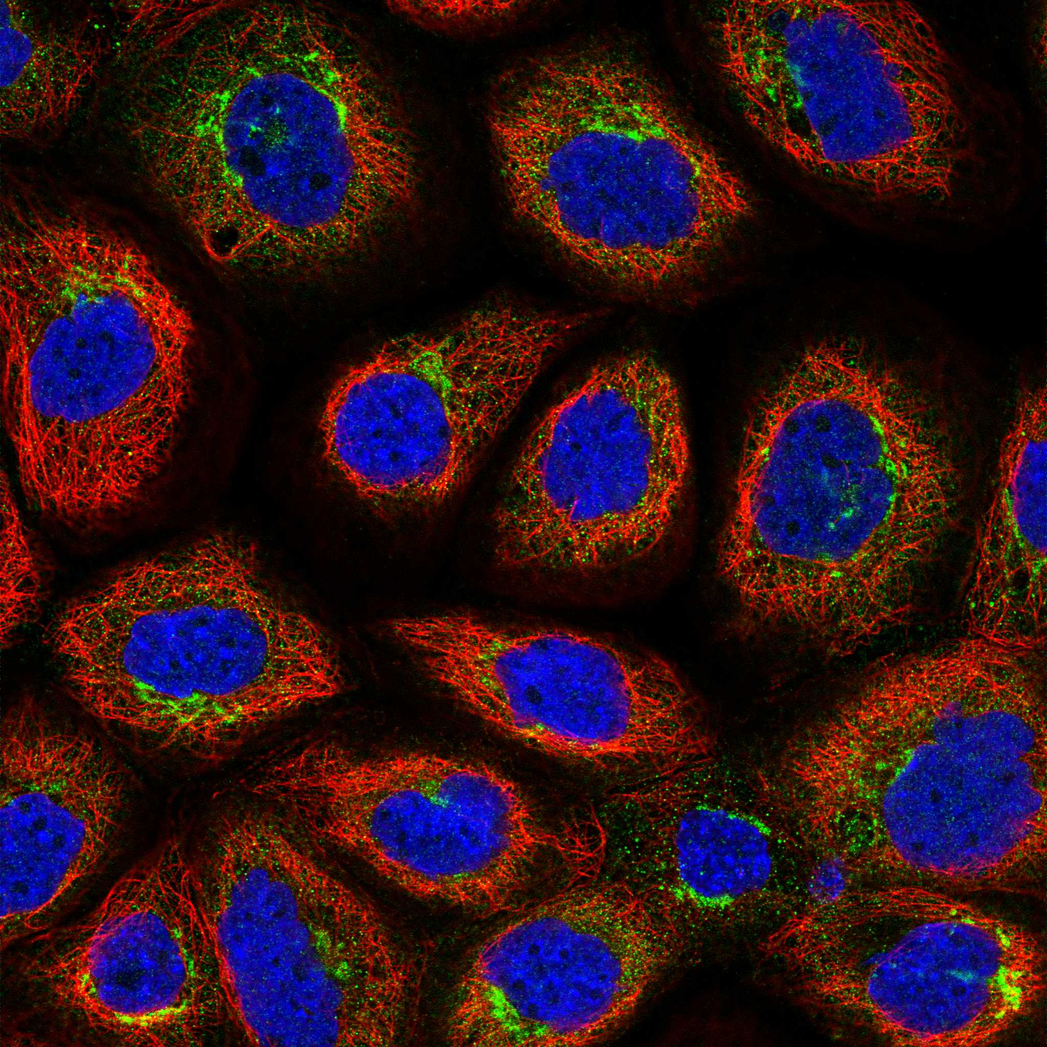
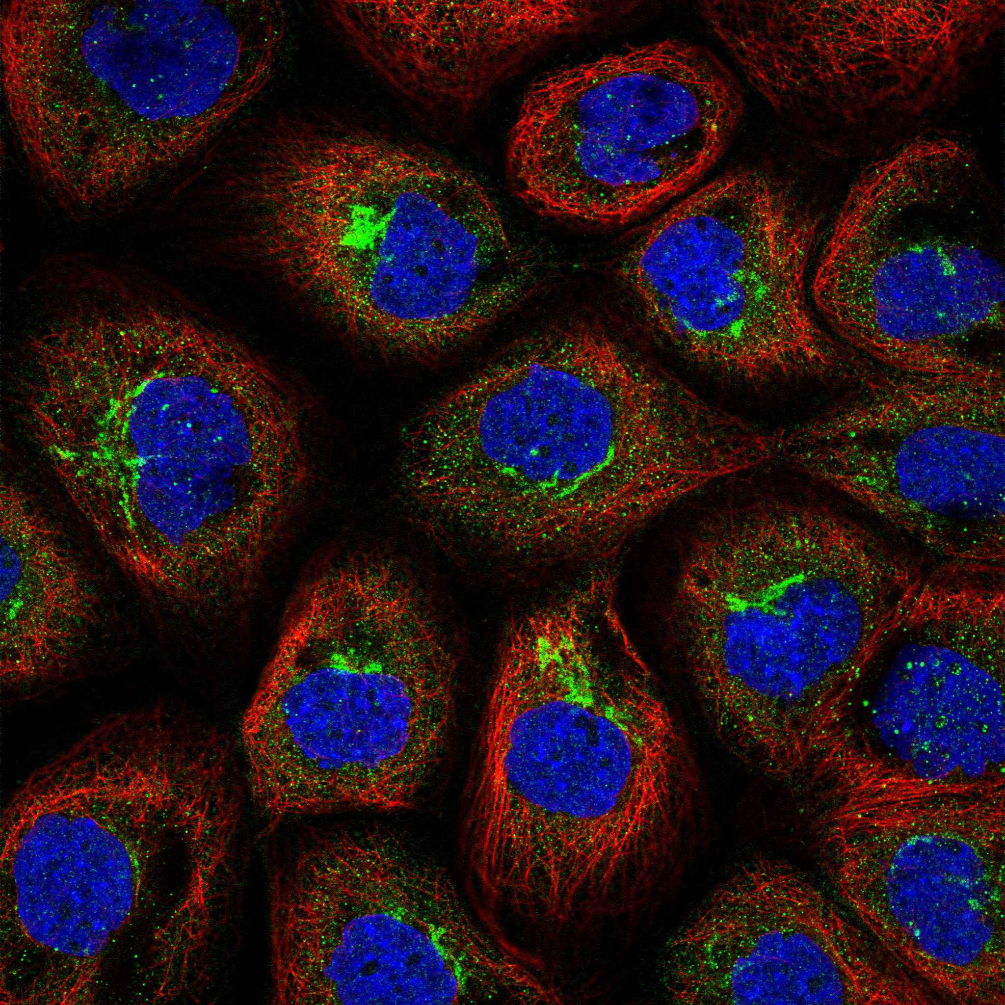
Comprehensive Human Protein Atlas IHC scoring per tissue (reliability: Approved — Medium consistency between antibody staining and RNA expression data.). Rows are taken directly from the HPA tissue chart — click any row's HPA link to view the source.
| Tissue | Cell type | Level | Evidence | Source |
|---|---|---|---|---|
| Adipose tissue | Adipocytes | Medium | Protein (IHC) | HPA → |
| Adrenal gland | Glandular cells | Medium | Protein (IHC) | HPA → |
| Breast | Adipocytes | Medium | Protein (IHC) | HPA → |
| Caudate | Neuronal cells | Medium | Protein (IHC) | HPA → |
| Cerebellum | Purkinje cells | Medium | Protein (IHC) | HPA → |
| Tissue | Cell type | Level | Evidence | Source |
|---|---|---|---|---|
| Cervix | Glandular cells | Not detected | Protein (IHC) | HPA → |
| Endometrium | Cells in endometrial stroma | Not detected | Protein (IHC) | HPA → |
| Esophagus | Squamous epithelial cells | Not detected | Protein (IHC) | HPA → |
| Oral mucosa | Squamous epithelial cells | Not detected | Protein (IHC) | HPA → |
| Ovary | Follicle cells | Not detected | Protein (IHC) | HPA → |
Troubleshoot PI4KB staining by checking retrieval, cellular distribution and controls against the catalog antibody’s tissue-IHC example and independent expression evidence.
A04249-2 has real IHC data from human paraffin-embedded skeletal muscle (image caption: A04249-2). Its listed reactivity covers human, mouse and rat (catalog: A04249-2 reactivity).
A04249-2 will render with its own IHC figure from human paraffin-embedded skeletal muscle (image caption: A04249-2). The catalog lists IHC as an application and human, mouse and rat reactivity; the figure documents human tissue only (catalog: A04249-2 applications/reactivity; image caption: A04249-2).
Which to pick: Choose A04249-2 for paraffin-section tissue IHC: its caption documents EDTA retrieval at pH 8.0, 10% goat-serum blocking, 2 μg/ml primary antibody overnight at 4°C, and HRP/DAB detection in human skeletal muscle (image caption: A04249-2). No IF/ICC application or IF figure is listed, so this payload supports no IF/ICC-validated choice (catalog: A04249-2 applications/IF images). For mouse or rat, A04249-2 has listed reactivity but only a human IHC figure; its host is rabbit, clonality is unreported, and the paraffin-section fixative is unreported (catalog: A04249-2 reactivity/host/clone; image caption: A04249-2).