Click image to see more details
Product Info Summary
| SKU: | A04249-2 |
|---|---|
| Size: | 100 µg/vial |
| Reactive Species: | Human, Mouse, Rat |
| Host: | Rabbit |
| Application: | ELISA, Flow Cytometry, IHC, WB |
Customers Who Bought This Also Bought

Product info
Product Name
Anti-PI4KB Antibody Picoband®
SKU/Catalog Number
A04249-2
Size
100 µg/vial
Form
Lyophilized
Description
Boster Bio Anti-PI4KB Antibody Picoband® catalog # A04249-2. Tested in ELISA, IHC, WB, Flow Cytometry applications. This antibody reacts with Human, Mouse, Rat. The brand Picoband indicates this is a premium antibody that guarantees superior quality, high affinity, and strong signals with minimal background in Western blot applications. Only our best-performing antibodies are designated as Picoband, ensuring unmatched performance.
Storage & Handling
At -20°C for one year from date of receipt. After reconstitution, at 4°C for one month. It can also be aliquotted and stored frozen at -20°C for six months. Avoid repeated freezing and thawing.
Cite This Product
Anti-PI4KB Antibody Picoband® (Boster Biological Technology, Pleasanton CA, USA, Catalog # A04249-2)
Host
Rabbit
Contents
Each vial contains 4 mg Trehalose, 0.9 mg NaCl, 0.2 mg Na2HPO4.
Clonality
Polyclonal
Isotype
IgG
Immunogen
E.coli-derived human PI4KB recombinant protein (Position: D375-D671). Human PI4KB shares 100% and 99.7% amino acid (aa) sequence identity with mouse and rat PI4KB, respectively
Cross-reactivity
No cross reactivity with other proteins.
Reactive Species
A04249-2 is reactive to PI4KB in Human, Mouse, Rat
Observed Molecular Weight
95 kDa
Calculated molecular weight
91.4 kDa
Background of PI4KB
Phosphatidylinositol 4-kinase beta is an enzyme that in humans is encoded by the PI4KB gene. Inositol phospholipids have an important role in intracellular signaling in response to hormones, growth factors and neurotransmitters. Phosphatidylinositol 4-kinase phosphorylates phosphatidylinositol (PI) to phosphatidylinositol-4-phosphate (PIP). In a second step, PIP is further phosphorylated to phosphatidylinositol-4,5-bisphosphate (PIP2), and PIP2 is subsequently hydrolyzed by phospholipase C, producing the two intracellular second messengers, inositol 1,4,5-triphosphate (IP3) and diacylglycerol (DAG). PI4K230, PI4K92 and PI4K55 are three PI4 kinase isoforms that have been characterized and classified according to their molecular weights of 230, 92 and 55 kD. Previously, PI4 kinases were classified into type II and III enzymes. All isoforms are located on distinct membranes and cellular compartments suggesting various tasks. PI4K230 is located at the endoplasmatic reticulum and outer membranes of mitochondria, PI4K92 at the Golgi apparatus and endoplasmatic reticulum, and PI4K55 at the plasma membrane and endosomes. PI4K230 is predominantly expressed in brain and moderately sensitive to wortmannin as well as specifically and irreversibly inhibited by cyclitol derivatives.
Antibody Validation
Boster validates all antibodies on WB, IHC, ICC, Immunofluorescence, and ELISA with known positive control and negative samples to ensure specificity and high affinity, including thorough antibody incubations.
Application & Images
Applications
A04249-2 is guaranteed for ELISA, Flow Cytometry, IHC, WB Boster Guarantee
Recommend Dilution
| Application | Dilution | Species |
|---|---|---|
| Western blot | 0.25-0.5 μg/ml | Human, Mouse, Rat |
| Immunohistochemistry(Paraffin-embedded Section) | 2-5 μg/ml | Human |
| Flow Cytometry (Fixed) | 1-3 μg/1x106 cells | Human |
| ELISA | 0.1-0.5 μg/ml | - |
Tested application
Suggested blocking solution with 5% non-fat milk or BSA; (*)Recommended protein loading: 20-40 µg per lane
Use TE buffer pH 9.0 for antigen retrieval; (*) citrate buffer pH 6.0 is an alternative.
Validation Images & Assay Conditions
Click image to see more details
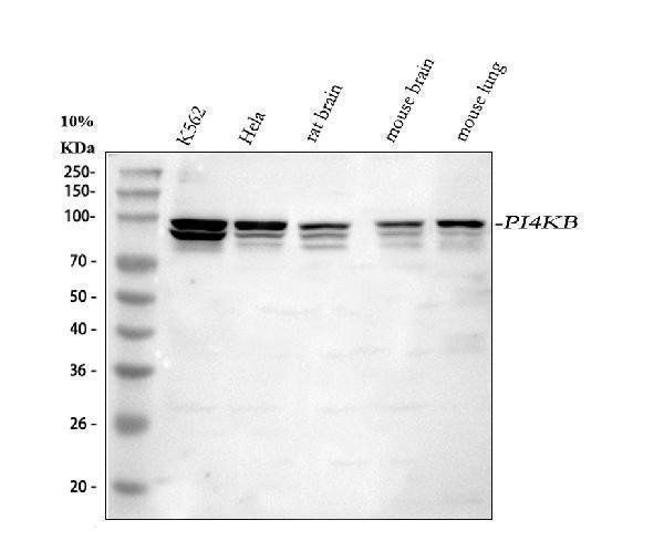
Western blot analysis of PI4KB using anti-PI4KB antibody (A04249-2).
Electrophoresis was performed on a 5-20% SDS-PAGE gel at 70V (Stacking gel) / 90V (Resolving gel) for 2-3 hours. The sample well of each lane was loaded with 30 ug of sample under reducing conditions.
Lane 1: human K562 whole cell lysates,
Lane 2: human Hela whole cell lysates,
Lane 3: rat brain tissue lysates,
Lane 4: mouse brain tissue lysates,
Lane 5: mouse lung tissue lysates.
After electrophoresis, proteins were transferred to a nitrocellulose membrane at 150 mA for 50-90 minutes. Blocked the membrane with 5% non-fat milk/TBS for 1.5 hour at RT. The membrane was incubated with rabbit anti-PI4KB antigen affinity purified polyclonal antibody (Catalog # A04249-2) at 0.5 μg/mL overnight at 4°C, then washed with TBS-0.1%Tween 3 times with 5 minutes each and probed with a goat anti-rabbit IgG-HRP secondary antibody at a dilution of 1:5000 for 1.5 hour at RT. The signal is developed using an Enhanced Chemiluminescent detection (ECL) kit (Catalog # EK1002) with Tanon 5200 system. A specific band was detected for PI4KB at approximately 95 kDa. The expected band size for PI4KB is at 91,91-110 kDa.

Click image to see more details
IHC analysis of PI4KB using anti-PI4KB antibody (A04249-2).
PI4KB was detected in a paraffin-embedded section of human skeletal muscle tissue. Heat mediated antigen retrieval was performed in EDTA buffer (pH 8.0, epitope retrieval solution). The tissue section was blocked with 10% goat serum. The tissue section was then incubated with 2 μg/ml rabbit anti-PI4KB Antibody (A04249-2) overnight at 4°C. Peroxidase Conjugated Goat Anti-rabbit IgG was used as secondary antibody and incubated for 30 minutes at 37°C. The tissue section was developed using HRP Conjugated Rabbit IgG Super Vision Assay Kit (Catalog # SV0002) with DAB as the chromogen.

Click image to see more details
Flow Cytometry analysis of JK cells using anti-PI4KB antibody (A04249-2).
Overlay histogram showing JK cells stained with A04249-2 (Blue line). To facilitate intracellular staining, cells were fixed with 4% paraformaldehyde and permeabilized with permeabilization buffer. The cells were blocked with 10% normal goat serum. And then incubated with rabbit anti-PI4KB Antibody (A04249-2, 1 μg/1x106 cells) for 30 min at 20°C. DyLight®488 conjugated goat anti-rabbit IgG (BA1127, 5-10 μg/1x106 cells) was used as secondary antibody for 30 minutes at 20°C. Isotype control antibody (Green line) was rabbit IgG (1 μg/1x106) used under the same conditions. Unlabelled sample (Red line) was also used as a control.

Click image to see more details
Flow Cytometry analysis of U251 cells using anti-PI4KB antibody (A04249-2).
Overlay histogram showing U251 cells stained with A04249-2 (Blue line). To facilitate intracellular staining, cells were fixed with 4% paraformaldehyde and permeabilized with permeabilization buffer. The cells were blocked with 10% normal goat serum. And then incubated with rabbit anti-PI4KB Antibody (A04249-2, 1 μg/1x106 cells) for 30 min at 20°C. DyLight®488 conjugated goat anti-rabbit IgG (BA1127, 5-10 μg/1x106 cells) was used as secondary antibody for 30 minutes at 20°C. Isotype control antibody (Green line) was rabbit IgG (1 μg/1x106) used under the same conditions. Unlabelled sample (Red line) was also used as a control.
Specific Publications For Anti-PI4KB Antibody Picoband® (A04249-2)
Loading publications
Recommended Resources
Here are featured tools and databases that you might find useful.
- Boster's Pathways Library
- Protein Databases
- Bioscience Research Protocol Resources
- Data Processing & Analysis Software
- Photo Editing Software
- Scientific Literature Resources
- Research Paper Management Tools
- Molecular Biology Software
- Primer Design Tools
- Bioinformatics Tools
- Phylogenetic Tree Analysis
Customer Reviews
Have you used Anti-PI4KB Antibody Picoband®?
Share your experimental results or join a short interview to earn up to $1,000 in product credits or other rewards.
0 Reviews For Anti-PI4KB Antibody Picoband®
Customer Q&As
Have a question?
Find answers in Q&As, reviews.
Can't find your answer?
Submit your question
