This website uses cookies to ensure you get the best experience on our website.


- Table of Contents
Expect nuclear PIAS1 staining in paraffin-section IHC (HPA tissue IHC). This guide covers fixation, controls and scoring, including the PIAS2/3 cross-reactivity of M01707 (datasheet M01707).

Expected localisation, validated protocols, controls and antibodies — the at-a-glance facts below, then the full design guide.
| Expected localisation | Ubiquitous nuclear tissue staining (HPA tissue IHC) | |
| Staining pattern | Widespread nuclear signal across tissue cell types (HPA tissue IHC) | |
| Antigen retrieval | EDTA pH 8.0 HIER, heat-mediated (datasheet M01707) | |
| Positive control | Adipose tissue+4 more · see all | |
| Negative control | None in HPA (detected in all 45 tissues); use no-primary + isotype controls |
| Fixation | Keep fixation consistent across sections (standard IHC practice; not target-specific) | |
| Caveat | M01707 also detects PIAS2/3; signal is not PIAS1-specific (datasheet M01707) | |
| Regulation | IFNA1-linked binding; intensity unknown (UniProt) | |
| Isoform / epitope | 3 isoforms; epitope coverage is unresolved (UniProt) |
The catalog antibody’s IHC-P protocol uses EDTA retrieval (datasheet: pH 8.0). These published PIAS1 protocols provide three chromogenic IHC examples (PMC4322998; PMC10184048; PMC5777733).
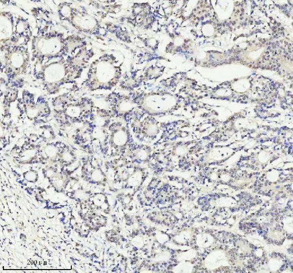
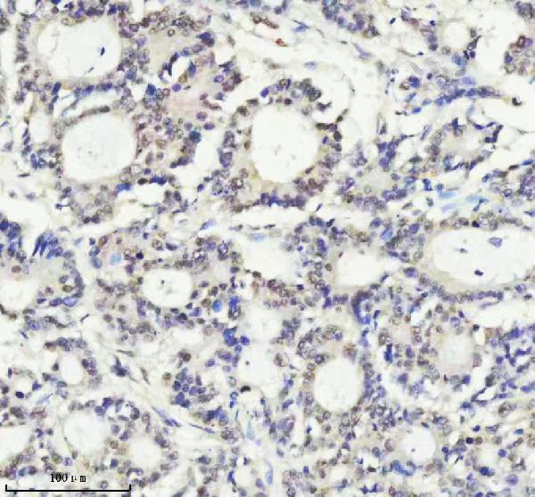
| Sample | Paraffin-embedded human colon adenocarcinoma tissue; fixative not specified (datasheet M01707) |
| Fixation | Image fixative and duration unreported (datasheet M01707); verify before use. |
| Sectioning | 4–5 µm sections on charged slides (standard) |
| Deparaffinisation | Xylene, graded ethanol series to water (standard) |
| Antigen retrieval | Heat retrieval: EDTA pH 8.0 (datasheet M01707); 20 min, 95–100 °C (standard) |
| Peroxidase block | 3% H2O2, 10 min, room temperature (standard) |
| Blocking | 10% goat serum (datasheet M01707) |
| Primary antibody | Rabbit monoclonal (clone ABFA-16) anti-PIAS1, 1:50 (datasheet M01707) |
| Primary incubation | Overnight at 4 °C (datasheet M01707) |
| Detection | HRP-conjugated secondary, DAB chromogen (datasheet M01707) |
| Counterstain | Hematoxylin, blue, dehydrate and mount (standard) |
| Expected result | PIAS1-positive staining in adipocytes of adipose tissue (HPA tissue IHC: High). HPA tissue profile: Ubiquitous nuclear expression. No signal in the no-primary control. |
PIAS1 should appear mainly in nuclei across many cell types in paraffin sections; HPA describes ubiquitous nuclear expression with Supported tissue IHC reliability, meaning staining is consistent with RNA expression (HPA tissue IHC). High staining is reported in adipocytes, glandular cells, hematopoietic cells and selected brain cells (HPA tissue IHC). PIAS1 has no transmembrane segment, so a membrane pattern is unexpected (UniProt O75925 topology).
| Nuclear chromogen in adipocytes, glandular cells or hematopoietic cells, with limited background. | This fits the reported ubiquitous nuclear pattern and High staining in these cell types (HPA tissue IHC). Judge each cell's nucleus against its surrounding cytoplasm and nearby unstained areas; intensity can vary across cells. A counterstain helps identify nuclei, while the chromogen shows where antibody-associated signal appears (general IHC practice). |
| Strong membrane-only or broadly cytoplasmic staining with little nuclear signal. | Treat this as a localisation mismatch: PIAS1 is nuclear, including nuclear speckles and PML bodies, and lacks a transmembrane segment (UniProt O75925). HPA tissue IHC reports a ubiquitous nuclear pattern. UniProt also lists conditional cytoskeletal redistribution, so limited extranuclear signal alone does not establish an artefact (UniProt O75925 subcellular location). |
| Strong signal in cells expected to stain weakly, especially smooth muscle cells. | HPA reports Low staining in smooth muscle cells and lists no negative tissue (HPA tissue IHC). Disproportionately strong signal there warrants a specificity check, including cross-reactivity; widespread chromogen outside cellular structures can also reflect endogenous detection activity (general IHC practice). Do not classify smooth muscle as a definitive negative control. |
| Diffuse haze across tissue and blank spaces, obscuring nuclear boundaries. | This does not match the nuclear distribution reported for PIAS1 (HPA tissue IHC). Possible general IHC causes include insufficient blocking, concentrated primary antibody, inadequate washing or endogenous enzyme activity. Compare a no-primary control and inspect whether the haze persists without primary antibody before assigning it to PIAS1 (general IHC practice). |
| No nuclear signal in a section containing a documented High-staining cell population. | Adipocytes and bone-marrow hematopoietic cells are among HPA's High examples (HPA tissue IHC). An absent signal there makes the run inconclusive; check tissue identity, primary-antibody application, retrieval and detection controls (general IHC practice). HPA's tissue pattern does not identify a PIAS1-specific fixation failure. |
| Nuclear localisation and topology | PIAS1 is nuclear, including speckles and PML bodies, with no transmembrane segment (UniProt O75925). Score nuclear signal first; cytoplasmic chromogen alone cannot confirm the expected tissue IHC pattern (HPA tissue IHC). |
| Cell population and comparator | HPA reports High staining in several named cell types, Low staining in smooth muscle cells, and no negative tissues (HPA tissue IHC). Use identified High populations as positive comparators; interpret a weak smooth-muscle result as relative, not absolute absence. |
| Antibody and application evidence | CAB012304 has Supported IHC status; HPA073610 has no listed IHC status in the supplied antibody record (HPA antibodies). These labels describe antibody-specific evidence and do not transfer validation from one antibody to another. |
| IF/ICC: should cytosolic signal be expected? | HPA reports mainly nucleoplasmic PIAS1 with additional cytosolic localisation, but marks both ICC-IF locations uncertain (HPA subcellular ICC-IF). That observation can guide interpretation of an IF image; it does not replace the nuclear expectation for tissue IHC. |
| Situation | Likely cause | Next action |
|---|---|---|
| High-population nuclei remain blank. | The run may have failed at tissue selection, primary-antibody application or detection; HPA reports High staining in adipocytes and hematopoietic cells (HPA tissue IHC). | Verify the cell population on the counterstained section, then check the run's positive control, reagent sequence and chromogen development (general IHC practice). |
| Only membranous staining appears. | A membrane-only pattern conflicts with PIAS1's lack of a transmembrane segment and its nuclear localisation (UniProt O75925). | Review morphology and the no-primary control, then repeat with an IHC-validated antibody if available; CAB012304 carries Supported IHC status in this record (HPA antibodies; general IHC practice). |
| Strong chromogen appears in smooth muscle. | This exceeds HPA's Low smooth-muscle observation and may reflect cross-reactivity or detection background (HPA tissue IHC; general IHC practice). | Compare nearby nuclear staining with a documented High cell population and inspect a no-primary control. A Low HPA call is a relative comparator, not proof that every smooth-muscle nucleus must be blank (HPA tissue IHC). |
| Diffuse staining persists across cells and empty areas. | Nonlocalized signal is inconsistent with the expected nuclear pattern (HPA tissue IHC); residual detection activity or nonspecific reagent binding is possible (general IHC practice). | Check the no-primary control, blocking and wash steps; adjust the primary concentration only after locating the background source (general IHC practice). |
| Nuclear detail is obscured despite visible chromogen. | Heavy chromogen or counterstain can conceal whether signal is truly nuclear (general IHC practice). | Review a lighter development or counterstain condition and compare cell boundaries with an adjacent section; call localisation only where nuclei remain identifiable (general IHC practice). |
| A proposed fix assumes PIAS1 is fixation-sensitive. | Neither the supplied HPA tissue IHC pattern nor UniProt topology establishes a target-specific fixation effect (HPA tissue IHC; UniProt O75925). | Record the fixation and retrieval conditions used, then compare controlled runs if optimisation is needed (general IHC practice). Describe any effect as observed in that workflow rather than established PIAS1 biology. |




Comprehensive Human Protein Atlas IHC scoring per tissue (reliability: Supported — High consistency between antibody staining and RNA expression data.). Rows are taken directly from the HPA tissue chart — click any row's HPA link to view the source.
| Tissue | Cell type | Level | Evidence | Source |
|---|---|---|---|---|
| Adipose tissue | Adipocytes | High | Protein (IHC) | HPA → |
| Adrenal gland | Glandular cells | High | Protein (IHC) | HPA → |
| Appendix | Glandular cells | High | Protein (IHC) | HPA → |
| Bone marrow | Hematopoietic cells | High | Protein (IHC) | HPA → |
| Breast | Adipocytes | High | Protein (IHC) | HPA → |
| Tissue | Cell type | Level | Evidence | Source |
|---|---|---|---|---|
| None in HPA: PIAS1 is detected in all 45 scored tissues. Use a no-primary (secondary-only) and an isotype control instead. | ||||
PIAS1 staining is mainly nuclear, but the catalog antibody also recognizes PIAS2 and PIAS3; interpret its signal accordingly (UniProt O75925 localization; M01707 caption).
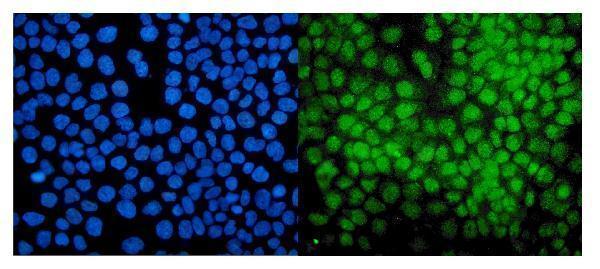
IHC figures cover human colon adenocarcinoma and spleen (M01707 captions), plus rat testis and brain and human placenta (PA2252 captions); IF covers human A431 cells (A01707 caption).
M01707 has paraffin-section IHC figures for human colon adenocarcinoma and spleen (M01707 captions), while PA2252 has IHC(P) figures for rat testis and brain and human placenta (PA2252 captions). A01707 has an IF figure for human A431 cells (A01707 caption).
Which to pick: For tissue IHC, choose PA2252 when a PIAS1-labeled reagent is required (PA2252 title and IHC(P) captions); M01707 has a documented 1:50 paraffin-section IHC workflow but recognizes PIAS1, PIAS2 and PIAS3 (M01707 caption). For IF/ICC, A01707 has an A431-cell IF figure at 2 μg/mL (A01707 caption). For human, mouse or rat IHC, both PA2252 and monoclonal M01707 list those species, although the pictured IHC samples establish only the species shown (catalog reactivity; M01707 and PA2252 captions); the paraffin-section captions do not report the fixative (M01707 and PA2252 captions).