This website uses cookies to ensure you get the best experience on our website.


- Table of Contents
Plan PIK3CG chromogenic IHC around cytoplasmic and membranous tissue staining (HPA tissue IHC). This guide covers the catalog antibody’s 2–5 μg/mL IHC range (datasheet A01517-2) and the presumed off-target staining caveat (HPA tissue IHC).

Expected localisation, validated protocols, controls and antibodies — the at-a-glance facts below, then the full design guide.
| Expected localisation | Cytoplasmic and membranous tissue staining (HPA tissue IHC) | |
| Staining pattern | Cytoplasmic and membranous staining, including lymphoid tissue (HPA tissue IHC) | |
| Antigen retrieval | EDTA pH 8.0 HIER, heat-mediated (datasheet A01517-2) | |
| Positive control | Adrenal gland+4 more · see all | |
| Negative control | Adipose tissue+4 more · see all |
| Fixation | Keep fixation consistent across samples (standard IHC practice; not target-specific) | |
| Caveat | Presumed off-target binding can confound staining (HPA tissue IHC) | |
| Regulation | Expression regulation is not established by this record (UniProt) | |
| Isoform / epitope | One full-length chain; no alternative isoforms listed (UniProt) |
The catalog antibody has an IHC-P protocol (datasheet: A01517-2). One published PIK3CG IHC method provides additional incubation and detection details (PMC8805872).
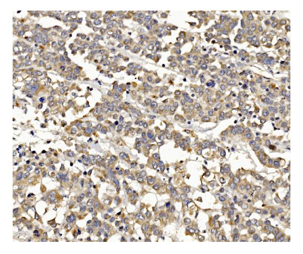
| Sample | Paraffin-embedded human breast cancer tissue; fixative not specified (datasheet A01517-2) |
| Fixation | Image fixative and duration unreported (datasheet A01517-2); verify before use. |
| Sectioning | 4–5 µm sections on charged slides (standard) |
| Deparaffinisation | Xylene, graded ethanol series to water (standard) |
| Antigen retrieval | Heat retrieval: EDTA pH 8.0 (datasheet A01517-2); 20 min, 95–100 °C (standard) |
| Peroxidase block | 3% H2O2, 10 min, room temperature (standard) |
| Blocking | 10% goat serum (datasheet A01517-2) |
| Primary antibody | Rabbit anti-PIK3CG, 2-5μg/ml (datasheet A01517-2) |
| Primary incubation | Overnight at 4 °C (datasheet A01517-2) |
| Detection | Streptavidin-biotin complex (SABC), DAB chromogen (datasheet A01517-2) |
| Counterstain | Hematoxylin, blue, dehydrate and mount (standard) |
| Expected result | PIK3CG-positive staining in glandular cells of adrenal gland (HPA tissue IHC: Medium). HPA tissue profile: Cytoplasmic and membranous expression in several different tissue types, including lymphoid tissues. No signal in the no-primary control. |
PIK3CG is a cytoplasmic and cell membrane protein with no transmembrane segment (UniProt P48736 topology). In paraffin section IHC, expect cytoplasmic and membranous staining in several tissue types, including lymphoid tissue; hematopoietic cells in bone marrow show medium staining (HPA tissue IHC). HPA rates the tissue profile Enhanced, while noting medium consistency with RNA data and presumed off target binding that was disregarded (HPA tissue IHC).
| Cytoplasmic staining with a membranous component in hematopoietic cells; bone marrow signal is around medium (HPA tissue IHC). | This fits the reported tissue pattern and PIK3CG localization (HPA tissue IHC; UniProt P48736 subcellular location). Judge staining in identified cells, since the HPA profile describes several tissue types and does not imply uniform staining throughout a section (HPA tissue IHC). |
| Glandular cells in adrenal gland, appendix, breast, duodenum, endometrium, fallopian tube or gallbladder show medium staining (HPA tissue IHC). | These are additional reported positive cell and tissue combinations (HPA tissue IHC). Score the glandular cells themselves; staining of adjacent cells does not establish that those cells share the reported pattern. Interpret both location and intensity before calling a section positive (general IHC practice). |
| Signal is predominantly nuclear, with little cytoplasmic or membranous staining. | That compartment does not match the reported PIK3CG locations (UniProt P48736 subcellular location; HPA tissue IHC). Recheck morphology, counterstain and antibody dependent staining before interpreting nuclear signal as target specific (general IHC practice). |
| Strong staining appears in adipocytes, cardiomyocytes or lung alveolar cells, despite their reported absence of staining (HPA tissue IHC). | Consider cross reactivity or endogenous detection activity and inspect cell identity and compartment (general IHC practice). HPA reports these specific cell types as not detected, but its tissue profile also notes presumed off target binding; an unexpected positive needs independent validation (HPA tissue IHC). |
| Broad, hazy color obscures cell boundaries, or a reported positive section has no discernible signal. | Diffuse background prevents a reliable compartment call (general IHC practice). For an absent signal, first verify that the section contains the reported positive cells, such as bone marrow hematopoietic cells, before changing the biological interpretation (HPA tissue IHC; general IHC practice). |
| Compartment and topology (UniProt P48736 topology; HPA tissue IHC) | PIK3CG has no transmembrane segment, yet both cytoplasmic and membranous locations are reported (UniProt P48736 topology and subcellular location; HPA tissue IHC). A membrane associated appearance is therefore plausible; a sharp membrane outline in every cell is not required by these sources. |
| Cell and tissue context (HPA tissue IHC) | HPA reports medium staining in bone marrow hematopoietic cells and several specified glandular cell populations, but not detected staining in selected other cell populations (HPA tissue IHC). Compare like cell types within a section; a whole tissue label alone is too coarse for scoring. |
| Evidence strength (HPA tissue IHC; HPA antibody validation) | HPA labels the tissue profile Enhanced and antibody HPA069976 IHC Enhanced, while describing medium agreement with RNA data and disregarded presumed off target binding (HPA tissue IHC; HPA antibody validation). Treat a matching pattern as supportive evidence, with discrepant staining requiring its own check. |
| Cardiac and liver interpretation (UniProt P48736 tissue specificity; HPA tissue IHC) | UniProt lists heart and liver in tissue specificity, whereas HPA reports cardiomyocytes not detected and hepatocytes low by IHC (UniProt P48736 tissue specificity; HPA tissue IHC). These entries should not be converted into an expectation of strong staining in either cell type. |
| IF/ICC localization (HPA subcellular ICC-IF) | HPA ICC-IF reports a supported main plasma membrane location with additional supported cytosolic localization (HPA subcellular ICC-IF). This helps interpret compartment agreement, but an ICC-IF image does not establish an IHC paraffin section protocol or a tissue staining intensity. |
| Situation | Likely cause | Next action |
|---|---|---|
| A reported positive cell population is absent or very weak in the stained section. | The section may lack the relevant cells, or the IHC detection run may have failed; HPA reports medium bone marrow hematopoietic and specified glandular cell staining (HPA tissue IHC; general IHC practice). | Confirm the cell population on the counterstained section and run an appropriately matched positive control. Review the catalog antibody's IHC-P instructions for the validated assay conditions before adjusting retrieval or dilution (general IHC practice). |
| Staining is mainly nuclear or has no credible cytoplasmic or membranous component. | The location conflicts with the reported target pattern (UniProt P48736 subcellular location; HPA tissue IHC). Counterstain overlap or nonspecific detection can complicate the reading (general IHC practice). | Inspect the same cell boundaries and nuclei at higher magnification; compare with an appropriate control section and the antibody validation pattern. Do not score isolated nuclear color as a matching PIK3CG pattern (general IHC practice; HPA tissue IHC). |
| A tissue is called positive solely because some cells in it stain. | HPA observations are cell specific: for example, its bone marrow entry names hematopoietic cells and its adrenal entry names glandular cells (HPA tissue IHC). | Record tissue, identified cell type, compartment and intensity together. Compare the scored cell type with HPA's reported cell type, and assess unexpected neighboring cell staining separately (HPA tissue IHC; general IHC practice). |
| Color appears broadly across the section or persists where primary antibody was omitted. | Nonspecific background or endogenous activity in the chromogenic detection system may contribute (general IHC practice). HPA also notes presumed off target binding in its tissue profile (HPA tissue IHC). | Check a primary antibody omission control and the detection system's recommended blocking steps; inspect whether color follows tissue structures and survives control conditions before assigning target staining (general IHC practice). |
| Strong staining occurs in cells HPA calls not detected, including adipocytes or cardiomyocytes. | The result differs from those cell specific HPA IHC observations; off target binding is a possible explanation, but a single discrepant section does not establish its cause (HPA tissue IHC; general IHC practice). | Reconfirm cell identity and compare controls and compartment pattern. Seek independent antibody or orthogonal evidence before reporting a new PIK3CG positive population; note the HPA discrepancy in the interpretation (HPA tissue IHC; general IHC practice). |
| Can an IF/ICC result resolve an IHC localization question? | HPA ICC-IF supports plasma membrane and cytosolic localization, while HPA tissue IHC reports cytoplasmic and membranous staining (HPA subcellular ICC-IF; HPA tissue IHC). The preparations answer related compartment questions under different assay conditions. | Use ICC-IF as a localization cross check and evaluate paraffin section staining against the tissue IHC pattern and its cell specific controls. Keep IF/ICC assay setup in its separate guide (HPA subcellular ICC-IF; HPA tissue IHC; general IHC practice). |


Comprehensive Human Protein Atlas IHC scoring per tissue (reliability: Enhanced — Medium consistency between antibody staining and RNA expression data. Presumed off target binding observed and disregarded.). Rows are taken directly from the HPA tissue chart — click any row's HPA link to view the source.
| Tissue | Cell type | Level | Evidence | Source |
|---|---|---|---|---|
| Adrenal gland | Glandular cells | Medium | Protein (IHC) | HPA → |
| Appendix | Glandular cells | Medium | Protein (IHC) | HPA → |
| Bone marrow | Hematopoietic cells | Medium | Protein (IHC) | HPA → |
| Breast | Glandular cells | Medium | Protein (IHC) | HPA → |
| Duodenum | Glandular cells | Medium | Protein (IHC) | HPA → |
| Tissue | Cell type | Level | Evidence | Source |
|---|---|---|---|---|
| Adipose tissue | Adipocytes | Not detected | Protein (IHC) | HPA → |
| Bronchus | Respiratory epithelial cells | Not detected | Protein (IHC) | HPA → |
| Cerebellum | Cells in granular layer | Not detected | Protein (IHC) | HPA → |
| Esophagus | Squamous epithelial cells | Not detected | Protein (IHC) | HPA → |
| Heart muscle | Cardiomyocytes | Not detected | Protein (IHC) | HPA → |
Troubleshoot PIK3CG staining by checking retrieval, compartment, cell identity and controls before comparing signal across paraffin sections (UniProt P48736; HPA tissue IHC).
A01517-2 has human paraffin-section IHC data (A01517-2 IHC caption). The catalog lists human, mouse and rat reactivity, with no IF/ICC validation (catalog applications/reactivity).
A01517-2 is the only SKU and shows staining in a paraffin-embedded human breast cancer section (A01517-2 IHC caption). Its listed applications include IHC but exclude IF/ICC; its listed reactivity is human, mouse and rat (catalog applications/reactivity).
Which to pick: For human tissue IHC, choose A01517-2: this rabbit antibody has a paraffin-section image and a human IHC dilution of 2–5 μg/ml (catalog host/dilution; A01517-2 IHC caption). No SKU has IF/ICC validation in the supplied catalog (catalog applications; IF image alts). For mouse or rat work, A01517-2 lists reactivity, but the supplied IHC dilution and tissue image cover human samples only; the caption does not report the fixative (catalog reactivity/dilution; A01517-2 IHC caption).