This website uses cookies to ensure you get the best experience on our website.


- Table of Contents
Plan chromogenic IHC for PIK3R3 in paraffin sections using cytoplasmic staining as the expected tissue pattern (HPA tissue IHC). Compare positive and negative cell populations reported in tissue IHC, and interpret staining cautiously because the HPA antibody may recognize proteins from more than one gene (HPA tissue IHC).

Expected localisation, validated protocols, controls and antibodies — the at-a-glance facts below, then the full design guide.
| Expected localisation | General cytoplasmic staining (HPA tissue IHC) | |
| Staining pattern | Cytoplasmic staining in breast glandular cells (HPA tissue IHC) | |
| Antigen retrieval | Citrate pH 6.0 HIER, 95–98 °C, 20 min (rule: cytoplasmic / membrane antigen) | |
| Positive control | Breast+4 more · see all | |
| Negative control | Esophagus+4 more · see all |
| Fixation | Keep formalin fixation consistent across sections. (standard IHC practice; not target-specific) | |
| Caveat | HPA antibody may recognize more than one gene (HPA tissue IHC) | |
| Regulation | Expression regulation unreported (UniProt) | |
| Isoform / epitope | 3 isoforms; verify epitope coverage (UniProt) |
The catalog antibody’s IHC-P protocol (datasheet) is accompanied by published PIK3R3 staining methods for colorectal tissue (PMC7735421), mouse skin (PMC5546695), and liver cancer tissue (PMC10358214).
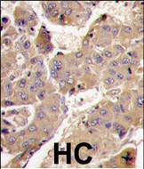
| Sample | Paraffin-embedded Human pituitary tumor tissue; fixative not specified (datasheet M06707) |
| Fixation | Image fixative and duration unreported (datasheet M06707); verify before use. |
| Sectioning | 4–5 µm sections on charged slides (standard) |
| Deparaffinisation | Xylene, graded ethanol series to water (standard) |
| Antigen retrieval | Heat-induced epitope retrieval in citrate buffer, pH 6.0, 20 min at 95–98 °C (standard rule: cytoplasmic / membrane antigen) |
| Peroxidase block | 3% H2O2, 10 min, room temperature (standard) |
| Blocking | 10% normal serum of the secondary host, 30 min, room temperature (standard) |
| Primary antibody | Rabbit monoclonal (clone DHC-16) anti-PIK3R3, 1:50 recommended; image 1:200 (datasheet M06707) |
| Primary incubation | Overnight at 4 °C (standard) |
| Detection | HRP-polymer secondary, DAB chromogen 5–10 min (standard) |
| Counterstain | Hematoxylin, blue, dehydrate and mount (standard) |
| Expected result | PIK3R3-positive staining in glandular cells of breast (HPA tissue IHC: High). HPA tissue profile: General cytoplasmic expression. No signal in the no-primary control. |
PIK3R3 is expected to show general cytoplasmic staining in tissue IHC, including strong staining in selected neuronal, epithelial, endocrine, and immune cell populations (HPA: tissue IHC). It has no transmembrane segment, so a membrane-only pattern is unexpected (UniProt Q92569 topology). Treat the pattern as provisional: HPA rates tissue IHC Approved but cautions that the antibody targets proteins from more than one gene and awaits external verification (HPA: tissue IHC reliability).
| Clear cytoplasmic staining in cerebral cortex or hippocampal neurons, bronchial respiratory epithelium, or pancreatic endocrine cells. | These cell populations are reported High by HPA and fit its general cytoplasmic IHC profile (HPA: tissue IHC). Compare staining within the identified cell population, rather than treating every cell in the section as an expected positive (general IHC practice). |
| Signal is restricted to cell borders or another unexpected compartment, with little cytoplasmic staining. | A border-only pattern conflicts with HPA's general cytoplasmic tissue profile and the absence of a transmembrane segment (HPA: tissue IHC; UniProt Q92569 topology). Check morphology and detection artefacts before assigning this signal to PIK3R3 (general IHC practice). |
| Strong staining appears in an unexpected cell population, such as cardiomyocytes or esophageal squamous epithelium. | HPA reports these cell populations as Not detected (HPA: heart muscle and esophagus tissue IHC). Investigate antibody cross-reactivity and endogenous detection activity; HPA specifically cautions that its antibody targets proteins from more than one gene (HPA: tissue IHC reliability; general IHC practice). |
| Brown signal spreads across extracellular space, tissue edges, or many cell types without clear cellular boundaries. | This does not provide a convincing cell-resolved cytoplasmic pattern (HPA: tissue IHC profile; general IHC practice). Background from detection reagents, inadequate blocking, or excessive antibody concentration can obscure interpretation (general IHC practice). |
| No convincing staining appears in an expected-positive cell population. | HPA reports High staining in breast glandular cells, kidney glomerular cells, skin keratinocytes, and lymph-node germinal-center cells (HPA: tissue IHC). First verify that the relevant cells are present and assess assay controls; an absent signal alone does not establish absent PIK3R3 (general IHC practice). |
| Cell population and tissue selection | HPA reports High staining in selected cells of breast, bronchus, cerebral cortex, hippocampus, kidney, lymph node, pancreas, and skin, while several other populations are Low or Not detected (HPA: tissue IHC). Score the named cell type, since a whole-tissue label can hide that distinction (general IHC practice). |
| Antibody evidence | HPA005751 is Approved for IHC, but HPA cautions that the tissue antibody targets proteins from more than one gene and awaits external verification (HPA: antibody status and tissue IHC reliability). A matching pattern supports interpretation without proving that every stained cell contains PIK3R3. |
| Topology and isoforms | PIK3R3 has no transmembrane segment and has 3 reported isoforms (UniProt Q92569 topology and isoforms). The supplied record gives no antibody epitope or isoform coverage, so neither membrane-only staining nor isoform-specific staining can be established from these facts. |
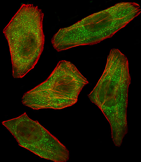
| IF/ICC Q: Where should fluorescence localize? | A: HPA reports mainly nucleoplasm and cytosol in ICC-IF, with additional locations in sperm structures (HPA: subcellular ICC-IF). Its subcellular summary cautions that the antibodies target proteins from multiple genes; this observation is context for IF interpretation, not an IHC compartment requirement (HPA: subcellular ICC-IF). |
| Situation | Likely cause | Next action |
|---|---|---|
| An expected-positive cell population has no chromogen. | The relevant cells may be absent from the section, or the IHC run may have failed (general IHC practice). HPA High is an observed pattern, not a guarantee for every specimen (HPA: tissue IHC). | Confirm cell identity and tissue preservation, then inspect the positive control and detection controls; optimize the assay using established antibody guidance (general IHC practice). |
| Signal is diffuse across the section. | Nonspecific binding or detection background can mask cell boundaries (general IHC practice). HPA instead describes general cytoplasmic expression (HPA: tissue IHC). | Compare a negative reagent control, review blocking and washes, and adjust antibody concentration within a validated range (general IHC practice). |
| Unexpected cells stain strongly. | Cross-reactivity is plausible because HPA cautions that its antibody targets proteins from more than one gene; endogenous detection activity is another possible IHC source (HPA: tissue IHC reliability; general IHC practice). | Check a reagent control and compare the cell type with HPA's named positive and Not detected populations; seek independent target validation before assigning PIK3R3 (HPA: tissue IHC; general IHC practice). |
| Staining appears only along membranes or tissue edges. | That pattern conflicts with general cytoplasmic tissue staining and PIK3R3's lack of a transmembrane segment (HPA: tissue IHC; UniProt Q92569 topology). Edge artefacts can occur in IHC (general IHC practice). | Examine intact interior cells, compare controls, and repeat staining if the compartment remains ambiguous (general IHC practice). |
| The candidate negative tissue shows some staining. | HPA's Not detected result applies to its specified cell population and assay; another population or assay background may account for the observed signal (HPA: tissue IHC; general IHC practice). | Identify the stained cells and compare them with the HPA cell-level entry; inspect reagent controls before using the section as a negative comparator (HPA: tissue IHC; general IHC practice). |
| IF/ICC shows nucleoplasmic signal that seems inconsistent with tissue IHC. | HPA reports nucleoplasm and cytosol in ICC-IF but a general cytoplasmic tissue IHC profile, both with antibody specificity cautions (HPA: subcellular ICC-IF and tissue IHC). | Interpret each observation within its assay and cell context; do not score nucleoplasmic fluorescence as proof of a nuclear IHC pattern (HPA: subcellular ICC-IF and tissue IHC; general IHC practice). |


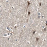


Comprehensive Human Protein Atlas IHC scoring per tissue (reliability: Approved — Caution, targets protein from more than one gene. Pending external verification.). Rows are taken directly from the HPA tissue chart — click any row's HPA link to view the source.
| Tissue | Cell type | Level | Evidence | Source |
|---|---|---|---|---|
| Breast | Glandular cells | High | Protein (IHC) | HPA → |
| Bronchus | Respiratory epithelial cells | High | Protein (IHC) | HPA → |
| Cerebral cortex | Neuronal cells | High | Protein (IHC) | HPA → |
| Hippocampus | Neuronal cells | High | Protein (IHC) | HPA → |
| Kidney | Cells in glomeruli | High | Protein (IHC) | HPA → |
| Tissue | Cell type | Level | Evidence | Source |
|---|---|---|---|---|
| Esophagus | Squamous epithelial cells | Not detected | Protein (IHC) | HPA → |
| Heart muscle | Cardiomyocytes | Not detected | Protein (IHC) | HPA → |
| Ovary | Ovarian stroma cells | Not detected | Protein (IHC) | HPA → |
| Prostate | Glandular cells | Not detected | Protein (IHC) | HPA → |
| Skeletal muscle | Myocytes | Not detected | Protein (IHC) | HPA → |
Troubleshoot PIK3R3 chromogenic IHC in paraffin sections using the catalog image, expression evidence, and matched controls.
The catalog shows human tissue IHC and cell IF images for PIK3R3 (catalog image captions). M06707 lists Human, Mouse and Rat reactivity; A06707-1 lists Human (catalog reactivity).
M06707 has IHC images of paraffin-embedded human pituitary tumor and lung adenocarcinoma, plus an IF image of C6 cells (M06707 image captions). A06707-1 has a DAB IHC image of formalin-fixed, paraffin-embedded human hepatocarcinoma and an IF image of U251 cells (A06707-1 image captions).
Which to pick: For tissue IHC, A06707-1 documents paraffin-section, paraffin-embedded tissue (A06707-1 IHC caption); M06707 documents paraffin sections, but its caption does not report the fixative (M06707 IHC caption). For IF/ICC, M06707 lists both applications and shows C6 IF, while A06707-1 lists IF and shows U251 IF (catalog applications; IF captions). Choose M06707 for listed Human, Mouse and Rat reactivity and a monoclonal format; A06707-1 is Human-reactive and polyclonal (catalog reactivity; catalog descriptions). The selected M06707 tissue-IHC caption documents paraffin sections, but does not specify the fixative (selected-SKU IHC image M06707).