This website uses cookies to ensure you get the best experience on our website.
- Table of Contents
2 Citations 1 Q&As
1 Citations

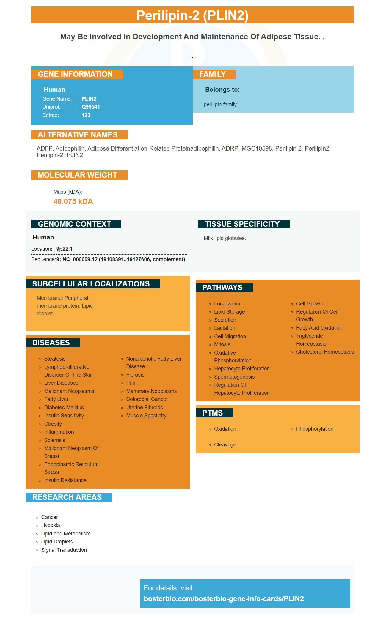
Facts about Perilipin-2.

.
| Human | |
|---|---|
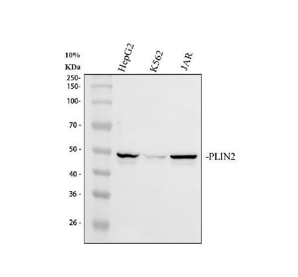
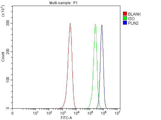
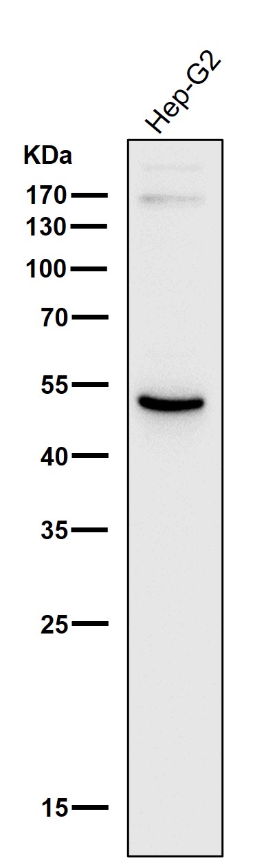
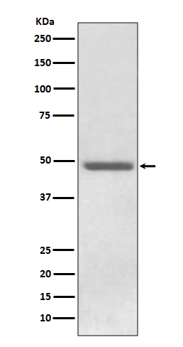
| Gene Name: | PLIN2 |
| Uniprot: | Q99541 |
| Entrez: | 123 |

| Belongs to: |
|---|
| perilipin family |

ADFP; Adipophilin; Adipose differentiation-related proteinadipophilin; ADRP; MGC10598; perilipin 2; Perilipin2; Perilipin-2; PLIN2
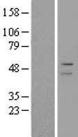
Mass (kDA):
48.075 kDA

| Human | |
|---|---|
| Location: | 9p22.1 |
| Sequence: | 9; NC_000009.12 (19108391..19127606, complement) |
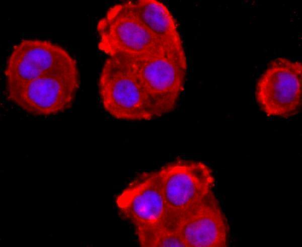
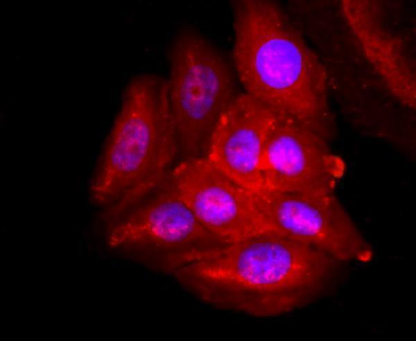
Milk lipid globules.
Membrane; Peripheral membrane protein. Lipid droplet.


PMID: 9003395 by Heid H.W., et al. Adipocyte differentiation-related protein is secreted into milk as a constituent of milk lipid globule membrane.
PMID: 26357594 by Chughtai A.A., et al. Perilipin-related protein regulates lipid metabolism in C. elegans.