Click image to see more details
Product Info Summary
| SKU: | PB10083 |
|---|---|
| Size: | 100 μg/vial |
| Reactive Species: | Human |
| Host: | Rabbit |
| Application: | Flow Cytometry, WB |
Customers Who Bought This Also Bought

Product info
Product Name
Anti-ADFP/PLIN2 Antibody Picoband®
SKU/Catalog Number
PB10083
Size
100 μg/vial
Form
Lyophilized
Description
Lipid-droplet coat protein PLIN2 involved in intracellular lipid storage; marker of lipid accumulation across multiple tissues/cell types (per product background). Assay context: WB/IHC on human/mouse/rat using rabbit polyclonal PB10083. Useful in metabolic/lipotoxicity studies; can be panelled with chromatin-state markers such as ARID2 for tumor biology contexts (putative).
Storage & Handling
Store at -20˚C for one year from date of receipt. After reconstitution, at 4˚C for one month. It can also be aliquotted and stored frozen at -20˚C for six months. Avoid repeated freeze-thaw cycles.
Cite This Product
Anti-ADFP/PLIN2 Antibody Picoband® (Boster Biological Technology, Pleasanton CA, USA, Catalog # PB10083)
Host
Rabbit
Contents
Each vial contains 4 mg Trehalose, 0.9 mg NaCl and 0.2 mg Na2HPO4.
Clonality
Polyclonal
Isotype
Rabbit IgG
Immunogen
E. coli-derived human ADFP recombinant protein (Position: K226-Q418). Human ADFP shares 88.4% amino acid (aa) sequence identity with mouse ADFP.
Cross-reactivity
No cross-reactivity with other proteins
Reactive Species
PB10083 is reactive to PLIN2 in Human
Observed Molecular Weight
48 kDa
Calculated molecular weight
48.1 kDa
Background of PLIN2
Adipose differentiation-related protein, also known as perilipin 2 (PLIN2), ADRP or adipophilin, is a protein which in humans is encoded by the ADFP gene. The protein encoded by this gene belongs to the perilipin family, members of which coat intracellular lipid storage droplets. This protein is associated with the lipid globule surface membrane material, and maybe involved in development and maintenance of adipose tissue. However, it is not restricted to adipocytes as previously thought, but is found in a wide range of cultured cell lines, including fibroblasts, endothelial and epithelial cells, and tissues, such as lactating mammary gland, adrenal cortex, Sertoli and Leydig cells, and hepatocytes in alcoholic liver cirrhosis, suggesting that it may serve as a marker of lipid accumulation in diverse cell types and diseases. Alternatively spliced transcript variants have been found for this gene.
Antibody Validation
Boster validates all antibodies on WB, IHC, ICC, Immunofluorescence, and ELISA with known positive control and negative samples to ensure specificity and high affinity, including thorough antibody incubations.
Application & Images
Applications
PB10083 is guaranteed for Flow Cytometry, WB Boster Guarantee
Recommend Dilution
| Application | Dilution | Species |
|---|---|---|
| Western blot | 0.1-0.5μg/ml | Human |
| Flow Cytometry(Fixed) | 1-3 μg/1x106 cells | Human |
Tested application
Suggested blocking solution with 5% non-fat milk or BSA; (*)Recommended protein loading: 20-40 µg per lane
Validation Images & Assay Conditions
Click image to see more details
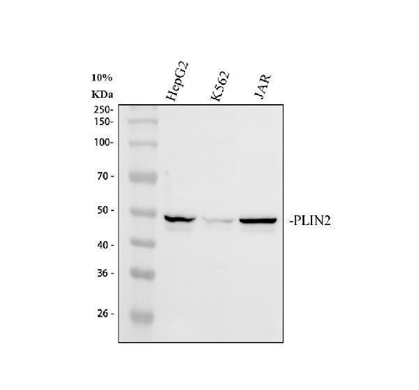
Western blot analysis of ADFP/PLIN2 using anti-ADFP/PLIN2 antibody (PB10083).
Electrophoresis was performed on a 10% SDS-PAGE gel at 80V (Stacking gel) / 120V (Resolving gel) for 2 hours. The sample well of each lane was loaded with 30 ug of sample under reducing conditions.
Lane 1: human HepG2 whole cell lysates,
Lane 2: human K562 whole cell lysates,
Lane 3: human JAR whole cell lysates.
After electrophoresis, proteins were transferred to a nitrocellulose membrane at 150 mA for 50-90 minutes. Blocked the membrane with 5% non-fat milk/TBS for 1.5 hour at RT. The membrane was incubated with rabbit anti-ADFP/PLIN2 antigen affinity purified polyclonal antibody (Catalog # PB10083) at 0.5 μg/mL overnight at 4°C, then washed with TBS-0.1%Tween 3 times with 5 minutes each and probed with a goat anti-rabbit IgG-HRP secondary antibody at a dilution of 1:5000 for 1.5 hour at RT. The signal is developed using an ECL Plus Western Blotting Substrate (Catalog # AR1196-200) with Tanon 5200 system. A specific band was detected for ADFP/PLIN2 at approximately 48 kDa. The expected band size for ADFP/PLIN2 is at 48 kDa.

Click image to see more details
Flow Cytometry analysis of HEL cells using anti-ADFP/PLIN2 antibody (PB10083).
Overlay histogram showing HEL cells stained with PB10083 (Blue line). To facilitate intracellular staining, cells were fixed with 4% paraformaldehyde and permeabilized with permeabilization buffer. The cells were blocked with 10% normal goat serum. And then incubated with rabbit anti-ADFP/PLIN2 Antibody (PB10083, 1 μg/1x106 cells) for 30 min at 20°C. DyLight®488 conjugated goat anti-rabbit IgG (BA1127, 5-10 μg/1x106 cells) was used as secondary antibody for 30 minutes at 20°C. Isotype control antibody (Green line) was rabbit IgG (1 μg/1x106) used under the same conditions. Unlabelled sample without incubation with primary antibody and secondary antibody (Red line) was used as a blank control.
Specific Publications For Anti-ADFP/PLIN2 Antibody Picoband® (PB10083)
Loading publications
Recommended Resources
Here are featured tools and databases that you might find useful.
- Boster's Pathways Library
- Protein Databases
- Bioscience Research Protocol Resources
- Data Processing & Analysis Software
- Photo Editing Software
- Scientific Literature Resources
- Research Paper Management Tools
- Molecular Biology Software
- Primer Design Tools
- Bioinformatics Tools
- Phylogenetic Tree Analysis
Customer Reviews
Have you used Anti-ADFP/PLIN2 Antibody Picoband®?
Share your experimental results or join a short interview to earn up to $1,000 in product credits or other rewards.
0 Reviews For Anti-ADFP/PLIN2 Antibody Picoband®
Customer Q&As
Have a question?
Find answers in Q&As, reviews.
Can't find your answer?
Submit your question
1 Customer Q&As for Anti-ADFP/PLIN2 Antibody Picoband®
Question
We are currently using anti-ADFP/PLIN2 antibody PB10083 for rat tissue, and we are well pleased with the IHC results. The species of reactivity given in the datasheet says human, mouse, rat. Is it likely that the antibody can work on pig tissues as well?
Verified Customer
Verified customer
Asked: 2017-09-21
Answer
The anti-ADFP/PLIN2 antibody (PB10083) has not been tested for cross reactivity specifically with pig tissues, but there is a good chance of cross reactivity. We have an innovator award program that if you test this antibody and show it works in pig you can get your next antibody for free. Please contact me if I can help you with anything.
Boster Scientific Support
Answered: 2017-09-21
