PLOD2 / Procollagen-lysine,2-oxoglutarate 5-dioxygenase 2 · IHC design guide

Design Immunohistochemistry for PLOD2

Plan paraffin-section PLOD2 IHC around cytoplasmic staining in most tissues (HPA tissue IHC). Use the catalog antibody’s documented IHC conditions and controls to assess cell-level signal; rough ER membrane localization is a molecular annotation (datasheet A04601-2; UniProt).

Evidence assembled Oct 2026 · For research use; verify linked source records and product datasheet before use
Immunohistochemistry protocol sheet for PLOD2 (IHC for PLOD2): expected localisation Cytoplasmic tissue staining; rough ER membrane molecularly (HPA tissue IHC; UniProt), antibody A04601-2, validated IHC image, and IHC protocol steps
Printable PLOD2 IHC protocol sheet — expected localisation Cytoplasmic tissue staining; rough ER membrane molecularly (HPA tissue IHC; UniProt), antibody A04601-2, controls and protocol steps. Open the full PLOD2 IHC guide →

PLOD2 Immunohistochemistry Experimental Design Guide

Expected localisation, validated protocols, controls and antibodies — the at-a-glance facts below, then the full design guide.

Must know before staining
Expected localisation Cytoplasmic tissue staining; rough ER membrane molecularly (HPA tissue IHC; UniProt)
Staining pattern Cytoplasmic staining in most tissues (HPA tissue IHC)
Antigen retrieval EDTA pH 8.0 HIER, heat-mediated (datasheet A04601-2)
Positive control ⓘ Adrenal gland+4 more · see all
Negative control ⓘ Ovary+1 more · see all
Important caveats
Reasons your staining may differ from the expected pattern.
Fixation Matched tissue-IHC evidence does not establish the fixation claim. Validate the specimen-specific method before use. (selected-SKU IHC image A04601-2)
Caveat Staining/RNA agreement is medium; verify in your tissue (HPA tissue IHC)
Regulation Isoform 2 shows tissue specificity (UniProt)
Isoform / epitope 3 isoforms; signal peptide 1–25; epitope map unspecified (UniProt)
Section 1

Recommended PLOD2 IHC & IF Protocols

Compare the catalog antibody’s IHC-P protocol with three published PLOD2 chromogenic IHC workflows (PMC5410313; PMC3656974; PMC6299564).

Recommended immunohistochemistry (IHC-P) protocol parameters
SampleParaffin-embedded human renal cancer tissue; fixative not specified (datasheet A04601-2)
FixationImage fixative and duration unreported (datasheet A04601-2); verify before use.
Sectioning4–5 µm sections on charged slides (standard)
DeparaffinisationXylene, graded ethanol series to water (standard)
Antigen retrievalHeat retrieval: EDTA pH 8.0 (datasheet A04601-2); 20 min, 95–100 °C (standard)
Peroxidase block3% H2O2, 10 min, room temperature (standard)
Blocking10% goat serum (datasheet A04601-2)
Primary antibodyRabbit anti-PLOD2, 1:50 recommended; image 1:100 (datasheet A04601-2)
Primary incubationOvernight at 4 °C (datasheet A04601-2)
DetectionHRP-conjugated secondary, DAB chromogen (datasheet A04601-2)
CounterstainHematoxylin, blue, dehydrate and mount (standard)
Expected resultPLOD2-positive staining in glandular cells of adrenal gland (HPA tissue IHC: Medium). HPA tissue profile: Cytoplasmic expression in most tissues. No signal in the no-primary control.
💡Decision noteStart with heat-mediated EDTA pH 8.0 retrieval because the catalog datasheet specifies it and a published PLOD2 IHC workflow used it (datasheet A04601-2; PMC5410313).
Section 2

What Is the Expected PLOD2 Staining Pattern?

PLOD2 is associated with the rough endoplasmic reticulum membrane and cytoplasm, with no transmembrane segment (UniProt O00469 topology). In paraffin sections, expect predominantly cytoplasmic staining in the cell populations identified by HPA, such as adrenal glandular cells and colon endothelial cells (HPA tissue IHC). HPA rates its tissue staining Approved, with medium consistency against RNA data and external verification pending (HPA tissue IHC).

What am I looking at on my slide?
Cytoplasmic staining in adrenal glandular cells or colon endothelial cells.Both populations show medium staining in the HPA tissue record (HPA tissue IHC). Judge the pattern within the identified cells; PLOD2 is reported in the cytoplasm and at the rough endoplasmic reticulum membrane (UniProt O00469 topology).
Strong nuclear-only or extracellular staining replaces the expected cytoplasmic pattern.That distribution conflicts with the HPA tissue IHC profile and should prompt an artefact check (HPA tissue IHC). HPA reports uncertain nucleolar staining in ICC-IF, so a nucleolar signal alone does not establish the expected paraffin-section pattern (HPA ICC-IF).
Staining appears mainly in an unexpected cell population within a reference tissue.Check cell identity against HPA’s population-specific observations before scoring it as PLOD2; for example, its colon entry identifies endothelial cells (HPA tissue IHC). Cross-reactivity or endogenous chromogenic activity are possible alternative explanations (general IHC practice).
Broad haze covers tissue and clear spaces, obscuring cell boundaries.This is difficult to interpret as the cell-associated cytoplasmic pattern reported by HPA (HPA tissue IHC). Uneven blocking, excess detection reagent, or inadequate washing can produce nonspecific background (general IHC practice).
No stain appears in a section containing an HPA-listed positive cell population.First confirm that the relevant cells are present: HPA records medium staining in adrenal glandular, duodenal glandular, and colon endothelial cells (HPA tissue IHC). An absent signal may reflect assay performance; it does not by itself overturn an Approved profile pending external verification (HPA tissue IHC).
💡Expected PLOD2 appearanceCall a result positive when identifiable HPA-listed cells show predominantly cytoplasmic staining, typically medium in the listed positive examples; nuclear-only, extracellular, or field-wide colour without cell definition calls for review (HPA tissue IHC; UniProt O00469 topology; general IHC practice).
How each factor affects the staining
Cell population and tissueUse cell-level comparisons: HPA reports medium staining in several listed populations, but no detected staining in ovarian stroma cells or splenic red-pulp cells (HPA tissue IHC). Those negative entries do not classify every cell in either organ.
Antibody validationThe mouse monoclonal CAB025898 has Approved IHC status, while HPA069126 has no listed IHC status (HPA antibodies). The broader tissue profile is Approved with medium RNA consistency and awaits external verification (HPA tissue IHC).
Compartment and processingUniProt lists rough endoplasmic reticulum membrane and cytoplasm, no transmembrane segment, a signal peptide at residues 1–25, and a chain at 26–737 (UniProt O00469). These annotations support a cellular pattern; they do not establish extracellular IHC staining.
Isoforms and modificationsUniProt lists three isoforms and seven glycosylation sites (UniProt O00469). With no epitope or isoform-specific validation supplied, do not infer which isoforms the IHC-validated antibody detects or predict a change in staining from those modifications.
IF/ICC Q: Should nucleolar signal define a positive IHC result?No. HPA lists nucleoli and cytosol as uncertain ICC-IF locations, whereas its tissue IHC profile is cytoplasmic (HPA ICC-IF; HPA tissue IHC). Interpret the IF finding on its own guide page; it supplies no IHC protocol option.
Why is my staining missing, weak or wrong?
SituationLikely causeNext action
A listed positive population has no visible signal.The relevant cells may be absent from the field, or an IHC step may have failed (HPA tissue IHC; general IHC practice).Confirm cell identity and run the IHC-validated antibody on a section containing an HPA-listed positive population; then check retrieval, primary incubation, detection, and counterstain against the established IHC workflow (HPA antibodies; HPA tissue IHC; general IHC practice).
Only nuclei show strong colour.The result does not match HPA’s cytoplasmic tissue profile; uncertain ICC-IF nucleolar localisation cannot confirm nuclear-only IHC staining (HPA tissue IHC; HPA ICC-IF).Review morphology and detection controls, then reassess whether cytoplasmic staining is present in the listed cell population (general IHC practice; HPA tissue IHC).
An unexpected population stains more strongly than the listed population.Cell misidentification, cross-reactivity, or endogenous detection activity can mimic target staining (general IHC practice).Compare the precise cell population with HPA’s tissue entries and use appropriate reagent controls to investigate the unexpected colour (HPA tissue IHC; general IHC practice).
Colour is diffuse across the section.Nonspecific background can obscure the cell-associated pattern (general IHC practice; HPA tissue IHC).Inspect a control without primary antibody; check blocking, reagent concentration, washes, and chromogen development using the established IHC workflow (general IHC practice).
Ovarian or splenic tissue appears negative.HPA reports PLOD2 as not detected specifically in ovarian stroma cells and splenic red-pulp cells (HPA tissue IHC).Check which cells were scored. Use an HPA-listed positive population to assess assay performance before interpreting a negative result in those populations (HPA tissue IHC; general IHC practice).
Staining differs between two antibodies.Their validation is unequal: CAB025898 is Approved for IHC, while HPA069126 has no listed IHC status (HPA antibodies).Identify each antibody and its IHC validation status, compare staining in the same HPA-listed cell population, and treat discordance as unresolved specificity rather than proof of an isoform difference (HPA antibodies; HPA tissue IHC; UniProt O00469).

Sample controls for PLOD2 IHC & IF

🧪Run adrenal gland first; its glandular cells should show PLOD2 staining (HPA: Medium in adrenal gland glandular cells). Use ovarian stroma as the biological tissue negative (HPA: Not detected in ovarian stroma cells); cells outside the glandular population on the positive slide should show only background, but their PLOD2-negative status is unverified by the supplied HPA rows.
Positive control tissue: Adrenal gland (Glandular cells, HPA Medium)
Negative control tissue: Ovary (HPA Not detected)
ICC-IF cell lines (HPA subcellular resource): HPA ICC-IF images show PLOD2 in BJ [Human fibroblast], SiHa, U2OS, with annotated localisation: Nucleoli (uncertain), Cytosol (uncertain) (HPA subcellular).
Technical controls: Include a no-primary, secondary-only control, a rabbit IgG isotype control matched to the primary antibody, and a PLOD2 knockout or validated peptide-block control (selected-SKU caption: rabbit primary; standard IHC practice). For chromogenic detection, quench endogenous peroxidase and check the adrenal section for background pigment before scoring (standard IHC practice).
⚠️Feasibility: A target-specific fixation window or fixation effect is unreported, and the selected-SKU paraffin-section caption does not state the fixative (selected-SKU caption: fixative unreported). The demonstrated IHC conditions use EDTA retrieval at pH 8.0 and a 1:100 primary dilution overnight at 4°C; retrieval dependence is unreported (selected-SKU caption). The supplied evidence does not establish that frozen sections or IF are easier, or identify a PLOD2-specific tissue artefact (supplied IHC caption and HPA rows).

HPA tissue IHC evidence for PLOD2

Comprehensive Human Protein Atlas IHC scoring per tissue (reliability: Approved — Medium consistency between antibody staining and RNA expression data. Pending external verification.). Rows are taken directly from the HPA tissue chart — click any row's HPA link to view the source.

Positive expression · recommended positive controls

TissueCell typeLevelEvidenceSource
Adrenal gland Glandular cells Medium Protein (IHC) HPA →
Caudate Neuronal cells Medium Protein (IHC) HPA →
Cerebral cortex Neuronal cells Medium Protein (IHC) HPA →
Cervix Squamous epithelial cells Medium Protein (IHC) HPA →
Colon Endothelial cells Medium Protein (IHC) HPA →

Undetected expression · recommended negative controls

TissueCell typeLevelEvidenceSource
Ovary Ovarian stroma cells Not detected Protein (IHC) HPA →
Spleen Cells in red pulp Not detected Protein (IHC) HPA →
Section 3

Advanced PLOD2 IHC Tips

Troubleshoot PLOD2 staining in paraffin sections by checking retrieval, cellular localisation, controls and scoring before interpreting differences in signal.

What should I change when PLOD2 staining is weak after antigen retrieval?
Start with heat-mediated retrieval in EDTA, pH 8.0, for paraffin sections (datasheet A04601-2). The selected tissue image used this retrieval, followed by 1:100 primary antibody overnight at 4°C; it does not establish a universal retrieval time (datasheet A04601-2). If signal remains weak, compare retrieval heating times on adjacent sections while keeping antibody concentration, detection and development constant (standard IHC practice). Check that tissue has stayed attached and morphology remains interpretable, since excessive heating can make apparent signal gains difficult to trust (standard IHC practice). Include a processed positive control in each comparison; PLOD2 is reported as cytoplasmic in most tissues (HPA tissue IHC).
Can fixation explain inconsistent PLOD2 staining between paraffin blocks?
The selected paraffin-section image does not report its fixative, so target-specific PLOD2 sensitivity to fixation is unknown (datasheet A04601-2). Record each block’s fixative, fixation duration and processing history before comparing staining intensity across blocks (standard IHC practice). Compare sections with the same retrieval in EDTA, pH 8.0 and the same antibody incubation, using a control block in each run (datasheet A04601-2; standard IHC practice). If only one block stains poorly, review its morphology and processing record before changing the primary antibody dilution (standard IHC practice). Do not infer a fixation effect from PLOD2’s reported rough endoplasmic reticulum localisation or glycosylation sites (UniProt O00469).
Which staining compartment should count as convincing PLOD2 signal?
Assess intracellular cytoplasmic staining first: tissue IHC reports cytoplasmic expression in most tissues, while UniProt places PLOD2 at the rough endoplasmic reticulum membrane and in cytoplasm (HPA tissue IHC; UniProt O00469). A granular perinuclear pattern can be examined alongside cytoplasmic signal, but morphology alone cannot prove organelle identity (standard IHC practice). HPA’s ICC/IF summary also lists nucleoli and cytosol, with both locations marked uncertain (HPA subcellular). Score cytoplasmic and nuclear or nucleolar staining separately rather than combining them into one positive call (standard IHC practice). Review suspicious nuclear-only staining against matched controls and the surrounding tissue morphology before assigning it to PLOD2 (standard IHC practice).
Can this antibody distinguish PLOD2 isoforms or detect a processed epitope?
PLOD2 has three listed isoforms, but the supplied antibody caption does not specify its epitope or establish isoform selectivity (UniProt O00469; datasheet A04601-2). Isoform 2 is reported in skin, lung, dura and aorta, so tissue distribution alone cannot identify which isoform produced a stain (UniProt O00469). The listed signal peptide spans residues 1–25, and the mature chain spans 26–737; the antibody’s binding region is unspecified (UniProt O00469; datasheet A04601-2). Seven glycosylation sites and reported modified residues add possible epitope considerations without demonstrating an effect on this antibody (UniProt O00469). Obtain epitope mapping or isoform-specific validation before making isoform claims from chromogenic IHC (standard IHC practice).
How should I investigate PLOD2 localisation with multiplex IF?
Treat IF/ICC as a separate assay: the selected antibody evidence is a paraffin-section chromogenic IHC image, with no IF protocol supplied (datasheet A04601-2). Pair PLOD2 with a validated marker for the cell population being examined, and inspect single-marker controls before interpreting overlap (standard IF practice). Choose a fluorophore channel with low measured tissue autofluorescence, using unstained tissue and single-stain controls to set acquisition and bleed-through limits (standard IF practice). Because UniProt reports rough endoplasmic reticulum membrane and cytoplasmic localisation without a transmembrane segment, test permeabilisation for access to the antibody’s unknown epitope rather than assuming its membrane-facing side (UniProt O00469; standard IF practice). HPA lists nucleoli and cytosol as uncertain ICC/IF locations, so verify either pattern independently (HPA subcellular).
How can I separate PLOD2 staining from diffuse DAB background?
The selected paraffin-section image used 10% goat serum, a peroxidase-conjugated secondary antibody and DAB development (datasheet A04601-2). For troubleshooting, include a primary-omission control and block endogenous peroxidase before DAB detection; these are general chromogenic IHC controls (standard IHC practice). If background persists, compare antibody dilution, wash stringency and DAB development time on adjacent sections while holding retrieval at EDTA, pH 8.0 (datasheet A04601-2; standard IHC practice). Inspect pigment, tissue folds and section edges separately from intact cell cytoplasm before scoring (standard IHC practice). PLOD2 is reported as cytoplasmic in most tissues, so uniform extracellular color warrants additional scrutiny (HPA tissue IHC; standard IHC practice).
What is a defensible way to quantify PLOD2 chromogenic IHC? ⚠ ANSWER MARKED FOR VERIFICATION
Define the cell population and compartment before scoring, using cytoplasmic staining as the primary tissue IHC readout (HPA tissue IHC; standard IHC practice). Report the percentage of positive cells and an H-score calculated as the sum of each intensity category’s percentage multiplied by its score, using 0–3 intensity categories (standard IHC practice). If counting positive cells per area, report density per mm² of evaluable tissue and keep the area definition consistent (standard IHC practice). Normalise comparisons to the same cell population and usable tissue area, excluding folds, necrosis and blank regions by a stated rule (standard IHC practice). Use the same retrieval, development and image-analysis thresholds across compared sections (standard IHC practice).
When is an apparent PLOD2-positive region more likely to be artefact?
Give greatest weight to intact-cell cytoplasmic signal: HPA reports cytoplasmic expression in most tissues, and UniProt lists cytoplasm and rough endoplasmic reticulum membrane (HPA tissue IHC; UniProt O00469). Check which cells carry the signal; HPA reports medium staining in colon endothelial cells and no detected staining in ovarian stromal cells, so a region label alone is insufficient (HPA tissue IHC). Treat isolated section-edge staining, necrotic debris and primary-omission-positive DAB deposits as possible artefacts requiring review (standard IHC practice). A nucleolar-only pattern needs independent validation because HPA marks nucleolar ICC/IF localisation uncertain (HPA subcellular). Compare the suspicious region with adjacent intact tissue, a run control and the omission control before calling it PLOD2-positive (standard IHC practice).
Boster reagents

Best PLOD2 / Procollagen-lysine,2-oxoglutarate 5-dioxygenase 2 IHC Antibodies

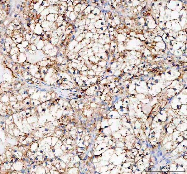
A04601-2 is listed for IHC and IF in human and rat samples (catalog applications; catalog reactivity). Its supplied image documents IHC in a paraffin-embedded human renal cancer section (catalog IHC caption).

Real IHC data IHC analysis of PLOD2 using anti-PLOD2 antibody (A04601-2). PLOD2 was detected in a paraffin-embedded section of human renal cancer tissue. Heat mediated antigen retrieval was performed in EDTA buffer (pH 8.0, epitope retrieval solution). The tissue section was blocked with 10% goat serum. The tissue section was then incubated with 1:100 rabbit anti-PLOD2 Antibody (A04601-2) overnight at 4°C. Peroxidase Conjugated Goat Anti-rabbit IgG was used as secondary antibody and incubated for 30 minutes at 37°C. The tissue section was developed using HRP Conjugated Rabbit IgG Super Vision Assay Kit (Catalog # SV0002) with DAB as the chromogen.
Anti-PLOD2 Antibody
Cat # A04601-2

A04601-2 has an IHC image from a paraffin-embedded human renal cancer section using EDTA retrieval at pH 8.0 and DAB detection (catalog IHC caption). IHC, IF, and ICC are listed applications, but no IF image is supplied (catalog applications; catalog IF image alts).

Which to pick: For tissue IHC, choose A04601-2: its image documents paraffin-embedded human renal cancer tissue, with the primary antibody incubated at 1:100 overnight; the fixative is unreported (catalog IHC caption). For IF/ICC, A04601-2 is the listed option, with an IF dilution of 1:50; no IF image is supplied (catalog applications; catalog IF dilution; catalog IF image alts). For rat samples, A04601-2 is a rabbit polyclonal with listed rat reactivity, while its supplied tissue image documents human tissue (catalog host; catalog dilution_raw; catalog reactivity; catalog IHC caption).

Each figure is that product's own IHC / IF validation image from its datasheet.

References

  1. UniProt Consortium. UniProt entry O00469 (PLOD2_HUMAN, Procollagen-lysine,2-oxoglutarate 5-dioxygenase 2).
  2. Human Protein Atlas. PLOD2 tissue IHC expression (reliability: Approved).
  3. Human Protein Atlas. PLOD2 subcellular location (ICC-IF): Localized to the nucleoli and cytosol..
  4. Human Protein Atlas. PLOD2 antibody validation summary (2 antibodies).
  5. Procollagen-lysine 2-oxoglutarate 5-dioxygenase 2 promotes hypoxia-induced glioma migration and invasion. Oncotarget 2017 — PMC5410313.
  6. Procollagen lysyl hydroxylase 2 is essential for hypoxia-induced breast cancer metastasis. Molecular cancer research : MCR 2013 — PMC3656974.
  7. Adipocyte-derived IL-6 and leptin promote breast Cancer metastasis via upregulation of Lysyl Hydroxylase-2 expression. Cell communication and signaling : CCS 2018 — PMC6299564.
  8. Pan-Cancer Analyses Reveal Oncogenic and Immunological Role of PLOD2. Frontiers in genetics 2022 — PMC9108334.
  9. PubMed PMID:9054364 — UniProt-cited evidence.
  10. PubMed PMID:14702039 — UniProt-cited evidence.
  11. PubMed PMID:16641997 — UniProt-cited evidence.