PLSCR1 / Phospholipid scramblase 1 · IHC design guide

Design Immunohistochemistry for PLSCR1

Use cytoplasmic staining in immune-cell subsets to interpret PLSCR1 paraffin-section IHC (HPA tissue IHC). This guide covers consistent fixation, the catalog antibody’s 0.5–1 μg/mL IHC range, chromogenic detection, and scoring (datasheet A02995-2).

Evidence assembled Oct 2026 · For research use; verify linked source records and product datasheet before use
Immunohistochemistry protocol sheet for PLSCR1 (IHC for PLSCR1): expected localisation Immune-cell cytoplasm (HPA tissue IHC); membrane/nucleus possible (UniProt), antibody A02995-2, validated IHC image, and IHC protocol steps
Printable PLSCR1 IHC protocol sheet — expected localisation Immune-cell cytoplasm (HPA tissue IHC); membrane/nucleus possible (UniProt), antibody A02995-2, controls and protocol steps. Open the full PLSCR1 IHC guide →

PLSCR1 Immunohistochemistry Experimental Design Guide

Expected localisation, validated protocols, controls and antibodies — the at-a-glance facts below, then the full design guide.

Must know before staining
Expected localisation Immune-cell cytoplasm (HPA tissue IHC); membrane/nucleus possible (UniProt)
Staining pattern Cytoplasmic staining in subsets of immune cells (HPA tissue IHC)
Antigen retrieval EDTA pH 8.0 HIER, heat-mediated (datasheet A02995-2)
Positive control ⓘ Bone marrow+1 more · see all
Negative control ⓘ Adipose tissue+4 more · see all
Important caveats
Reasons your staining may differ from the expected pattern.
Fixation Matched tissue-IHC evidence does not establish the fixation claim. Validate the specimen-specific method before use. (selected-SKU IHC image A02995-2)
Caveat Low staining–RNA consistency limits tissue prediction (HPA tissue IHC)
Regulation Expression regulation not established (UniProt)
Isoform / epitope 2 isoforms; map epitopes to cytoplasmic 1–288 or external 306–318 (UniProt)
Section 1

Recommended PLSCR1 IHC & IF Protocols

The catalog antibody has an IHC-P protocol (datasheet: A02995-2); three published PLSCR1 IHC studies provide additional tissue-array, breast-tumor, and liver-tissue procedures (PMC3551821; PMC13347015; PMC7590428).

Recommended immunohistochemistry (IHC-P) protocol parameters
SampleParaffin-embedded mouse spleen tissue; fixative not specified (datasheet A02995-2)
FixationImage fixative and duration unreported (datasheet A02995-2); verify before use.
Sectioning4–5 µm sections on charged slides (standard)
DeparaffinisationXylene, graded ethanol series to water (standard)
Antigen retrievalHeat retrieval: EDTA pH 8.0 (datasheet A02995-2); 20 min, 95–100 °C (standard)
Peroxidase block3% H2O2, 10 min, room temperature (standard)
Blocking10% goat serum (datasheet A02995-2)
Primary antibodyRabbit anti-PLSCR1, 0.5-1μg/ml (datasheet A02995-2)
Primary incubationOvernight at 4 °C (datasheet A02995-2)
DetectionStreptavidin-biotin complex (SABC), DAB chromogen (datasheet A02995-2)
CounterstainHematoxylin, blue, dehydrate and mount (standard)
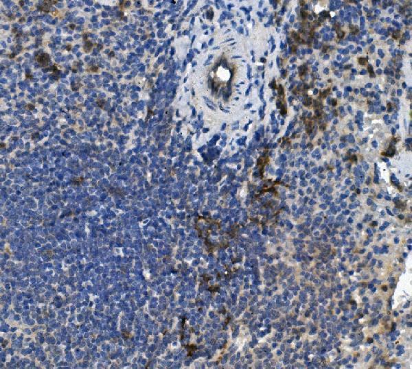
Expected resultPLSCR1-positive staining in hematopoietic cells of bone marrow (HPA tissue IHC: Medium). HPA tissue profile: Cytoplasmic expression in subsets of immune cells in several tissues, incuding lymphoid organs. No signal in the no-primary control.
💡Decision noteStart with heat-mediated EDTA pH 8.0 retrieval for the catalog antibody (datasheet: A02995-2). Use the published retrieval conditions when reproducing a study (PMC13347015; PMC7590428).
Section 2

What Is the Expected PLSCR1 Staining Pattern?

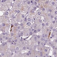
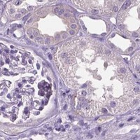
In paraffin-section IHC, expect predominantly cytoplasmic staining in subsets of immune cells, including hematopoietic cells in bone marrow and cells in splenic red pulp (HPA tissue IHC). PLSCR1 can also localize to the plasma membrane or nucleus; residues 289–305 form its transmembrane segment (UniProt O15162 topology and subcellular location). HPA rates tissue IHC “Approved,” while reporting low consistency between antibody staining and RNA expression (HPA tissue IHC).

What am I looking at on my slide?
Selective cytoplasmic staining in bone marrow hematopoietic cells or splenic red-pulp cells, with unstained neighboring cells (HPA tissue IHC).This matches the reported cell distribution. HPA scores both examples Medium, so assess which cells stain and where, rather than requiring every cell to stain strongly (HPA tissue IHC).
A sharply defined membrane or nuclear component accompanies staining in a subset of cells (UniProt O15162 subcellular location).These compartments are plausible for PLSCR1; palmitoylation regulates membrane versus nuclear localization (UniProt O15162). Staining confined instead to extracellular deposits or unrelated slide structures does not fit the annotated cellular locations and warrants control review (UniProt O15162 subcellular location).
Broad staining appears in adipocytes or adrenal glandular cells, where HPA reports PLSCR1 as not detected (HPA tissue IHC).Treat this as unexpected for those cell types, especially if staining is uniform. Check for antibody cross-reactivity and endogenous chromogenic detection activity using appropriate controls; an HPA “not detected” result is a reference pattern, not proof that every positive cell is false (HPA tissue IHC; general IHC practice).
Brown color spreads across tissue, stroma, or blank areas without a clear cellular boundary.Diffuse deposit is difficult to score as PLSCR1 localization. Review the no-primary control, blocking, detection reagents, and washing for general IHC background; HPA’s selective immune-cell pattern provides the tissue-level comparison (general IHC practice; HPA tissue IHC).
No staining is visible in bone marrow hematopoietic cells or splenic red-pulp cells (HPA tissue IHC).A blank reference tissue leaves assay performance unresolved. Confirm the tissue contains the expected cells, then check the catalog antibody’s IHC-P conditions and a positive control before calling a test specimen negative; the HPA examples are Medium, not universal positives (HPA tissue IHC; general IHC practice).
💡Expected PLSCR1 appearanceA convincing positive is selective, chiefly cytoplasmic staining of the reported immune-cell populations at approximately Medium intensity in HPA’s bone marrow or spleen examples; uniform staining of HPA-negative cell types or diffuse extracellular color is suspect (HPA tissue IHC; general IHC practice).
How each factor affects the staining
What does PLSCR1 topology imply for interpretation?PLSCR1 has a cytoplasmic region at residues 1–288, one transmembrane segment at 289–305, and an extracellular region at 306–318 (UniProt O15162 topology). These locations make membrane-associated staining biologically plausible. The antibody epitope is not supplied, so topology alone cannot predict retrieval requirements or which compartment this assay will emphasize.
Can nuclear or perinuclear staining be genuine?Yes, as a localization possibility: UniProt lists nucleus and cytoplasm, with perinuclear localization in the presence of RELT; palmitoylation regulates membrane versus nuclear localization (UniProt O15162 subcellular location). Interpret a nuclear or perinuclear pattern alongside cellular specificity and controls, without assuming it must appear in every tissue.
How strong is the tissue IHC reference?HPA calls tissue IHC Approved but notes low consistency between antibody staining and RNA expression; its listed IHC antibody is approved, not marked enhanced (HPA tissue IHC; HPA antibodies). Use the reported cell pattern as a benchmark while resolving discordant findings with controls rather than RNA abundance alone.
IF/ICC Q&A: Where might PLSCR1 fluorescence appear?HPA reports nucleoplasm and plasma membrane as supported locations and Golgi apparatus as approved in ICC-IF (HPA subcellular). This answers localization interpretation only; IF/ICC has its own guide and no IF protocol option is supplied here.
Why is my staining missing, weak or wrong?
SituationLikely causeNext action
Expected bone marrow or spleen cells are unstained (HPA tissue IHC).The target cells may be absent from the examined field, or the IHC run may lack detectable signal (HPA tissue IHC; general IHC practice).Locate hematopoietic or red-pulp cells on the counterstained section, confirm the positive control, and review the catalog antibody’s IHC-P instructions for retrieval, dilution, and detection (general IHC practice).
Every cell shows similarly strong brown staining, including HPA-negative cell types (HPA tissue IHC).Non-specific antibody binding or endogenous detection activity is possible; broad staining does not match HPA’s selective pattern (general IHC practice; HPA tissue IHC).Compare no-primary and other appropriate controls, check blocking for the detection system, and reassess antibody dilution under the catalog IHC-P instructions (general IHC practice).
Background obscures the boundary between positive immune cells and neighboring cells.Diffuse chromogen or insufficient washing can prevent cellular localization from being scored (general IHC practice).Inspect blank tissue areas and the no-primary control; review washing, detection time, and counterstain so cellular signal can be distinguished from background (general IHC practice).
Signal is mainly nuclear or perinuclear, rather than cytoplasmic.PLSCR1 can localize to the nucleus and, with RELT, the perinuclear region; the compartment alone does not establish an artefact (UniProt O15162 subcellular location).Check whether staining remains cell-selective, compare controls, and report the observed compartment. Do not force a cytoplasmic-only score from HPA’s tissue summary (HPA tissue IHC; general IHC practice).
Membrane staining is present but cytoplasmic staining is weak.Membrane localization is compatible with PLSCR1 topology and palmitoylation-dependent trafficking; the supplied record gives no antibody epitope (UniProt O15162 topology and subcellular location).Record membrane and cytoplasmic components separately, confirm the signal follows cell boundaries, and compare with the HPA tissue pattern and assay controls (HPA tissue IHC; general IHC practice).
A test tissue disagrees with HPA staining or an RNA-based expectation.HPA explicitly reports low consistency between antibody staining and RNA expression; its listed tissue positives are specific cell populations (HPA tissue IHC).Verify cell identity and assay controls, document the compartment and proportion of positive cells, and qualify the discrepancy instead of assigning a tissue-wide positive or negative from RNA alone (HPA tissue IHC; general IHC practice).

Sample controls for PLSCR1 IHC & IF

🧪Run spleen first: cells in the red pulp should stain (HPA: Medium in spleen red-pulp cells; catalog caption: PLSCR1 detected in mouse spleen). Use adipose tissue adipocytes as the negative comparison (HPA: Not detected in adipocytes). Within the spleen slide, cells without specific staining should remain at counterstain or background levels; the supplied HPA row does not identify a specific negative spleen cell type.
Positive control tissue: Bone marrow (Hematopoietic cells, HPA Medium)
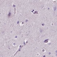
Negative control tissue: Adipose tissue (HPA Not detected)
ICC-IF cell lines (HPA subcellular resource): HPA ICC-IF images show PLSCR1 in A-431, SK-MEL-30, U2OS, RPTEC/TERT1, hTERT-RPE1 (serum starved), with annotated localisation: Nucleoplasm (supported), Golgi apparatus (approved), Plasma membrane (supported) (HPA subcellular).
Technical controls: Include a no-primary (secondary-only) control, a concentration-matched rabbit immunoglobulin isotype control (catalog caption: rabbit primary antibody), and PLSCR1-knockout tissue as a biological negative. Block endogenous peroxidase and check endogenous biotin in spleen when using the caption’s biotin-based SABC/DAB detection (catalog caption: biotinylated secondary, SABC and DAB).
⚠️Feasibility: A target-specific fixation window or fixation effect is unreported in the supplied evidence; the exact A02995-2 tissue-IHC caption does not state the fixative. The caption uses heat retrieval in EDTA at pH 8.0, but does not establish that retrieval is required (catalog caption: EDTA heat retrieval). Neither frozen sections nor IF are established as easier by the supplied evidence; for spleen IHC, endogenous peroxidase and biotin can complicate interpretation with the caption’s SABC/DAB method.

HPA tissue IHC evidence for PLSCR1

Comprehensive Human Protein Atlas IHC scoring per tissue (reliability: Approved — Low consistency between antibody staining and RNA expression data.). Rows are taken directly from the HPA tissue chart — click any row's HPA link to view the source.

Positive expression · recommended positive controls

TissueCell typeLevelEvidenceSource
Bone marrow Hematopoietic cells Medium Protein (IHC) HPA →
Spleen Cells in red pulp Medium Protein (IHC) HPA →

Undetected expression · recommended negative controls

TissueCell typeLevelEvidenceSource
Adipose tissue Adipocytes Not detected Protein (IHC) HPA →
Adrenal gland Glandular cells Not detected Protein (IHC) HPA →
Appendix Glandular cells Not detected Protein (IHC) HPA →
Breast Adipocytes Not detected Protein (IHC) HPA →
Bronchus Respiratory epithelial cells Not detected Protein (IHC) HPA →
Section 3

Advanced PLSCR1 IHC Tips

Troubleshoot PLSCR1 staining in paraffin sections by checking retrieval, cellular compartment, immune-cell distribution, and assay controls.

Which retrieval condition should I try first for weak PLSCR1 staining?
Start with heat-mediated antigen retrieval in EDTA at pH 8.0 for paraffin sections (datasheet A02995-2). The selected mouse spleen image used that condition, followed by 10% goat-serum blocking and 1 μg/ml primary antibody overnight at 4°C (datasheet A02995-2). If staining remains weak, compare retrieval duration on adjacent sections while holding antibody concentration and detection constant, and watch for damaged tissue morphology (standard IHC practice). Include a known positive section in each comparison; the selected mouse spleen image provides an assay reference, although its caption does not state the fixative (datasheet A02995-2).
Could fixation explain a weak or uneven PLSCR1 signal?
Target-specific fixation sensitivity is unknown: the selected paraffin-section caption does not report a fixative, so it cannot establish how fixation affects PLSCR1 staining (datasheet A02995-2). Compare sections with documented fixation histories using the same EDTA pH 8.0 retrieval, 1 μg/ml primary concentration, and detection method (datasheet A02995-2; standard IHC practice). Uneven staining near tissue edges can reflect processing variation, so assess morphology and the distribution of staining before changing the primary concentration (standard IHC practice). Record fixation and processing conditions alongside staining results; neither tissue-expression patterns nor PLSCR1 topology demonstrate a fixation-specific effect (HPA tissue IHC; UniProt O15162 topology).
How should I assess membrane, cytoplasmic, and nuclear PLSCR1 staining?
Score membrane, cytoplasmic, and nuclear staining separately rather than treating every positive pixel as equivalent (standard IHC practice). PLSCR1 has a cytoplasmic region at residues 1–288 and a transmembrane segment at 289–305; UniProt reports membrane, nuclear, cytoplasmic, and perinuclear localisation (UniProt O15162 topology and subcellular location). Palmitoylation is associated with membrane trafficking, while its absence favours nuclear localisation; RELT is associated with perinuclear localisation (UniProt O15162 subcellular location). HPA tissue IHC describes cytoplasmic staining in subsets of immune cells, so compare compartment calls with cell identity and morphology on the same section (HPA tissue IHC; standard IHC practice).
Can the antibody distinguish PLSCR1 isoforms or an inaccessible epitope?
Do not assign an isoform from chromogenic staining alone: PLSCR1 has 2 annotated isoforms, but the supplied antibody caption does not specify an epitope or isoform selectivity (UniProt O15162 isoforms; datasheet A02995-2). The protein spans the membrane at residues 289–305, with residues 1–288 cytoplasmic and 306–318 extracellular (UniProt O15162 topology). Its annotated modifications include phosphotyrosines at 69 and 74 and phosphothreonine at 161, without evidence here that they alter this antibody's staining (UniProt O15162 modified residues; datasheet A02995-2). Check the antibody's epitope documentation before interpreting differences in retrieval response or compartment staining as isoform-specific (standard IHC practice).
How can I check PLSCR1 localisation with multiplex IF?
Pair PLSCR1 with a validated marker of the expected immune-cell population, since tissue IHC reports cytoplasmic expression in immune-cell subsets (HPA tissue IHC; standard IF practice). Select spectrally separated fluorophores and place a weaker signal in a far-red channel when tissue autofluorescence interferes with shorter wavelengths (standard IF practice). Plan permeabilisation around the mapped epitope: residues 1–288 face the cytoplasm, whereas 306–318 face the extracellular space; the supplied caption does not identify this antibody's epitope (UniProt O15162 topology; datasheet A02995-2). Include single-colour and no-primary controls when assessing nuclear, membrane, or perinuclear overlap (standard IF practice; UniProt O15162 subcellular location).
What should I check when PLSCR1 staining is diffuse or widespread?
Inspect a no-primary control and the tissue morphology before calling diffuse DAB deposition PLSCR1-positive (standard IHC practice). The selected workflow used 10% goat serum, biotinylated goat anti-rabbit secondary antibody, a streptavidin-biotin complex, and DAB; each detection stage warrants its own background check (datasheet A02995-2; standard IHC practice). Use a peroxidase block and assess endogenous biotin when troubleshooting this chromogenic workflow (standard IHC practice). Compare the signal with the expected cytoplasmic immune-cell subsets and separately note plausible membrane or nuclear staining, rather than scoring uniform haze across unrelated cells (HPA tissue IHC; UniProt O15162 subcellular location).
How should I quantify PLSCR1 staining across paraffin sections? ⚠ ANSWER MARKED FOR VERIFICATION
Define the cell population and compartment before scoring, because HPA reports PLSCR1 in subsets of immune cells and UniProt lists several cellular locations (HPA tissue IHC; UniProt O15162 subcellular location). Report the percentage of positive cells and an intensity-weighted H-score from 0–300, or count positive cells per mm² when spatial density matters (standard IHC practice). Normalise counts to the number of eligible cells or the analysed viable tissue area, and apply one staining threshold across compared sections (standard IHC practice). Record cytoplasmic, membrane, and nuclear results separately and keep retrieval, antibody concentration, and detection conditions matched (standard IHC practice; datasheet A02995-2).
How can I distinguish convincing PLSCR1 staining from artefact?
Give greatest weight to reproducible staining in morphologically intact cells with a plausible compartment and distribution (standard IHC practice; UniProt O15162 subcellular location). HPA reports cytoplasmic staining in immune-cell subsets, including medium staining in splenic red-pulp cells, but notes low consistency between antibody staining and RNA expression (HPA tissue IHC). Treat staining confined to section edges, necrotic areas, or structures also positive in a no-primary control as suspect; check endogenous peroxidase contribution to DAB signal (standard IHC practice). Nuclear or membrane staining can be biologically plausible for PLSCR1, but verify unexpected cell populations and compartments with controls before assigning biological meaning (UniProt O15162 subcellular location; standard IHC practice).
Boster reagents

Best PLSCR1 / Phospholipid scramblase 1 IHC Antibodies

A02995-2 has a mouse spleen paraffin-section IHC image (catalog image caption) and lists human and mouse reactivity (catalog: reactivity). M02995-1 lists human ICC/IF (catalog: applications and reactivity); no IF figure is supplied (catalog: image data).

Real IHC data IHC analysis of PLSCR1 using anti-PLSCR1 antibody (A02995-2). PLSCR1 was detected in paraffin-embedded section of mouse spleen tissue. Heat mediated antigen retrieval was performed in EDTA buffer (pH8.0, epitope retrieval solution). The tissue section was blocked with 10% goat serum. The tissue section was then incubated with 1μg/ml rabbit anti-PLSCR1 Antibody (A02995-2) overnight at 4°C. Biotinylated goat anti-rabbit IgG was used as secondary antibody and incubated for 30 minutes at 37°C. The tissue section was developed using Strepavidin-Biotin-Complex (SABC) (Catalog # SA1022) with DAB as the chromogen.
Anti-Scramblase 1/PLSCR1 Antibody ®
Cat # A02995-2

A02995-2 is shown staining a paraffin-embedded mouse spleen section by IHC (catalog image caption) and lists human and mouse reactivity (catalog: reactivity). M02995-1 lists human ICC/IF (catalog: applications and reactivity), but has no IF image (catalog: image data).

Which to pick: Choose A02995-2 for paraffin-section IHC: its own image shows staining in mouse spleen after EDTA pH 8 retrieval at 1 μg/ml (catalog image caption); the fixative is unreported (catalog image caption). Choose M02995-1 for human IF/ICC because it is a rabbit monoclonal listed for those applications, although no IF figure is supplied (catalog: title, applications, reactivity and image data). For human and mouse work, A02995-2 lists both species, but its IHC image demonstrates mouse tissue only (catalog: reactivity and A02995-2 image caption).

Each figure is that product's own IHC / IF validation image from its datasheet.

References

  1. UniProt Consortium. UniProt entry O15162 (PLS1_HUMAN, Phospholipid scramblase 1).
  2. Human Protein Atlas. PLSCR1 tissue IHC expression (reliability: Approved).
  3. Human Protein Atlas. PLSCR1 subcellular location (ICC-IF): Localized to the nucleoplasm, plasma membrane and the Golgi apparatus..
  4. Human Protein Atlas. PLSCR1 antibody validation summary (2 antibodies).
  5. Phospholipid scramblase 1 as a critical node at the crossroad between autophagy and apoptosis in mantle cell lymphoma. Oncotarget 2016 — PMC5173105.
  6. Blockade of phospholipid scramblase 1 with its N-terminal domain antibody reduces tumorigenesis of colorectal carcinomas in vitro and in vivo. Journal of translational medicine 2012 — PMC3551821.
  7. PLSCR1 drives chemoresistance in TNBC via METTL3/IGF2BP3-mediated mRNA stabilization and EGFR-MAPK pathway activation. Cell death & disease 2026 — PMC13347015.
  8. RNA interference-mediated downregulation of phospholipid scramblase 1 expression in primary liver cancer in vitro. Oncology letters 2020 — PMC7590428.
  9. PubMed PMID:9218461 — UniProt-cited evidence.
  10. PubMed PMID:9712717 — UniProt-cited evidence.
  11. PubMed PMID:10930526 — UniProt-cited evidence.