PMVK / Phosphomevalonate kinase · IHC design guide

Design Immunohistochemistry for PMVK

Plan PMVK staining in paraffin sections with the IHC-validated antibody PA1067 (datasheet: IHC-P). Use documented positive cell populations and score predominantly cytoplasmic staining while allowing for nuclear signal in some tissues (HPA tissue IHC).

Evidence assembled Oct 2026 · For research use; verify linked source records and product datasheet before use
Immunohistochemistry protocol sheet for PMVK (IHC for PMVK): expected localisation Generally cytoplasmic; nuclear in some tissues (HPA tissue IHC), antibody PA1067, validated IHC image, and IHC protocol steps
Printable PMVK IHC protocol sheet — expected localisation Generally cytoplasmic; nuclear in some tissues (HPA tissue IHC), antibody PA1067, controls and protocol steps. Open the full PMVK IHC guide →

PMVK Immunohistochemistry Experimental Design Guide

Expected localisation, validated protocols, controls and antibodies — the at-a-glance facts below, then the full design guide.

Must know before staining
Expected localisation Generally cytoplasmic; nuclear in some tissues (HPA tissue IHC)
Staining pattern High in glandular cells; generally cytoplasmic (HPA tissue IHC)
Antigen retrieval Citrate pH 6 HIER, heat-mediated (datasheet PA1067)
Positive control ⓘ Colon+4 more · see all
Negative control ⓘ None in HPA (detected in all 45 tissues); use no-primary + isotype controls
Important caveats
Reasons your staining may differ from the expected pattern.
Fixation Keep fixation consistent across sections. (standard IHC practice; not target-specific)
Caveat Nuclear signal in some tissues can complicate cytoplasmic scoring (HPA tissue IHC)
Regulation Staining intensity varies by cell type (HPA tissue IHC)
Isoform / epitope One 1–192 chain; no isoforms annotated (UniProt)
Section 1

Recommended PMVK IHC & IF Protocols

Compare the catalog antibody’s citrate pH 6 IHC-P protocol (datasheet PA1067) with published PMVK IHC methods in ovarian carcinoma TMAs (PMC7226362) and human lung tissue (PMC10475767).

Recommended immunohistochemistry (IHC-P) protocol parameters
SampleParaffin-embedded rat skeletal muscle tissues; fixative not specified (datasheet PA1067)
FixationImage fixative and duration unreported (datasheet PA1067); verify before use.
Sectioning4–5 µm sections on charged slides (standard)
DeparaffinisationXylene, graded ethanol series to water (standard)
Antigen retrievalHeat retrieval: Citrate pH 6, 20 min (datasheet PA1067)
Peroxidase block3% H2O2, 10 min, room temperature (standard)
Blocking10% goat serum (datasheet PA1067)
Primary antibodyRabbit anti-PMVK, 0.5-1μg/ml (datasheet PA1067)
Primary incubationOvernight at 4 °C (datasheet PA1067)
DetectionStreptavidin-biotin complex (SABC), DAB chromogen (datasheet PA1067)
CounterstainHematoxylin, blue, dehydrate and mount (standard)
Expected resultPMVK-positive staining in glandular cells of colon (HPA tissue IHC: High). HPA tissue profile: General cytoplasmic expression, in some tissues additional nuclear expression. No signal in the no-primary control.
💡Decision noteStart with citrate pH 6 heat retrieval: it matches the catalog protocol (datasheet PA1067) and the published lung IHC method (PMC10475767).
Section 2

What Is the Expected PMVK Staining Pattern?

PMVK is a cytosolic enzyme with no transmembrane segment (UniProt Q15126). In paraffin-section IHC, expect predominantly cytoplasmic staining, with additional nuclear staining possible in some tissues (HPA tissue IHC). Strong examples include colon and duodenal glandular cells, kidney tubular cells, and pancreatic endocrine cells (HPA: High). HPA rates its tissue staining Approved, with medium consistency between antibody staining and RNA expression data (HPA tissue IHC).

What am I looking at on my slide?
Clear cytoplasmic staining in colon or duodenal glandular cells, kidney tubular cells, or pancreatic endocrine cells.This matches the reported compartment and cell types (HPA tissue IHC: general cytoplasmic expression; High in these cells). Compare signal with adjacent structures and the control slide before scoring (general IHC practice).
Predominantly nuclear staining, with little cytoplasmic signal.Additional nuclear staining can occur in some tissues, so nuclear signal alone is not automatically false (HPA tissue IHC). A nuclear-only pattern departs from the general cytoplasmic profile; check morphology and controls before interpreting it as PMVK (HPA tissue IHC; general IHC practice).
Strong staining in an unexpected cell population while the expected cells are weak.Cell-type discordance raises possible nonspecific binding or endogenous chromogenic activity (general IHC practice). HPA reports High staining in defined populations, but its list is not an exhaustive map of every positive cell (HPA tissue IHC).
Uniform brown haze across cells, stroma, and tissue-free areas.A haze without cell boundaries is difficult to score as intracellular PMVK (general IHC practice). Assess the background control and detection steps; PMVK’s reported distribution is chiefly cytoplasmic within cells (HPA tissue IHC).
No staining in a morphologically intact known-positive tissue.A negative run in a reported High cell population calls for a technical check before a biological conclusion (HPA tissue IHC; general IHC practice). Confirm the expected cells are present, then review controls and the IHC workflow (general IHC practice).
💡Expected PMVK appearanceCall the slide positive when expected cells show distinct, predominantly cytoplasmic staining, strongest in HPA High populations; a uniform haze or isolated stain in unexpected structures warrants control review (HPA tissue IHC; general IHC practice).
How each factor affects the staining
Compartment and topologyPMVK is cytosolic and has no transmembrane segment (UniProt Q15126). Score staining within identifiable cells against the predominantly cytoplasmic HPA pattern; additional nuclear staining is reported in some tissues (HPA tissue IHC).
Choice of comparison tissueColon and duodenal glandular cells, kidney tubular cells, and pancreatic endocrine cells are reported High (HPA tissue IHC). Adipocytes and chondrocytes are reported Low, which does not make them validated negative controls (HPA tissue IHC).
Strength of IHC evidenceThe tissue profile is Approved but has medium staining-to-RNA consistency (HPA tissue IHC). HPA lists HPA029900 as IHC Approved; the supplied record does not establish enhanced validation or a negative tissue (HPA antibodies; HPA tissue IHC).
Chromogenic detection backgroundEndogenous detection activity can mimic specific stain in chromogenic IHC (general IHC practice). Use an appropriate detection control to distinguish it from a cell-shaped PMVK pattern; no target-specific fixation sensitivity is supplied (general IHC practice; supplied UniProt/HPA record).
Why is my staining missing, weak or wrong?
SituationLikely causeNext action
Known-positive glandular or tubular cells are unstained (HPA tissue IHC: High).Missing target cells or a failed staining run are possibilities (general IHC practice); absence of signal alone does not establish absent PMVK.Confirm morphology and tissue identity, then inspect the run controls, antibody dilution, retrieval, and detection steps (general IHC practice).
Nuclear signal dominates and cytoplasmic signal is absent.Some nuclear expression is reported, but the general HPA profile is cytoplasmic (HPA tissue IHC); background remains possible (general IHC practice).Compare with the tissue-specific HPA image and control slide; score the nuclear pattern cautiously if it lacks convincing cytoplasmic staining (HPA tissue IHC; general IHC practice).
Signal appears mainly in cells outside the expected population.Possible cross-reactivity or endogenous chromogenic activity can imitate a positive result (general IHC practice); HPA High labels identify particular cells, not exclusive expression (HPA tissue IHC).Check cell identity by morphology and review detection controls before assigning that signal to PMVK (general IHC practice).
Brown stain covers tissue broadly without cellular detail.Diffuse background can arise from nonspecific staining or detection activity (general IHC practice); it does not resolve the reported cytoplasmic pattern (HPA tissue IHC).Compare a detection control and assess blocking, washing, and antibody dilution under the laboratory’s IHC procedure (general IHC practice).
A Low tissue has faint staining, or a putative negative control stains.HPA reports Low staining in adipocytes, chondrocytes, splenic red-pulp cells, and marrow hematopoietic cells; it supplies no negative tissue (HPA tissue IHC).Treat faint cell-associated signal as potentially compatible with Low expression; judge specificity with controls and a reported High population (HPA tissue IHC; general IHC practice).
Q: Does this IHC pattern predict an IF/ICC result?HPA provides no main subcellular location or ICC-IF images here, and HPA029900 has no listed ICC status (HPA subcellular; HPA antibodies).A: Use cytosolic localization as a hypothesis (UniProt Q15126); validate IF/ICC independently with suitable controls rather than assuming the IHC result transfers (general IF practice).

Sample controls for PMVK IHC & IF

🧪Run kidney first: tubular cells should stain strongly (HPA: High in cells in tubules). HPA lists no negative tissue because PMVK is detected in all 45 scored tissues, so use no-primary and isotype controls; on the positive slide, treat cells without visible signal as background comparators only after confirming they lack specific staining, since HPA does not identify an internal negative cell type (HPA: no negative rows; standard IHC practice).
Positive control tissue: Colon (Glandular cells, HPA High)
Negative control tissue: None in HPA: PMVK is detected in all 45 scored tissues. Use a no-primary (secondary-only) and an isotype control instead.
ICC-IF cell lines (HPA subcellular resource): HPA carries no ICC-IF cell line for PMVK; derive a cell-line control from the positive tissue's cell type (Glandular cells) and confirm it by RNA or western blot first.
Technical controls: Include a no-primary, secondary-only control, a rabbit IgG isotype control matched to the catalog primary, and a PMVK knockout specimen or validated immunogen-peptide block as a biological specificity control (selected PA1067 caption: rabbit primary and goat anti-rabbit secondary; standard IHC practice). For kidney, check endogenous peroxidase and biotin background when using the caption’s biotin–SABC/DAB detection (selected PA1067 caption; standard IHC practice).
⚠️Feasibility: No supplied source reports a PMVK-specific fixation window or fixation effect; the selected PA1067 paraffin-section caption leaves the fixative unreported. That caption documents citrate retrieval at pH 6 for 20 minutes, but does not establish whether retrieval is required for PMVK staining (selected PA1067 caption). A frozen-section or IF advantage is unreported; the supplied HPA record lists no ICC-IF image lines, while endogenous biotin in kidney tubules warrants attention with biotin-based detection (HPA subcellular record; selected PA1067 caption; standard IHC practice).

HPA tissue IHC evidence for PMVK

Comprehensive Human Protein Atlas IHC scoring per tissue (reliability: Approved — Medium consistency between antibody staining and RNA expression data.). Rows are taken directly from the HPA tissue chart — click any row's HPA link to view the source.

Positive expression · recommended positive controls

TissueCell typeLevelEvidenceSource
Colon Glandular cells High Protein (IHC) HPA →
Duodenum Glandular cells High Protein (IHC) HPA →
Fallopian tube Glandular cells High Protein (IHC) HPA →
Gallbladder Glandular cells High Protein (IHC) HPA →
Kidney Cells in tubules High Protein (IHC) HPA →

Undetected expression · recommended negative controls

TissueCell typeLevelEvidenceSource
None in HPA: PMVK is detected in all 45 scored tissues. Use a no-primary (secondary-only) and an isotype control instead.
Section 3

Advanced PMVK IHC Tips

Troubleshoot PMVK staining in paraffin sections using the documented IHC conditions, expected cytosolic localisation, and cell-specific tissue patterns.

How should I retrieve PMVK antigen when paraffin sections stain weakly?
Start with heat-mediated citrate retrieval at pH 6 for 20 minutes (datasheet PA1067). The documented rat skeletal muscle image used that treatment before an overnight primary incubation at 4°C (caption PA1067). If staining is weak, compare a modestly longer retrieval with the documented condition on adjacent sections, keeping antibody concentration and detection identical (standard IHC practice). Inspect tissue integrity and staining in an expected positive cell population before increasing retrieval further (standard IHC practice; HPA: High in kidney tubular cells). Record heating and cooling conditions because inconsistent temperature exposure can change staining across a slide (standard IHC practice).
Could fixation explain variable PMVK staining between paraffin blocks?
Target-specific PMVK sensitivity to fixation is unknown: the selected paraffin-section caption does not state a fixative (caption PA1067). Compare blocks with documented fixation histories using the same pH 6 citrate retrieval and detection conditions (datasheet PA1067; standard IHC practice). Review section morphology and include an expected positive cell population in each comparison, since poor preservation can complicate staining assessment (standard IHC practice; HPA: High in colon glandular cells). Avoid assigning a weak result to overfixation without a controlled comparison of processing histories (standard IHC practice). Record fixation and processing details alongside staining results so differences can be investigated (standard IHC practice).
Where should PMVK staining appear, and how should nuclear signal be assessed?
Expect predominantly cytoplasmic staining because PMVK is annotated as cytosolic and has no transmembrane segment (UniProt Q15126 localisation and topology). HPA tissue IHC also describes general cytoplasmic expression, with additional nuclear expression in some tissues (HPA: tissue IHC profile). Assess nuclear colour separately from cytoplasmic colour using a counterstain and matched negative controls (standard IHC practice). If nuclear staining predominates, compare its cell distribution with the cytoplasmic pattern and inspect for diffuse background before assigning it to PMVK (standard IHC practice; HPA: tissue IHC profile). Keep compartment scores separate when reporting heterogeneous sections (standard IHC practice).
How should I investigate unexpectedly selective staining across cell populations?
First confirm the antibody's immunogen or mapped epitope, because the supplied PMVK record does not identify the catalog antibody's epitope (record: PMVK Q15126; caption PA1067). The record lists one 1–192 chain, no annotated isoforms, and no annotated glycosylation or modified residues (UniProt Q15126 processing, isoforms and PTMs). Those annotations do not establish that every cell will expose the antibody epitope equally after paraffin processing (standard IHC practice). Compare adjacent sections under the documented pH 6 retrieval condition while holding detection constant (datasheet PA1067; standard IHC practice). Use expected positive cells to distinguish a broad technical loss from a cell-restricted pattern (HPA: High in kidney tubular cells; standard IHC practice).
How can I examine PMVK by IF while interpreting this IHC result?
Treat IF as a separate assay requiring its own antibody and staining validation; the selected PA1067 evidence documents chromogenic IHC in paraffin rat skeletal muscle (caption PA1067). Multiplex PMVK with a marker identifying the cell population under study, then assess signal within those cells (standard IF practice). Choose a fluorophore channel after checking tissue autofluorescence, and include single-stain controls to evaluate bleed-through (standard IF practice). Because PMVK is annotated as cytosolic with no transmembrane segment, use permeabilisation sufficient to expose its intracellular epitope, then optimise detergent conditions for the specimen (UniProt Q15126 localisation and topology; standard IF practice). Compare compartment patterns with IHC cautiously (HPA: tissue IHC profile; standard IF practice).
How can I reduce diffuse or misleading DAB staining?
Start from the documented 10% goat serum block and 1 µg/mL primary antibody concentration, then compare changes one at a time (caption PA1067; standard IHC practice). Include a no-primary control to reveal colour arising from the secondary or detection system (standard IHC practice). Check peroxidase blocking and DAB development time when colour appears broadly, especially where tissue pigmentation or endogenous enzyme activity complicates assessment (standard IHC practice). The selected image used a biotinylated secondary, streptavidin–biotin complex and DAB, so assess background from that detection sequence (caption PA1067; standard IHC practice). Judge improvements against expected cytoplasmic staining in positive cells (UniProt Q15126 localisation; HPA: High in kidney tubular cells).
What is a defensible way to quantify PMVK in heterogeneous tissue? ⚠ ANSWER MARKED FOR VERIFICATION
Define the cell compartment and population before scoring, since PMVK is primarily cytosolic while HPA reports additional nuclear expression in some tissues (UniProt Q15126 localisation; HPA: tissue IHC profile). For chromogenic IHC, record the percentage of positive cells and staining intensity, or calculate an H-score from intensity categories 0–3 (standard IHC practice). Score nuclear and cytoplasmic staining separately if both are present (standard IHC practice; HPA: tissue IHC profile). Normalise positive counts to all evaluable cells of the same type, or positive-cell density to analysed tissue area in mm² (standard IHC practice). Exclude folds, edges and necrotic regions consistently across sections (standard IHC practice).
What findings support true PMVK staining rather than artefact?
A credible result shows reproducible cytoplasmic staining in morphologically intact cells, consistent with the annotated cytosolic location (UniProt Q15126 localisation; standard IHC practice). Kidney tubular cells and pancreatic endocrine cells provide examples of high HPA tissue IHC staining, while HPA also reports additional nuclear expression in some tissues (HPA: High in kidney tubular cells and pancreatic endocrine cells; HPA: tissue IHC profile). Compare suspected signal with a no-primary control and neighbouring intact regions (standard IHC practice). Treat staining confined to section edges or necrosis, or colour reproduced without primary antibody, as a technical warning (standard IHC practice). Review peroxidase blocking if DAB colour persists in the control (standard IHC practice).
Boster reagents

Best PMVK / Phosphomevalonate kinase IHC Antibodies

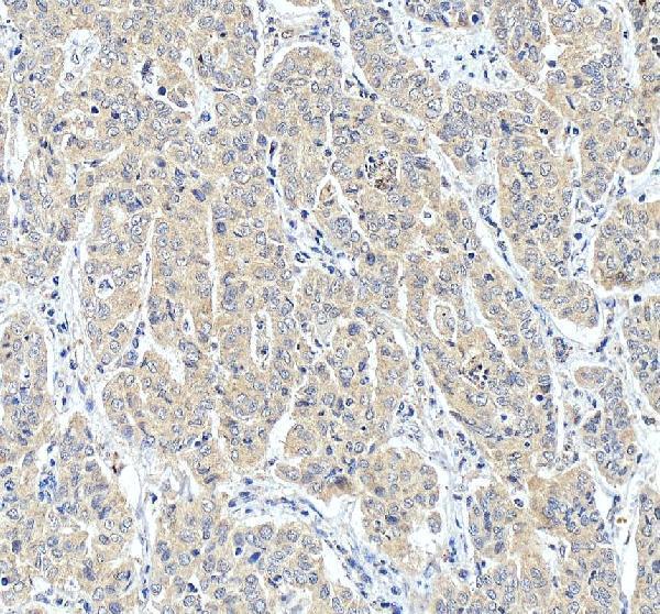
PA1067 has IHC data from paraffin sections of rat skeletal muscle and human liver tissue, including liver cancer (PA1067 image captions). A10103 lists human ICC/IF use; no IF image is supplied (A10103 catalog payload).

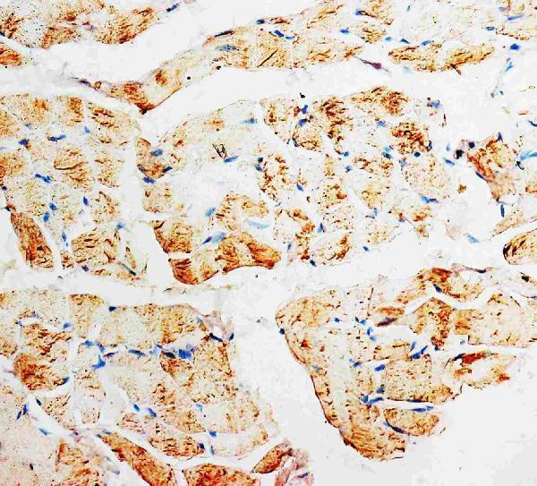
Real IHC data IHC analysis of PMVK using anti-PMVK antibody (PA1067). PMVK was detected in paraffin-embedded section of rat skeletal muscle tissues. Heat mediated antigen retrieval was performed in citrate buffer (pH6, epitope retrieval solution) for 20 mins. The tissue section was blocked with 10% goat serum. The tissue section was then incubated with 1μg/ml rabbit anti-PMVK Antibody (PA1067) overnight at 4°C. Biotinylated goat anti-rabbit IgG was used as secondary antibody and incubated for 30 minutes at 37°C. The tissue section was developed using Strepavidin-Biotin-Complex (SABC)(Catalog # SA1022) with DAB as the chromogen.
Anti-Phosphomevalonate kinase PMVK Antibody ®
Cat # PA1067

PA1067 is the sole rendered card, with an IHC figure from a paraffin section of rat skeletal muscle (rendered card caption). Its additional captions show IHC in paraffin sections of human liver and human liver cancer tissue (PA1067 image captions).

Which to pick: Choose PA1067 for paraffin-section IHC: its rat skeletal muscle caption specifies citrate retrieval at pH 6, while its human liver captions specify EDTA retrieval at pH 8; the fixative is unreported (PA1067 image captions). Choose A10103 for human IF/ICC at 5 μg/ml (A10103 catalog payload). Both list human, mouse and rat reactivity, but only PA1067 lists IHC and only A10103 lists IF/ICC (catalog application and reactivity lists).

Each figure is that product's own IHC / IF validation image from its datasheet.

References

  1. UniProt Consortium. UniProt entry Q15126 (PMVK_HUMAN, Phosphomevalonate kinase).
  2. Human Protein Atlas. PMVK tissue IHC expression (reliability: Approved).
  3. Human Protein Atlas. PMVK subcellular location (ICC-IF): Highest expression in OE19: 105.1 nTPM.
  4. Human Protein Atlas. PMVK antibody validation summary (1 antibodies).
  5. Phosphomevalonate Kinase Controls β-Catenin Signaling via the Metabolite 5-Diphosphomevalonate. Advanced science (Weinheim, Baden-Wurttemberg, Germany) 2023 — PMC10131864.
  6. Proteomic Discovery of Biomarkers to Predict Prognosis of High-Grade Serous Ovarian Carcinoma. Cancers 2020 — PMC7226362.
  7. Targeting phosphomevalonate kinase enhances radiosensitivity via ubiquitination of the replication protein A1 in lung cancer cells. Cancer science 2023 — PMC10475767.
  8. PubMed PMID:8663599 — UniProt-cited evidence.
  9. PubMed PMID:15489334 — UniProt-cited evidence.
  10. PubMed PMID:10191291 — UniProt-cited evidence.