POMC / Pro-opiomelanocortin · IHC design guide

Design Immunohistochemistry for POMC

Plan paraffin IHC with anterior pituitary cells as a positive reference (HPA tissue IHC). Use adipose tissue adipocytes as a negative reference (HPA tissue IHC), and consider POMC processing when interpreting the signal (UniProt).

Evidence assembled Oct 2026 · For research use; verify linked source records and product datasheet before use
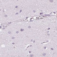
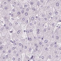
Immunohistochemistry protocol sheet for POMC (IHC for POMC): expected localisation Secreted; tissue subcellular pattern unreported (UniProt; HPA tissue IHC), antibody PB9039, validated IHC image, and IHC protocol steps
Printable POMC IHC protocol sheet — expected localisation Secreted; tissue subcellular pattern unreported (UniProt; HPA tissue IHC), antibody PB9039, controls and protocol steps. Open the full POMC IHC guide →

POMC Immunohistochemistry Experimental Design Guide

Expected localisation, validated protocols, controls and antibodies — the at-a-glance facts below, then the full design guide.

Must know before staining
Expected localisation Secreted; tissue subcellular pattern unreported (UniProt; HPA tissue IHC)
Staining pattern High staining in anterior pituitary cells; compartment unreported (HPA tissue IHC)
Antigen retrieval Citrate pH 6 HIER, heat-mediated (datasheet PB9039)
Positive control ⓘ Pituitary gland
Negative control ⓘ Adipose tissue+4 more · see all
Important caveats
Reasons your staining may differ from the expected pattern.
Fixation Keep fixation consistent across sections. (standard IHC practice; not target-specific)
Caveat Cleavage may change which POMC-derived peptides are detected (UniProt)
Regulation Expression regulator unreported (UniProt)
Isoform / epitope No annotated isoforms; processing can alter epitope availability (UniProt)
Section 1

Recommended POMC IHC & IF Protocols

Start with the catalog antibody’s paraffin-section IHC protocol (datasheet PB9039); the published options describe POMC staining in brain or sheep tissue (PMC8228876; PMC3583363; PMC13217381).

Recommended immunohistochemistry (IHC-P) protocol parameters
SampleParaffin-embedded mouse kidney tissues; fixative not specified (datasheet PB9039)
FixationImage fixative and duration unreported (datasheet PB9039); verify before use.
Sectioning4–5 µm sections on charged slides (standard)
DeparaffinisationXylene, graded ethanol series to water (standard)
Antigen retrievalHeat retrieval: Citrate pH 6, 20 min (datasheet PB9039)
Peroxidase block3% H2O2, 10 min, room temperature (standard)
Blocking10% goat serum (datasheet PB9039)
Primary antibodyRabbit anti-POMC, 0.5-1μg/ml (datasheet PB9039)
Primary incubationOvernight at 4 °C (datasheet PB9039)
DetectionStreptavidin-biotin complex (SABC), DAB chromogen (datasheet PB9039)
CounterstainHematoxylin, blue, dehydrate and mount (standard)
Expected resultPOMC-positive staining in cells in anterior of pituitary gland (HPA tissue IHC: High). HPA tissue profile: Highly expressed in the anterior lobe of the pituitary gland. No signal in the no-primary control.
💡Decision noteFor the catalog antibody, start with heat-mediated citrate retrieval at pH 6 (datasheet PB9039). The published excerpts do not specify antigen retrieval (PMC8228876; PMC3583363; PMC13217381).
Section 2

What Is the Expected POMC Staining Pattern?

POMC is a secreted, processed hormone precursor with no transmembrane segment (UniProt P01189 topology and processing). For chromogenic IHC on paraffin sections, expect the strongest staining in cells of the anterior pituitary (HPA: High in anterior pituitary cells). HPA rates the tissue pattern Enhanced, while cautioning that a secreted protein’s location may differ from its RNA source (HPA: tissue IHC reliability).

What am I looking at on my slide?
Strong staining in anterior pituitary cells, with a cellular or punctate cytoplasmic appearance.This fits the reported high anterior pituitary signal (HPA: High in cells in anterior). A secretory pattern is plausible from POMC biology, but HPA supplies no validated subcellular IHC compartment (UniProt P01189: secreted; HPA: tissue IHC).
Predominantly crisp nuclear staining or a continuous cell-surface outline.Neither is the expected pattern for a secreted precursor without a transmembrane segment (UniProt P01189 topology). Treat it as suspect and compare controls; the supplied HPA tissue record does not establish a precise intracellular pattern.
Strong signal in adipocytes or adrenal glandular cells.These cell types are reported as not detected (HPA: adipocytes; adrenal glandular cells). Consider cross-reactivity or endogenous chromogen-forming activity (general IHC practice), especially if the same signal persists without primary antibody.
Diffuse color across cells and surrounding tissue, with little distinction between positive and negative areas.The distribution does not resemble the enriched anterior pituitary pattern (HPA: tissue IHC). Nonspecific binding, excess detection signal, or endogenous activity are possible workflow causes (general IHC practice); extracellular signal alone is not proof of artifact for a secreted protein (UniProt P01189: secreted).
No signal in an otherwise interpretable anterior pituitary section.A negative result conflicts with the reported high staining in anterior cells (HPA: tissue IHC). Check the antibody’s IHC-P validation and the assay controls before interpreting the specimen as biologically negative (general IHC practice).
💡Expected POMC appearanceCall a convincing positive when anterior pituitary cells show strong, cell-associated staining, often plausibly cytoplasmic or punctate; isolated nuclear staining or equally strong staining in reported negative cell types warrants investigation (HPA: tissue IHC; UniProt P01189: secreted).
How each factor affects the staining
Tissue and cell choice (HPA: tissue IHC).Anterior pituitary cells are the supported positive comparison; adipocytes and adrenal glandular cells are reported not detected (HPA: High in cells in anterior; Not detected in adipocytes and adrenal glandular cells).
Secretory biology (UniProt P01189).POMC has a signal peptide and no transmembrane segment; released material may not remain confined to its producing cell (UniProt P01189 topology; HPA: secreted-protein location caveat). HPA does not specify an IHC organelle pattern.
Processing and epitope choice (UniProt P01189 processing).POMC is a precursor of pituitary hormones, including ACTH and MSH (UniProt P01189 function and tissue specificity). Without a supplied epitope map, the stain cannot identify which POMC-derived product the antibody detects.
Molecular modifications (UniProt P01189).UniProt lists glycosylation at residues 71 and 91 and several modified residues (UniProt P01189 PTMs). The payload gives no evidence that these alter IHC staining or antigen retrieval, so do not adjust interpretation on that basis.
Evidence strength and application (HPA: antibody validation).Four listed antibodies have Enhanced IHC validation (HPA: HPA046135, HPA063644, CAB002762, CAB079881). Their ICC entries are empty, and HPA provides no ICC-IF images; the IHC pattern does not validate an IF/ICC pattern (HPA: antibodies; subcellular record).
Why is my staining missing, weak or wrong?
SituationLikely causeNext action
Anterior pituitary positive control is blank (HPA: High in anterior cells).The assay may have failed, or the antibody may not detect the available epitope; the supplied record cannot distinguish these causes (general IHC practice; UniProt P01189 processing).Check section integrity, primary-antibody identity and IHC-P instructions, detection reagents, and a run control before changing retrieval or dilution (general IHC practice).
Strong signal appears in reported negative cells (HPA: adipocytes or adrenal glandular cells Not detected).Cross-reactivity or endogenous detection activity can mimic specific staining (general IHC practice).Compare a no-primary control and the positive tissue on the same run; investigate persistent signal before assigning POMC expression (general IHC practice; HPA: tissue IHC).
Staining is mainly nuclear or forms a sharp membrane rim.That pattern is difficult to reconcile with a secreted protein lacking a transmembrane segment (UniProt P01189 topology), although HPA provides no validated IHC subcellular location.Review morphology and no-primary controls, then compare with an independently IHC-validated antibody if available (general IHC practice; HPA: Enhanced IHC validation).
Background obscures the anterior pituitary cell pattern (HPA: High in cells in anterior).Nonspecific binding or excess chromogenic detection can reduce contrast (general IHC practice).Review blocking, antibody concentration, washes, detection time, and counterstain against the run controls (general IHC practice); no target-specific optimal settings are supplied.
Extracellular color is interpreted as evidence that every adjacent cell produces POMC.POMC is secreted, and HPA cautions that protein and RNA tissue locations may differ (UniProt P01189: secreted; HPA: reliability description).Score cell-associated staining separately from extracellular deposit and anchor cell-type calls to morphology and controls (general IHC practice; HPA: tissue IHC).
Can the IHC result establish an IF/ICC staining pattern?HPA lists no ICC validation or ICC-IF images and gives no main subcellular location (HPA: antibodies; subcellular record).No. Treat IF/ICC as unvalidated by this payload and consult its separate guide; do not transfer the IHC pattern or invent an IF protocol (HPA: ICC and subcellular records).

Sample controls for POMC IHC & IF

🧪Run pituitary gland first: cells in the anterior pituitary should stain strongly (HPA: High in cells in anterior). Use adipose tissue as a negative comparator, focusing on adipocytes (HPA: Not detected in adipocytes); on the pituitary slide, cells outside the stained population should show only background, but their negative status requires validation.
Positive control tissue: Pituitary gland (Cells in anterior, HPA High)
Negative control tissue: Adipose tissue (HPA Not detected)
ICC-IF cell lines (HPA subcellular resource): HPA carries no ICC-IF cell line for POMC; derive a cell-line control from the positive tissue's cell type (Cells in anterior) and confirm it by RNA or western blot first.
Technical controls: Include a no-primary, secondary-only control and an isotype control matched to the primary antibody’s host species and immunoglobulin class; use POMC knockout tissue or cognate peptide competition, where available, as a biological specificity control. Check endogenous peroxidase and biotin background in the pituitary section when using the caption’s biotin-based DAB detection (PB9039 tissue-IHC caption: biotinylated secondary, streptavidin-biotin complex and DAB).
⚠️Feasibility: No target-specific fixation window or fixation effect is reported in the supplied evidence, and the exact PB9039 tissue-IHC caption does not state a fixative. That caption reports citrate retrieval at pH 6 for 20 minutes in a paraffin-embedded mouse kidney section, but does not establish that retrieval is required for pituitary tissue (PB9039 tissue-IHC caption). The supplied evidence does not establish whether frozen sections or IF are easier, or identify a pituitary-specific artefact (HPA subcellular: no ICC-IF images; PB9039 tissue-IHC caption: mouse kidney).

HPA tissue IHC evidence for POMC

Comprehensive Human Protein Atlas IHC scoring per tissue (reliability: Enhanced — High consistency between antibody staining and RNA expression data. Secreted protein, tissue location of RNA and protein is expected to differ. Antibody staining consistent with GTEx and FANTOM5 RNA expression data.). Rows are taken directly from the HPA tissue chart — click any row's HPA link to view the source.

Positive expression · recommended positive controls

TissueCell typeLevelEvidenceSource
Pituitary gland Cells in anterior High Protein (IHC) HPA →

Undetected expression · recommended negative controls

TissueCell typeLevelEvidenceSource
Adipose tissue Adipocytes Not detected Protein (IHC) HPA →
Adrenal gland Glandular cells Not detected Protein (IHC) HPA →
Appendix Glandular cells Not detected Protein (IHC) HPA →
Bone marrow Hematopoietic cells Not detected Protein (IHC) HPA →
Breast Adipocytes Not detected Protein (IHC) HPA →
Section 3

Advanced POMC IHC Tips

Troubleshoot paraffin section POMC IHC with the ACTH directed catalog antibody, while distinguishing precursor staining from processed peptide detection (UniProt P01189; datasheet PB9039).

Which retrieval condition should I start with for POMC IHC?
Start with heat mediated citrate retrieval at pH 6 for 20 minutes in paraffin sections (datasheet PB9039). The selected image used this condition with the ACTH directed antibody at 1 μg/ml, followed by overnight incubation at 4°C (datasheet PB9039). If staining is weak, compare retrieval duration on adjacent sections while holding antibody concentration and detection constant, and inspect morphology after heating (standard IHC practice). Include anterior pituitary cells as a reference for expected strong staining, and compare sections processed together before attributing a change to retrieval (HPA: High in anterior pituitary cells; standard IHC practice).
How should I troubleshoot possible fixation effects on POMC staining?
Target specific sensitivity to fixation is unknown: the selected paraffin section caption does not state a fixative (datasheet PB9039). Record the actual fixative and processing history for each specimen, then compare sections with different histories using the same citrate pH 6 retrieval and detection conditions (datasheet PB9039; standard IHC practice). Inspect tissue preservation and compare signal in a concurrently processed positive reference before increasing antibody concentration (standard IHC practice). Do not infer a POMC fixation effect from its secreted topology, processing, or pituitary staining pattern; those records establish biological context rather than fixation performance (UniProt P01189; HPA: High in anterior pituitary cells).
Where should convincing POMC related staining appear in a section?
Assess staining in cells and their secretory compartment rather than requiring a membrane outline: POMC is a secreted precursor without a transmembrane segment (UniProt P01189 topology). Anterior pituitary cells provide the strongest tissue reference in the supplied IHC profile, with High staining reported there (HPA: High in anterior pituitary cells). The catalog image shows ACTH antibody staining in a paraffin embedded mouse kidney section, but its caption does not establish that every stained structure contains intact POMC (datasheet PB9039). Compare the cellular pattern, neighboring unstained areas, and negative controls before assigning extracellular or diffuse chromogen to specific POMC related signal (standard IHC practice).
Can this ACTH antibody distinguish intact POMC from processed peptides?
Do not interpret ACTH directed immunostaining as a measurement of intact POMC without epitope and cross reactivity evidence (datasheet PB9039; UniProt P01189 processing). POMC is a hormone precursor, and the record lists no annotated isoforms; cleavage and modification can change which molecular forms expose an epitope (UniProt P01189). The selected protocol establishes staining with this ACTH antibody at 1 μg/ml in paraffin sections, but supplies no peptide discrimination experiment (datasheet PB9039). If biological conclusions require a particular product, compare antibodies against documented distinct epitopes and use an appropriate peptide or genetic specificity control where available (standard IHC practice).
How would I adapt this POMC question to multiplex IF?
Treat IF as a separate assay requiring its own validation; the supplied catalog example documents chromogenic paraffin section IHC, and HPA lists no ICC/IF images (datasheet PB9039; HPA subcellular). Multiplex an ACTH directed signal with a validated marker for the expected anterior pituitary cell population, using single stain controls to assess overlap and spectral bleed through (HPA: High in anterior pituitary cells; standard IF practice). Choose fluorophores in channels with low specimen autofluorescence, assessed on an unstained section (standard IF practice). For an intracellular secretory epitope, compare mild permeabilisation with an unpermeabilised control; POMC has no transmembrane segment, so a membrane side assignment is unsupported (UniProt P01189 topology; standard IF practice).
What should I check when DAB background obscures POMC staining?
Check a no primary control and inspect whether diffuse deposit follows tissue edges, damaged regions, or endogenous enzyme activity (standard IHC practice). Include a peroxidase block before DAB development and check the biotin based detection system for tissue associated background with its appropriate control (standard IHC practice; datasheet PB9039: biotinylated secondary, SABC and DAB). The selected protocol used 10% goat serum blocking and 1 μg/ml primary antibody overnight at 4°C; match those conditions before adjusting them systematically (datasheet PB9039). Interpret low level background against a concurrently stained anterior pituitary reference rather than against chromogen intensity alone (HPA: High in anterior pituitary cells; standard IHC practice).
How should I score POMC related DAB staining across sections? ⚠ ANSWER MARKED FOR VERIFICATION
Define the anatomical region and eligible cells before scoring, then report the percentage of positive cells and staining intensity or an H-score on a 0–300 scale (standard IHC practice). Normalise positive cell counts to all eligible cells in that region, or report positive cell density per mm² when cell number varies substantially (standard IHC practice). Keep retrieval, antibody concentration, chromogen development, imaging, and threshold settings consistent across the comparison set (standard IHC practice). Label the result ACTH directed immunoreactivity rather than intact POMC abundance, because this staining protocol does not establish which processed form contributes to the signal (datasheet PB9039; UniProt P01189 processing).
How can I separate true POMC related signal from artefact?
Give greatest weight to reproducible cellular staining in anterior pituitary cells, where the supplied tissue profile reports High staining (HPA: High in anterior pituitary cells). Treat an isolated membrane outline cautiously because POMC is secreted and lacks a transmembrane segment (UniProt P01189 topology). Compare suspected positives with a no primary control and inspect edges, necrotic regions, and endogenous peroxidase signal before calling DAB deposit specific (standard IHC practice). The paraffin section image documents an ACTH antibody staining workflow in mouse kidney, but does not by itself identify intact POMC or establish a general kidney expression pattern (datasheet PB9039; UniProt P01189 processing).
Boster reagents

Best POMC / Pro-opiomelanocortin IHC Antibodies

Both antibodies list IHC reactivity with human, mouse, and rat (catalog applications/reactivity); PB9039 has paraffin-section IHC images from mouse kidney and rat brain and kidney (PB9039 image captions).

Real IHC data IHC analysis of ACTH using anti-ACTH antibody (PB9039). ACTH was detected in paraffin-embedded section of mouse kidney tissues. Heat mediated antigen retrieval was performed in citrate buffer (pH6, epitope retrieval solution) for 20 mins. The tissue section was blocked with 10% goat serum. The tissue section was then incubated with 1μg/ml rabbit anti-ACTH Antibody (PB9039) overnight at 4°C. Biotinylated goat anti-rabbit IgG was used as secondary antibody and incubated for 30 minutes at 37°C. The tissue section was developed using Strepavidin-Biotin-Complex (SABC)(Catalog # SA1022) with DAB as the chromogen.
Anti-ACTH/POMC Antibody
Cat # PB9039

PB9039 lists IHC for human, mouse, and rat and shows staining in paraffin sections of mouse kidney and rat brain and kidney (PB9039 applications/reactivity and image captions). M00305-1 lists IHC for human, mouse, and rat, but has no IHC image in the payload (M00305-1 applications/reactivity and image alts).

Which to pick: Choose PB9039 for tissue IHC when a demonstrated paraffin-section procedure matters; its captions specify citrate retrieval and 1 μg/ml primary antibody, but do not report the fixative (PB9039 image captions). M00305-1 is a rabbit monoclonal option for IHC across the three listed species, without a supplied IHC figure (M00305-1 clone, applications/reactivity and image alts). Both list human, mouse, and rat reactivity, but neither lists IF/ICC, so neither has catalog support here for an IF/ICC recommendation (catalog applications/reactivity).

Each figure is that product's own IHC / IF validation image from its datasheet.

References

  1. UniProt Consortium. UniProt entry P01189 (COLI_HUMAN, Pro-opiomelanocortin).
  2. Human Protein Atlas. POMC tissue IHC expression (reliability: Enhanced).
  3. Human Protein Atlas. POMC subcellular location (ICC-IF): Secreted.
  4. Human Protein Atlas. POMC antibody validation summary (4 antibodies).
  5. Hypothalamic Expression of Neuropeptide Y (NPY) and Pro-OpioMelanoCortin (POMC) in Adult Male Mice Is Affected by Chronic Exposure to Endocrine Disruptors. Metabolites 2021 — PMC8228876.
  6. Infrared heat treatment reduces food intake and modifies expressions of TRPV3-POMC in the dorsal medulla of obesity prone rats. International journal of hyperthermia : the official journal of European Society for Hyperthermic Oncology, North American Hyperthermia Group 2011 — PMC3583363.
  7. Activity of arcuate kisspeptin/neurokinin B neurons and the melanocortin system during pubertal development in female sheep†. Biology of reproduction 2025 — PMC13217381.
  8. Role of proopiomelanocortin neuron Stat3 in regulating arterial pressure and mediating the chronic effects of leptin. Hypertension (Dallas, Tex. : 1979) 2013 — PMC3678380.
  9. PubMed PMID:6274691 — UniProt-cited evidence.
  10. PubMed PMID:6299668 — UniProt-cited evidence.
  11. PubMed PMID:6314261 — UniProt-cited evidence.