This website uses cookies to ensure you get the best experience on our website.


- Table of Contents
Plan chromogenic POR IHC in paraffin sections using a catalog antibody with documented human tissue staining (datasheet M02166-2). Compare the observed cytoplasmic pattern with HPA tissue IHC, while using POR’s ER membrane location as molecular context (HPA tissue IHC; UniProt).

Expected localisation, validated protocols, controls and antibodies — the at-a-glance facts below, then the full design guide.
| Expected localisation | Cytoplasmic staining (HPA tissue IHC); ER membrane (UniProt) | |
| Staining pattern | General cytoplasmic staining, including glandular cells (HPA tissue IHC) | |
| Antigen retrieval | EDTA pH 8.0 HIER, heat-mediated (datasheet M02166-2) | |
| Positive control | Adrenal gland+4 more · see all | |
| Negative control | Heart muscle+1 more · see all |
| Fixation | Keep fixation consistent across sections (standard IHC practice; not target-specific); Matched tissue-IHC evidence does not establish the fixation claim. Validate the specimen-specific method before use. (selected-SKU IHC image M02166-2) | |
| Caveat | Medium antibody–RNA concordance complicates interpretation (HPA tissue IHC) | |
| Regulation | Liver-enhanced RNA expression (HPA tissue RNA) | |
| Isoform / epitope | No annotated isoforms; residues 43–677 are cytoplasmic (UniProt) |
The catalog antibody’s IHC-P protocol is accompanied by one published POR immunohistochemistry protocol in humanized mouse liver (PMC9437039).
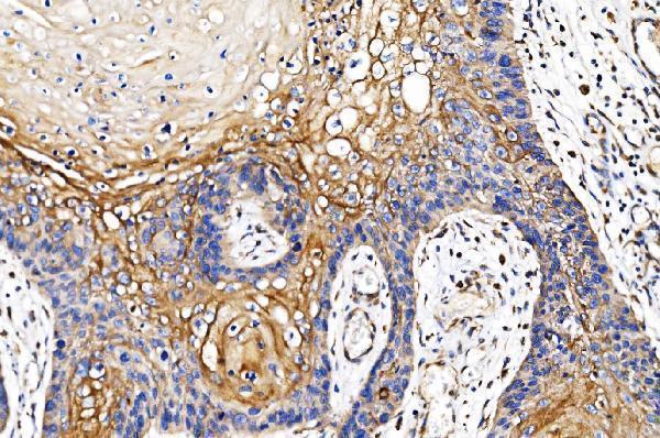
| Sample | Paraffin-embedded human esophageal squamous carcinoma tissue; fixative not specified (datasheet M02166-2) |
| Fixation | Image fixative and duration unreported (datasheet M02166-2); verify before use. |
| Sectioning | 4–5 µm sections on charged slides (standard) |
| Deparaffinisation | Xylene, graded ethanol series to water (standard) |
| Antigen retrieval | Heat retrieval: EDTA pH 8.0 (datasheet M02166-2); 20 min, 95–100 °C (standard) |
| Peroxidase block | 3% H2O2, 10 min, room temperature (standard) |
| Blocking | 10% goat serum (datasheet M02166-2) |
| Primary antibody | Mouse monoclonal (clone 7F5) anti-POR, 2 μg/ml (datasheet M02166-2) |
| Primary incubation | Overnight at 4 °C (datasheet M02166-2) |
| Detection | Streptavidin-biotin complex (SABC), DAB chromogen (datasheet M02166-2) |
| Counterstain | Hematoxylin, blue, dehydrate and mount (standard) |
| Expected result | POR-positive staining in glandular cells of adrenal gland (HPA tissue IHC: High). HPA tissue profile: General cytoplasmic expression. No signal in the no-primary control. |
POR is an endoplasmic reticulum membrane protein with a short lumenal segment and a predominantly cytoplasmic domain (UniProt P16435 topology). In paraffin tissue sections, expect mainly cytoplasmic staining, including strong staining in adrenal glandular cells and colon glandular cells (HPA tissue IHC). HPA rates the tissue IHC pattern Enhanced, while reporting medium consistency between antibody staining and RNA expression (HPA tissue IHC).
| Cytoplasmic staining in adrenal or colon glandular cells, with a clear counterstained nucleus. | This matches the reported high staining in those cell types and the general cytoplasmic tissue pattern (HPA tissue IHC). The ER membrane assignment supports an intracellular, largely cytoplasmic interpretation (UniProt P16435 topology). Judge intensity against controls processed in the same run (general IHC practice). |
| Predominantly nuclear staining in a paraffin section, with little convincing cytoplasmic signal. | This is discordant with the general tissue IHC pattern and warrants checking controls and staining distribution (HPA tissue IHC; general IHC practice). Do not treat all nuclear signal as disproving POR: HPA also reports supported nucleoplasmic localisation in ICC-IF, a different preparation (HPA subcellular ICC-IF). |
| Strong staining in cardiomyocytes or smooth muscle cells, especially when expected positive cells are weak. | HPA reports POR as not detected in those cell types, so the result raises concern about nonspecific binding or endogenous detection activity (HPA tissue IHC; general IHC practice). It does not alone identify which cause applies; examine reagent controls and morphology (general IHC practice). |
| Broad, uniform colour across cells, stroma, or empty areas that obscures cell boundaries. | This is background rather than a readable cell-specific pattern (general IHC practice). Compare a no-primary control and inspect blocking, washes, and chromogen development before scoring POR; HPA's high and not-detected calls apply to interpretable cellular staining (general IHC practice; HPA tissue IHC). |
| No detectable staining in an adrenal or colon section expected to contain glandular cells. | The absent signal conflicts with HPA's high staining in those cells, but a single negative run cannot establish absent POR (HPA tissue IHC; general IHC practice). Check that the expected cells are present, then assess the positive control and each detection step (general IHC practice). |
| Compartment and epitope location | POR has one membrane segment at residues 22–42 and a cytoplasmic region at 43–677 (UniProt P16435 topology). An antibody's epitope is not given here, so topology cannot specify its antigen-retrieval response or predict a unique punctate IHC pattern. |
| Tissue and cell context | HPA reports High staining in adrenal and colon glandular cells, Not detected in cardiomyocytes and smooth muscle cells, and low staining in skeletal myocytes (HPA tissue IHC). Use these as relative interpretation anchors; the HPA summary does not make every cell in a tissue a positive or negative control. |
| Evidence strength | HPA rates the tissue IHC pattern Enhanced and describes medium consistency with RNA expression; HPA010136 and CAB004372 have Enhanced IHC status (HPA tissue IHC; HPA antibodies). These support pattern comparison, while leaving room for sample-to-sample or assay differences. |
| Processing and variants | UniProt lists no isoforms, signal peptide, propeptide, or glycosylation sites, and annotates a 2–677 chain (UniProt P16435). These annotations do not establish a fixation effect or justify attributing an unexpected IHC pattern to a known alternative isoform or shedding event. |
| Situation | Likely cause | Next action |
|---|---|---|
| Expected glandular cells are negative in the same run as other stained tissue. | A failed primary, retrieval, or detection step is possible; the HPA High category does not establish a POR-specific retrieval requirement (HPA tissue IHC; general IHC practice). | Confirm cell identity and section quality, inspect the run's positive control, then check primary dilution, retrieval conditions, and detection reagents against the validated IHC procedure (general IHC practice). |
| Cardiomyocytes or smooth muscle cells show strong colour. | Nonspecific antibody binding or endogenous chromogenic detection activity could mimic a positive signal; HPA reports POR as Not detected in these cells (general IHC practice; HPA tissue IHC). | Compare no-primary and detection-only controls, review morphology, and optimise blocking or endogenous-enzyme quenching where appropriate for the detection system (general IHC practice). |
| Colour covers the section without distinct positive cells. | Excess background can follow insufficient blocking or washing, concentrated reagents, or prolonged chromogen development (general IHC practice). | Use a no-primary control to locate background, then adjust blocking, washes, reagent concentration, or development time one variable at a time (general IHC practice). |
| Signal appears confined to nuclei in paraffin tissue. | The result differs from HPA's general cytoplasmic tissue IHC profile, although supported nucleoplasmic localisation is reported for ICC-IF (HPA tissue IHC; HPA subcellular ICC-IF). | Check cytoplasmic staining in a known positive cell population and review no-primary controls before assigning nuclear IHC signal to POR (HPA tissue IHC; general IHC practice). |
| A negative comparison tissue has faint staining. | Low signal need not equal a failed control: HPA reports low staining in skeletal myocytes, while cardiomyocytes and smooth muscle cells are Not detected (HPA tissue IHC). | Identify the cell type before scoring, compare intensity with the run's positive tissue, and report faint signal separately from strong cell-specific staining (HPA tissue IHC; general IHC practice). |
| Q: Should IF/ICC show the same compartment pattern as paraffin IHC? | HPA tissue IHC reports general cytoplasmic staining; HPA ICC-IF reports mainly vesicles, with additional nucleoplasm and cytosol (HPA tissue IHC; HPA subcellular ICC-IF). | A: Interpret each preparation against its own HPA localisation evidence. Do not use the ICC-IF vesicle call as a required chromogenic IHC pattern (HPA tissue IHC; HPA subcellular ICC-IF). |


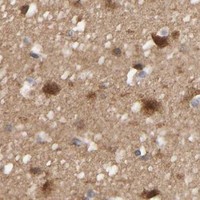
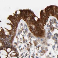

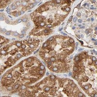


Comprehensive Human Protein Atlas IHC scoring per tissue (reliability: Enhanced — Medium consistency between antibody staining and RNA expression data.). Rows are taken directly from the HPA tissue chart — click any row's HPA link to view the source.
| Tissue | Cell type | Level | Evidence | Source |
|---|---|---|---|---|
| Adrenal gland | Glandular cells | High | Protein (IHC) | HPA → |
| Appendix | Glandular cells | High | Protein (IHC) | HPA → |
| Bone marrow | Hematopoietic cells | High | Protein (IHC) | HPA → |
| Bronchus | Respiratory epithelial cells | High | Protein (IHC) | HPA → |
| Cerebral cortex | Neuronal cells | High | Protein (IHC) | HPA → |
Troubleshoot POR staining in paraffin sections by checking retrieval, cell type, compartment, and controls before interpreting signal intensity.
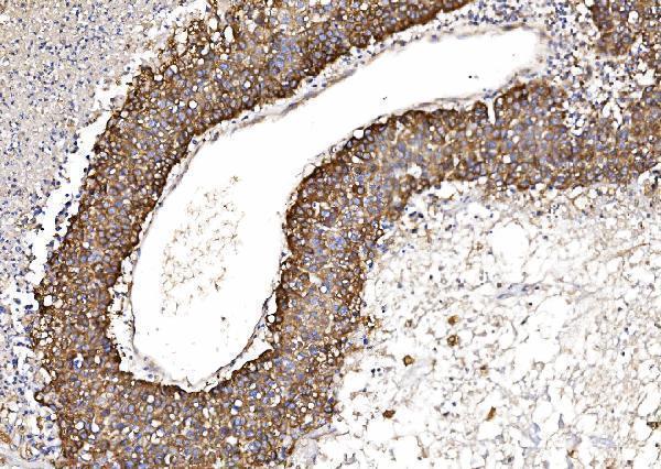
Anti-POR IHC images cover paraffin-embedded human carcinoma and placenta sections, plus mouse and rat tissues (catalog image captions: M02166-2, PB9736, PA1952); PA1952 also has a HeLa ICC image (catalog image caption).
M02166-2 has IHC images from human esophageal squamous carcinoma, liver cancer, lung cancer and placenta sections; PB9736 has images from human mammary cancer and mouse and rat intestine sections (catalog IHC image captions). PA1952 has IHC images from human placenta and rat ovary sections, plus a HeLa ICC image (catalog image captions).
Which to pick: For human paraffin-section IHC, M02166-2 is a mouse monoclonal with human tissue images (catalog host, clone and IHC image captions). For cross-species IHC, PB9736 lists human, mouse and rat reactivity and has a paraffin-section image for each species (catalog reactivity and IHC image captions). For cell imaging, PA1952 lists ICC and has a HeLa ICC image (catalog applications and image caption); the rendered cards have no IF figure (catalog image captions). The IHC captions document paraffin sections but do not report the fixative (catalog IHC image captions).