This website uses cookies to ensure you get the best experience on our website.


- Table of Contents
Plan PPARD paraffin-section IHC around the nuclear staining reported in most tissues (HPA tissue IHC). Compare staining with tissue controls while accounting for the reported low consistency between antibody staining and RNA levels (HPA tissue IHC).

Expected localisation, validated protocols, controls and antibodies — the at-a-glance facts below, then the full design guide.
| Expected localisation | Nuclear staining in most tissues (HPA tissue IHC) | |
| Staining pattern | Nuclear signal in most tissues; high in several glandular cell types (HPA tissue IHC) | |
| Antigen retrieval | Tris-EDTA pH 9.0 HIER, 95–98 °C, 20 min (rule: nuclear antigen) | |
| Positive control | Adrenal gland+4 more · see all | |
| Negative control | Heart muscle+3 more · see all |
| Fixation | Keep fixation conditions consistent across sections. (standard IHC practice; not target-specific) | |
| Caveat | Antibody staining and RNA levels show low consistency (HPA tissue IHC) | |
| Regulation | Expression regulation unspecified (UniProt) | |
| Isoform / epitope | 4 isoforms; epitope coverage is unresolved (UniProt) |
The catalog antibody IHC-P protocol is accompanied by published PPARD staining methods for bovine corpus luteum and yak tissue sections (PMC7297635; PMC11855600).
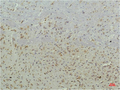
| Sample | Paraffin-embedded Mouse Brain tissue; fixative not specified (datasheet M01557) |
| Fixation | Image fixative and duration unreported (datasheet M01557); verify before use. |
| Sectioning | 4–5 µm sections on charged slides (standard) |
| Deparaffinisation | Xylene, graded ethanol series to water (standard) |
| Antigen retrieval | Heat-induced epitope retrieval in Tris-EDTA buffer, pH 9.0, 20 min at 95–98 °C (standard rule: nuclear antigen) |
| Peroxidase block | 3% H2O2, 10 min, room temperature (standard) |
| Blocking | 10% normal serum of the secondary host, 30 min, room temperature (standard) |
| Primary antibody | Mouse monoclonal (clone 2F9) anti-PPARD, 1:100 recommended; image 1:200 (datasheet M01557) |
| Primary incubation | Overnight at 4 °C (standard) |
| Detection | HRP-polymer secondary, DAB chromogen 5–10 min (standard) |
| Counterstain | Hematoxylin, blue, dehydrate and mount (standard) |
| Expected result | PPARD-positive staining in glandular cells of adrenal gland (HPA tissue IHC: High). HPA tissue profile: Nuclear expression in most tissues. No signal in the no-primary control. |
PPARD is a nuclear receptor without a transmembrane segment (UniProt Q03181: subcellular location and topology). In paraffin sections, expect nuclear staining in many tissues, including glandular cells of the colon and breast and squamous epithelial cells of the cervix (HPA: tissue IHC). HPA rates the tissue profile Approved but reports low consistency between antibody staining and RNA expression; interpret any single tissue result with that limitation (HPA: reliability description).
| Distinct nuclear staining in colon glandular cells, with nuclei identifiable against the counterstain. | This fits PPARD's nuclear location and an HPA High staining example (UniProt Q03181: subcellular location; HPA: High in colon glandular cells). Judge the cellular pattern as well as signal strength; a brown deposit alone does not identify a positive nucleus (general IHC practice). |
| Predominantly cytoplasmic staining, especially when nuclei remain unstained. | That distribution conflicts with the reported nuclear location (UniProt Q03181: subcellular location; HPA: nuclear expression in most tissues). Review controls and staining conditions before calling it PPARD; morphology, counterstain and primary omission can help identify misplaced or nonspecific signal (general IHC practice). |
| Strong staining in an unexpected cell population while the expected cells remain pale. | Compare the named cell population, rather than the organ alone, with HPA's observations (HPA: tissue IHC). Cross-reactivity or endogenous chromogen activity is possible; use a primary-omission control and inspect where deposit sits in the section (general IHC practice). |
| Diffuse brown haze across tissue and spaces between cells. | Diffuse deposit lacks the nuclear, cell-associated pattern reported for PPARD (UniProt Q03181: subcellular location; HPA: tissue IHC). Background can arise during blocking, antibody incubation, washing or detection; assess a primary-omission section alongside the stained slide (general IHC practice). |
| No nuclear signal in colon glandular cells or another HPA High cell population. | An absent result in a reported High population calls for checking the assay before inferring absent PPARD (HPA: High in colon glandular cells; general IHC practice). HPA's Approved profile has low antibody–RNA consistency, so its High calls are useful comparators, not guarantees for every specimen (HPA: reliability description). |
| Subcellular location | PPARD is reported in the nucleus and has no transmembrane segment; score nuclei within identifiable cells (UniProt Q03181: subcellular location and topology; general IHC practice). |
| Cell-specific tissue contrast | HPA reports High staining in colon glandular cells but Not detected in prostate glandular cells. This is an observed IHC contrast, not a universal absence claim (HPA: tissue IHC). |
| RNA–protein agreement | The tissue profile is Approved despite low consistency between antibody staining and RNA expression. Do not use RNA alone to settle an ambiguous slide (HPA: reliability description; general IHC practice). |
| Antibody evidence | CAB017635 is a mouse monoclonal antibody with Approved IHC status; the payload does not report an Enhanced IHC rating or ICC validation for it (HPA: antibody validation). |
| Isoforms and epitope uncertainty | UniProt lists 4 isoforms and an NR ligand-binding domain at residues 211–439. The supplied evidence does not locate this antibody's epitope or establish isoform coverage (UniProt Q03181: isoforms and domains; HPA: antibody listing). |
| Situation | Likely cause | Next action |
|---|---|---|
| Colon glandular nuclei appear unstained despite a working chromogenic run. | The result differs from HPA's High example; the supplied sources do not identify a PPARD-specific fixation or retrieval failure (HPA: High in colon glandular cells). | Check section integrity, counterstain, detection controls and the catalog antibody's IHC-P instructions; review routine retrieval only within that validated workflow (general IHC practice). |
| Brown signal appears mainly in cytoplasm or along cell edges. | The compartment conflicts with the reported nuclear pattern; morphology or nonspecific deposit may explain it (UniProt Q03181: nucleus; HPA: nuclear expression in most tissues; general IHC practice). | Compare stained and primary-omission sections at the same magnification, confirm intact nuclei, and avoid scoring cytoplasmic colour as nuclear positivity (general IHC practice). |
| A suspected negative control stains strongly. | HPA's Not detected calls describe observed cells, not an absolute absence across specimens; unwanted detection signal is another possibility (HPA: tissue IHC; general IHC practice). | Confirm the exact cell type, assess nuclear localisation, and use a primary-omission control to investigate endogenous detection activity (HPA: tissue IHC; general IHC practice). |
| Skeletal-muscle myocytes lack staining even though UniProt says skeletal-muscle expression is maximal. | These source statements measure different things: UniProt reports tissue specificity, while HPA reports myocyte IHC as Not detected (UniProt Q03181: tissue specificity; HPA: skeletal muscle myocytes). | Record the discrepancy and judge the slide against additional HPA High cell populations and assay controls; do not force a positive myocyte call (HPA: tissue IHC; general IHC practice). |
| Patchy nuclear staining is hard to distinguish from uneven background. | HPA reports nuclear expression in most tissues, but its Approved tissue profile has low antibody–RNA consistency; patchiness alone cannot resolve specificity (HPA: profile and reliability description). | Score only identifiable nuclei in a stated cell population, compare a primary-omission section, and document distribution separately from intensity (general IHC practice). |
| Q: Can this IHC pattern establish the expected IF/ICC result? | HPA provides no ICC-IF images or main subcellular location in this payload, and CAB017635 has no listed ICC validation (HPA: subcellular and antibody records). | A: Use nuclear PPARD localisation as a biological expectation, but assess IF/ICC validation on its own guide page; this IHC evidence does not establish an IF/ICC staining pattern (UniProt Q03181: nucleus; HPA: ICC-IF and antibody records). |
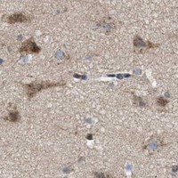


Comprehensive Human Protein Atlas IHC scoring per tissue (reliability: Approved — Low consistency between antibody staining and RNA expression data.). Rows are taken directly from the HPA tissue chart — click any row's HPA link to view the source.
| Tissue | Cell type | Level | Evidence | Source |
|---|---|---|---|---|
| Adrenal gland | Glandular cells | High | Protein (IHC) | HPA → |
| Breast | Glandular cells | High | Protein (IHC) | HPA → |
| Cervix | Squamous epithelial cells | High | Protein (IHC) | HPA → |
| Colon | Glandular cells | High | Protein (IHC) | HPA → |
| Epididymis | Glandular cells | High | Protein (IHC) | HPA → |
| Tissue | Cell type | Level | Evidence | Source |
|---|---|---|---|---|
| Heart muscle | Cardiomyocytes | Not detected | Protein (IHC) | HPA → |
| Liver | Cholangiocytes | Not detected | Protein (IHC) | HPA → |
| Prostate | Glandular cells | Not detected | Protein (IHC) | HPA → |
| Skeletal muscle | Myocytes | Not detected | Protein (IHC) | HPA → |
Troubleshoot PPARD staining by checking nuclear localisation, retrieval, controls and cell-specific scoring alongside the available tissue evidence (UniProt Q03181; HPA tissue IHC).
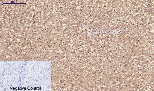
M01557 has IHC images of paraffin-embedded mouse brain and human liver, lung, and uterus (IHC image captions); IF is listed, but no IF image is supplied (catalog applications; IF image alts).
M01557 is the only SKU shown, with IHC images of paraffin-embedded mouse brain and human liver, lung, and uterus (IHC image captions). Its listed applications are IHC and IF, and its listed reactivity is Human, Mouse, and Rat (catalog applications; catalog reactivity).
Which to pick: Choose M01557 for tissue IHC: its mouse brain image specifies paraffin-embedded tissue, while its human liver, lung, and uterus captions report IHC without specifying processing; the fixative is unreported (IHC image captions). M01557 is also the IF option because IF is listed, although no IF image or separate ICC validation is supplied (catalog applications; IF image alts). For work across Human, Mouse, and Rat, M01557 is the listed mouse monoclonal option; the supplied IHC images cover Mouse and Human (catalog host and clone; catalog reactivity; IHC image captions).