This website uses cookies to ensure you get the best experience on our website.
- Table of Contents
2 Citations 1 Q&As
1 Citations 6 Q&As
Facts about Protein phosphatase 1 regulatory subunit 12A.
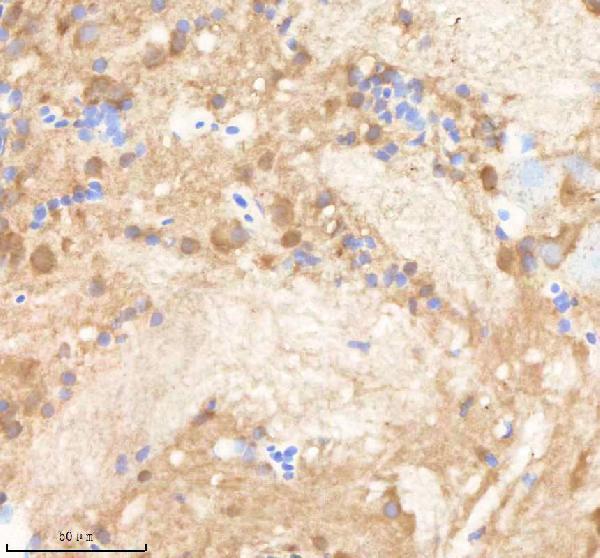
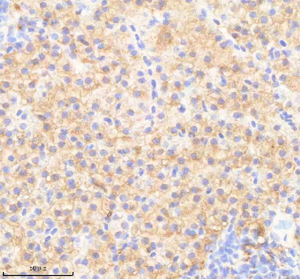
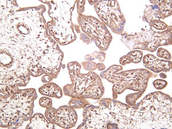
As part of the PPP1C complex, involved in dephosphorylation of PLK1. Capable of inhibiting HIF1AN- dependent suppression of HIF1A activity.
Human | |
---|---|
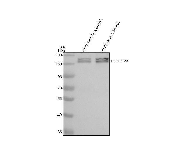
Gene Name: | PPP1R12A |
Uniprot: | O14974 |
Entrez: | 4659 |
Belongs to: |
---|
No superfamily |
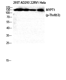
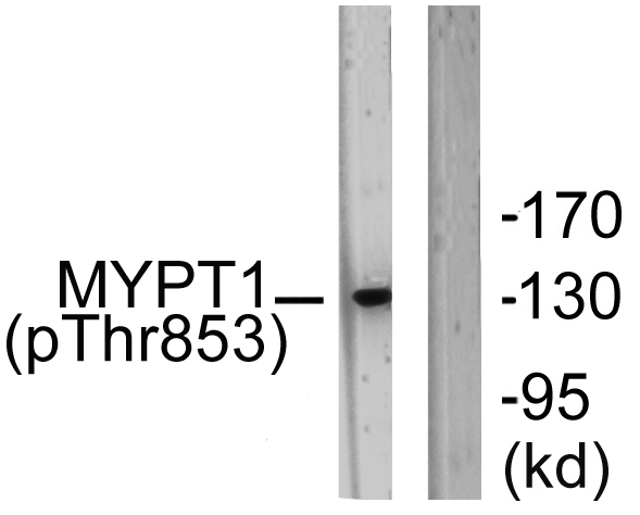
MBSmyosin phosphatase, target subunit 1; MGC133042; Myosin phosphatase target subunit 1; Myosin phosphatase-targeting subunit 1; MYPT1protein phosphatase 1 regulatory subunit 12A; protein phosphatase 1, regulatory (inhibitor) subunit 12A; Protein phosphatase myosin-binding subunit
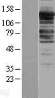
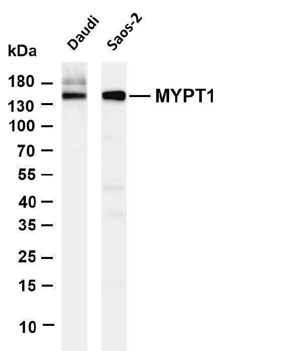
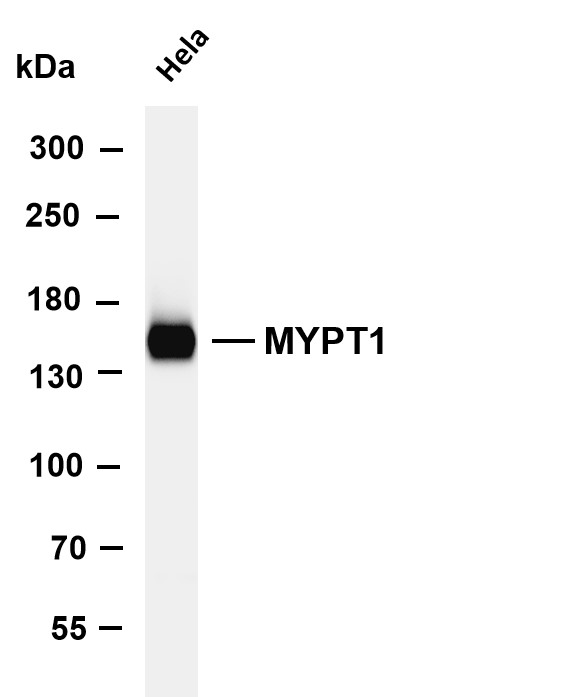
Mass (kDA):
115.281 kDA
Human | |
---|---|
Location: | 12q21.2-q21.31 |
Sequence: | 12; NC_000012.12 (79773563..79935455, complement) |
Expressed in striated muscles, specifically in type 2a fibers (at protein level).
Cytoplasm. Cytoplasm, cytoskeleton, stress fiber. Also along actomyosin filaments.
If you are seeking information about the Boster Bio Anti-Myosin Phosphatase/PPP1R12A Marker Read this article. This article will discuss specific biological processes and applications for this product. Whether you're an individual researcher, an organization, or a business, there's an Boster Bio product for you.
This antibody recognizes N-terminal regions of the human Myosin Phosphataser (MP). The catalytic domain of MP is very similar to the one found in the related proteins of rats and mice. In addition to myosin phosphorylation PPC also interacts with the transcription and the actin cytoskeleton.
This antibody recognizes myosin the process of phosphorylation that is necessary for muscle contraction. It binds myosin . It can be utilized in muscle tissue to observe the activity of muscle cells and also to monitor muscle contractility. The antibody is available as a soluble powder for blood and tissue samples. It is highly specific for myosin, a protein found in human blood.
There are many applications for the PPP1R12A indicator. It can be used to determine the levels of proteins in samples, as well as the levels of antibodies to specific proteins and even disease. The PPP1R12A gene closely resembles MP, a protein found in a variety of cells of the body. This gene can help detect autoimmune disease and its manifestations. It is also used for studying the immune response to chemotherapy and other drugs.
PSMC5 expression and PPP1R12A mRNA expression were observed in biliary, colon and pancreatic Adenocarcinomas. In addition to breast cancer These markers were also found in both HER-2+ and ER+ pancreatic cancers. It was also detected in lung, liver and biliary cancers. These findings may be a clue to the future of immunotherapy for solid malignancies even though they are preliminary.
Molecular Biology and the PPP1R12.A marker are both associated with prostate cancer. The PPP1R12A gene is present in both tumors and normal tissues and is involved in the formation of prostate cancer, is present in both tumors as well as normal tissue. TCGA identified five promising genes from three datasets. We also conducted a subgroup analysis in the TCGA cohort and assessed the levels of protein expression for PPP1R12A.
The Hippo pathway regulates the growth and fate of cells in the body. The Hippo pathway is composed of a complex network proteins which include MST1/2, an MST1/2 kinase-like kinase, and LATS1/2, a major tumor suppressor kinase. The pathway is activated by NF2 phosphorylating MST1/2, while the PPP1R12A marker dephosphorylates NF2.
The five-gene profile associated with PPP1R12A and metabolism in PCa is a highly effective predictor of future health. It has been proven to predict biochemical relapse-free and disease-free survival. These findings, verified by two datasets from TCGA and one from Gene Expression Omnibus, can aid in identifying patients at high risk of developing PCa. They may also assist in developing new treatment strategies for those with PCa.
The TCGA PRAD dataset contains 338 DEGs that are associated with PPP1R12A. These genes are expressed in different ways in different tissues. The top 350 DEGs with PPP1R12A expression are shown on the heatmap graphs. The heatmap plots display the top 340 DEGs that have high PPP1R12A levels. The red shade represents tissues with low levels of PPP1R12A. The KEGG pathway analysis shows the KEGG pathways that are closely related to PPP1R12A.
These are the most effective uses for PPP1R12A. This marker belongs to the myosin-phosphatase proteins family. It is involved in the activation of other proteins in the cytosol. These include actin. This protein plays a role in regulating the cell cycle. It also plays a role in preventing formation of tumors.
PMID: 9286714 by Takahashi N., et al. Localization of the gene coding for myosin phosphatase, target subunit 1 (MYPT1) to human chromosome 12q15-q21.
PMID: 11342221 by Machida H., et al. Molecular cloning and analysis of the 5'-flanking region of human MYPT1 gene.
*More publications can be found for each product on its corresponding product page