This website uses cookies to ensure you get the best experience on our website.
- Table of Contents
2 Citations 7 Q&As
1 Citations 7 Q&As
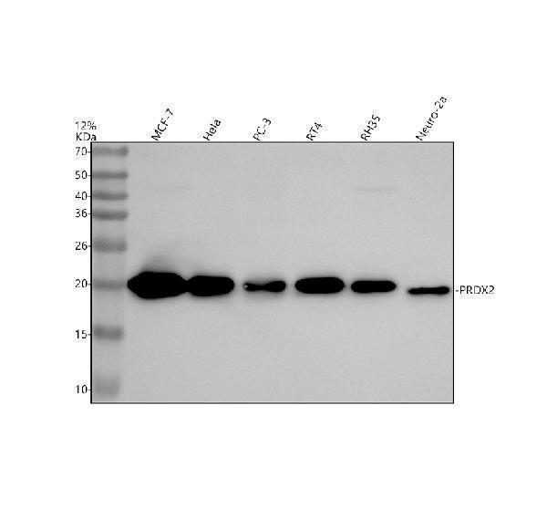
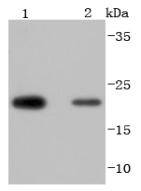
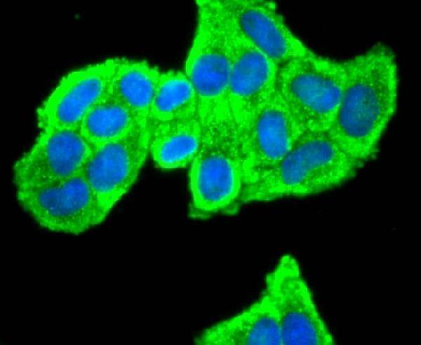
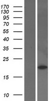
Facts about Peroxiredoxin-2.
Might participate in the signaling cascades of growth factors and tumor necrosis factor-alpha by regulating the intracellular concentrations of H(2)O(2). .
Human | |
---|---|
Gene Name: | PRDX2 |
Uniprot: | P32119 |
Entrez: | 7001 |
Belongs to: |
---|
peroxiredoxin family |
EC 1.11.1; MGC4104; Natural killer cell-enhancing factor B; natural killer-enhancing factor B; NKEF-B; NKEFBNKEF-B; Peroxiredoxin 2; peroxiredoxin-2; PRDX2; PRPTDPX1; PRX2; PRXII; TDPX1; thiol-specific antioxidant 1; Thiol-specific antioxidant protein; Thioredoxin peroxidase 1; Thioredoxin-dependent peroxide reductase 1; torin; TPX1; TSA; TSAEC 1.11.1.15
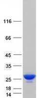
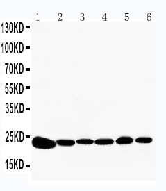
Mass (kDA):
21.892 kDA
Human | |
---|---|
Location: | 19p13.13 |
Sequence: | 19; NC_000019.10 (12796823..12801800, complement) |
Cytoplasm.
Steven Boster created a new antibody for the detection peroxiredoxin-2. This antibody is approved for use in Flow Cytometry. This article reviews its Historie and Efficacy in Histology. You can also learn more about his research on the PRDX2 protein.
Both the Historie gene PRDX2 (and its gene product, PRDX2) are important for cardiovascular diseases. Their diverse functions include regulation of TLR4, VEGF, myocyte hypertrophy, acute myocardial infarction, and inflammatory responses. We will discuss the role of PRDX2 and review the various research on PRDX2.
A representative study on bladder and colorectal carcinomas revealed an increased expression level of PRDX1 & PRDX2 in the tumors. Both markers were found to be significantly elevated in the urines of patients, but not to be associated with the recurrence or progression of disease. These studies revealed the need for further research to determine which marker may be useful in identifying patients suffering from invasive bladder cancer.
The PRDX2 nuclear/cytosolic relationship remained constant when there was circadian regulation. PRDX2 nuclear levels were stable under normal conditions. They did however show a detrended oscillation of Bmal1 promoter activation (Fig. 1A). The AUCs of PRDX 2 and 4 for the PRDX1 gene product are also significantly different.
To determine the serum PRDX2 concentration in urine, enzyme-linked immunosorbent assays were used. These tests use a microtiter plate coated with biotin-conjugated antibody. The standards are then added in the ordered. The plates are then filled with 100 uL of standards and samples. Afterwards, avidin-conjugated horseradish peroxidase and a sulfuric acid solution are added to the wells, and the color changes are monitored at 450 nm.
This study showed that Prx2 is linked to b-2 globulins, but not with the GN. These findings suggest that Prx2 may have a greater impact on the consumption of peroxides. This is not surprising, given the fact that Prx2 is a very effective H2O2 scavenger. Prx2 may also be an important molecule to prevent cancer.
It is not clear what role PRDX2 plays in gastric cancer. The enzyme is involved in gastric tumorigenesis and is associated with decreased progression-free and overall survival. Infection by H. pilori activates and increases PRDX2 gene expression. Thus, future therapeutic approaches may benefit patients with gastric cancer. Further research is needed in order to determine if PRDX2 is involved with the development of gastric carcinoma.
In vitro studies show that overexpression of the PRDX2 marker enhances the migration and invasion of NSCLC cells. These results suggest PRDX2 as a potential target of the circDIDO1.
Although the biological role of Prdx2 is still unknown in trophoblasts, it may play an important role in RM. Prdx2 regulates cell death and also inhibits cell proliferation. It may also play an important role in the pathogenesis of RM. Prdx2-targeted techniques may offer new insights into the disease.
The PRDX2 genes is a protumor regulatory gene. It is involved in tumor initiation and progression. Multiple studies have investigated the role PRDX2 plays in cancer. PRDX2 expression was highest among A549 cells, and lowest in NCI-h2299 cell lines. Further research is needed to evaluate the biological significance of this gene in the development of cancer. Improvements in treatment are needed.
It is interesting to note that drugs can inhibit cell proliferation by downregulating the PRDX2 protein gene. While PRDX2 knockdown results in cell proliferation inhibition, upregulation increases expression of p21. It did not affect total expression of p53. Treatment with 5mM NAC almost restored the expression of p-p53. Prdx2 expression was also reduced in vitro by treatment with 5 mM NAC. This is a result of apoptosis.
Using a qRTPCR method, the PRDX2 mRNA levels were measured. The mRNA data were more reliable than the proteins results, since the latter method measures both levels of mRNA. The mRNA results are more precise and sensitive than the protein data, which gives a new perspective to the protein results. These methods can be useful tools in cancer research. These methods can be applied to cancer cell lines, or used in clinical trials to test patients.
It isn't clear whether the PRDX2 Gene directly affects chemosensitivity of patients. If a tumor has a high PRDX2 concentration, knocking this gene down will inhibit colony growth and decrease tumor weight in a mouse-xenograft model. In vitro and vivo, REV7 overexpression results in radioresistance. The presence and activity of nuclear PRDX2 are reduced when REV7 is knocked down.
Histology as well as cytology make up the majority of the Histology/cytology market. In this report, you'll learn about key players, revenue forecasts, and market sizes for each region and Type. The report also discusses key players in the United States market and focuses on application-specific market size. Here are some frequently asked questions regarding cytology and histology. Download the report to learn more!
The process of staining cell tissues with antibodies is called immunohistochemistry. The antibodies used in immunohistochemistry can be used to detect antigens (or haptens) in cells. These are molecules found in the human body. Boster Bio optimizes its antigens, dilution and cell concentrations for optimal detection. In addition, it offers Multiplex Immunohistochemistry services, which enable detection of multiple targets with a single assay. This allows for high throughput during screening and fast assay processing.
While antibodies are vital for histological research they are just one piece of the puzzle. Boster Bio has over 12,000 antibody samples, many of which have been validated to work in multiple applications. Boster Bio antibodies have also been optimized for multiplex IHC/WB and validated against panels of 250 tissue samples. In addition, Boster Bio's antibodies are tested quantitatively, against known quantities of recombinant proteins and untransfected cell lines.
ELISA - a simple technique for detecting antigens - can also be used for immunofluorescence. In this method, antibodies target specific antigens like DNA. ELISA is a simple way to measure the concentrations of proteins and antigens within cells. The immune system can also detect the presence of foreign materials, such as bacteria. It is important to understand the method before you use it to analyze the results.
PMID: 8144038 by Lim Y.-S., et al. The thiol-specific antioxidant protein from human brain: gene cloning and analysis of conserved cysteine regions.
PMID: 8026862 by Shau H., et al. Cloning and sequence analysis of candidate human natural killer- enhancing factor genes.
*More publications can be found for each product on its corresponding product page