PRDX6 / Peroxiredoxin-6 · IHC design guide

Design Immunohistochemistry for PRDX6

Plan PRDX6 paraffin-section IHC around the broad cytoplasmic staining reported in tissue (HPA tissue IHC). This guide covers fixation consistency, antigen retrieval, antibody dilution and chromogenic detection, with lower-staining cell types in mind (HPA tissue IHC).

Evidence assembled Oct 2026 · For research use; verify linked source records and product datasheet before use
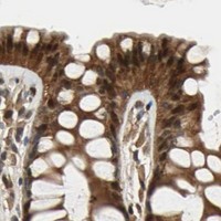
Immunohistochemistry protocol sheet for PRDX6 (IHC for PRDX6): expected localisation Cytoplasmic staining (HPA tissue IHC), antibody PB9350, validated IHC image, and IHC protocol steps
Printable PRDX6 IHC protocol sheet — expected localisation Cytoplasmic staining (HPA tissue IHC), antibody PB9350, controls and protocol steps. Open the full PRDX6 IHC guide →

PRDX6 Immunohistochemistry Experimental Design Guide

Expected localisation, validated protocols, controls and antibodies — the at-a-glance facts below, then the full design guide.

Must know before staining
Expected localisation Cytoplasmic staining (HPA tissue IHC)
Staining pattern Broad cytoplasmic staining across tissue cell types (HPA tissue IHC)
Antigen retrieval Citrate pH 6 HIER, heat-mediated (datasheet PB9350)
Positive control ⓘ Adipose tissue+4 more · see all
Negative control ⓘ None in HPA (detected in all 45 tissues); use no-primary + isotype controls
Important caveats
Reasons your staining may differ from the expected pattern.
Fixation Keep fixation consistent across sections (standard IHC practice; not target-specific); Matched tissue-IHC evidence does not establish the fixation claim. Validate the specimen-specific method before use. (selected-SKU IHC image PB9350)
Caveat Urothelial cells may stain weakly despite broad expression (HPA tissue IHC)
Regulation Broad expression; cell levels vary (HPA tissue IHC)
Isoform / epitope No annotated isoforms; no extracellular epitope expected (UniProt)
Section 1

Recommended PRDX6 IHC & IF Protocols

The catalog antibody protocol is followed by published PRDX6 IHC methods for myocardial infarction and abdominal aortic aneurysm samples (PMC3459299; PMC5146935).

Recommended immunohistochemistry (IHC-P) protocol parameters
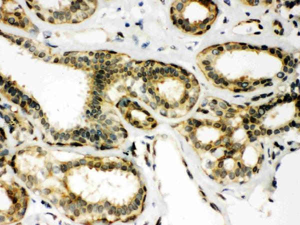
SampleParaffin-embedded Human Mammary Cancer tissues; fixative not specified (datasheet PB9350)
FixationImage fixative and duration unreported (datasheet PB9350); verify before use.
Sectioning4–5 µm sections on charged slides (standard)
DeparaffinisationXylene, graded ethanol series to water (standard)
Antigen retrievalHeat retrieval: Citrate pH 6, 20 min (datasheet PB9350)
Peroxidase block3% H2O2, 10 min, room temperature (standard)
Blocking10% goat serum (datasheet PB9350)
Primary antibodyRabbit anti-PRDX6, 0.5-1μg/ml (datasheet PB9350)
Primary incubationOvernight at 4 °C (datasheet PB9350)
DetectionStreptavidin-biotin complex (SABC), DAB chromogen (datasheet PB9350)
CounterstainHematoxylin, blue, dehydrate and mount (standard)
Expected resultPRDX6-positive staining in adipocytes of adipose tissue (HPA tissue IHC: High). HPA tissue profile: Ubiquitous cytoplasmic expression. No signal in the no-primary control.
💡Decision noteStart with heat-mediated citrate retrieval at pH 6 for the catalog antibody (datasheet: PB9350); the included articles do not state retrieval conditions (PMC3459299; PMC5146935).
Section 2

What Is the Expected PRDX6 Staining Pattern?

PRDX6 should show predominantly cytoplasmic staining in paraffin sections, consistent with its cytoplasmic and lysosomal localization and lack of a transmembrane segment (UniProt P30041). High staining is reported in adipocytes, bronchial respiratory epithelial cells, bone marrow hematopoietic cells, and several other cell populations (HPA tissue IHC). HPA describes the tissue pattern as ubiquitous cytoplasmic expression, with Supported reliability and medium agreement between staining and RNA expression (HPA tissue IHC).

What am I looking at on my slide?
Cytoplasmic staining is strong in bronchial respiratory epithelial cells or adipocytes.This matches reported High staining in those cells (HPA tissue IHC) and cytoplasmic localization (UniProt P30041). Compare identifiable cells within the section; staining across several cell types can also fit HPA’s ubiquitous profile (HPA tissue IHC).
Signal is confined to nuclei or extracellular material, with little cytoplasmic staining.A nucleus-only or extracellular-only result conflicts with the reported cytoplasmic tissue pattern (HPA tissue IHC; UniProt P30041). Treat it as a localization warning and check controls, morphology, and detection conditions before interpreting it as PRDX6 (standard IHC practice).
Intense staining appears mainly in fibroblasts while expected high-staining cells are weak.HPA reports Low staining in soft-tissue fibroblasts and High staining in several other cell populations (HPA tissue IHC). This mismatch raises concern for cross-reactivity or detection background; cell identity and a no-primary control help distinguish them (standard IHC practice).
A broad brown haze obscures cell borders and compartment boundaries.The slide cannot establish the reported cytoplasmic pattern while diffuse background dominates (HPA tissue IHC). Check the no-primary control and review blocking, washes, and chromogen development as general IHC troubleshooting steps (standard IHC practice).
A known high-staining tissue has no detectable signal in its expected cells.Absence in bronchial respiratory epithelial cells, for example, conflicts with HPA’s High tissue IHC result (HPA tissue IHC). First check that the relevant cells are present and that positive-control tissue and detection reagents worked (standard IHC practice); one negative section alone does not establish biological absence.
💡Expected PRDX6 appearanceCall positive staining when identifiable high-staining cells show a predominantly cytoplasmic chromogenic signal (HPA tissue IHC; UniProt P30041); a nucleus-only, extracellular-only, or cell-independent brown deposit is suspect (HPA tissue IHC; standard IHC practice).
How each factor affects the staining
Choice of tissue and cell populationHPA reports High staining in adipocytes, bronchial respiratory epithelial cells, and bone marrow hematopoietic cells, but Low staining in esophageal squamous epithelial cells, urothelial cells, and soft-tissue fibroblasts (HPA tissue IHC). Score the specified cell population rather than the section as a whole (standard IHC practice).
Expected compartmentUniProt places PRDX6 in the cytoplasm and lysosome, including lung lamellar bodies, and reports no transmembrane segment (UniProt P30041). HPA tissue IHC describes ubiquitous cytoplasmic expression (HPA tissue IHC). These annotations support a cytoplasmic IHC call; they do not require every positive cell to show resolvable lysosomal puncta.
Strength of antibody evidenceBoth listed antibodies have Supported IHC status, while HPA rates the overall tissue profile Supported with medium staining–RNA consistency (HPA antibodies; HPA tissue IHC). Use the reported pattern as a reference, with controls and morphology when assessing an unexpected result (standard IHC practice).
IF/ICC Q&A: where is PRDX6 seen?HPA reports mainly plasma membrane and cytosol signal in ICC-IF, with plasma membrane approved and cytosol supported (HPA subcellular). That IF observation can inform compartment interpretation; it does not replace the cytoplasmic tissue IHC reference (HPA tissue IHC).
Chromogenic detection backgroundEndogenous enzyme activity can produce chromogen signal independent of primary-antibody binding (standard IHC practice). A no-primary control helps test that possibility; the supplied HPA and UniProt records do not identify PRDX6-specific endogenous-detection interference.
Why is my staining missing, weak or wrong?
SituationLikely causeNext action
Bronchial respiratory epithelial cells appear unstained.They are reported as High in HPA tissue IHC; missing cells or a failed IHC run can account for an uninformative result (HPA tissue IHC; standard IHC practice).Confirm epithelial morphology and inspect a positive-control section; then review the documented retrieval, antibody dilution, and detection steps for the antibody in use (standard IHC practice).
Only nuclei show strong signal.Nucleus-only staining disagrees with cytoplasmic and lysosomal localization (UniProt P30041) and HPA’s cytoplasmic tissue profile (HPA tissue IHC).Compare with a no-primary control and a known high-staining cell population; reassess localization before assigning a positive PRDX6 score (standard IHC practice).
Fibroblasts stain more strongly than expected high-staining cells.HPA lists soft-tissue fibroblasts as Low, so that reversal raises concern for nonspecific binding or detection background (HPA tissue IHC; standard IHC practice).Verify cell identity and compare controls and adjacent cell populations; report the unexpected distribution rather than treating fibroblast intensity alone as validation (standard IHC practice).
Brown color covers cells and surrounding tissue uniformly.Diffuse deposit can obscure the cytoplasmic distribution needed for interpretation (HPA tissue IHC; standard IHC practice).Check no-primary staining, washes, blocking, and chromogen development; repeat with conditions that preserve clear cellular boundaries (standard IHC practice).
Granular cytoplasmic staining is difficult to classify.UniProt includes lysosomes among PRDX6 locations, but the tissue IHC reference specifies a broader cytoplasmic profile (UniProt P30041; HPA tissue IHC).Score whether granules lie within identifiable cytoplasm and compare the overall cell distribution with HPA’s reported high-staining populations (HPA tissue IHC; standard IHC practice).
A low-staining population is used as the sole negative control.HPA reports Low staining in some populations but lists no negative tissue population (HPA tissue IHC). Low expression cannot serve as an established absence control.Use a no-primary control to assess detection background and a reported high-staining population to assess the positive pattern (HPA tissue IHC; standard IHC practice).

Sample controls for PRDX6 IHC & IF

🧪Run adipose tissue first and require staining in adipocytes (HPA: High in adipocytes). HPA detects PRDX6 in all 45 scored tissues, so use no-primary and isotype controls in place of a negative tissue; cells without specific signal on the positive slide should show only counterstain or background and should not be treated as validated PRDX6-negative cells (HPA: all 45 tissues detected; standard IHC practice).
Positive control tissue: Adipose tissue (Adipocytes, HPA High)
Negative control tissue: None in HPA: PRDX6 is detected in all 45 scored tissues. Use a no-primary (secondary-only) and an isotype control instead.
ICC-IF cell lines (HPA subcellular resource): HPA ICC-IF images show PRDX6 in A-431, U-251MG, U2OS, with annotated localisation: Plasma membrane (approved), Cytosol (supported) (HPA subcellular).
Technical controls: Run a no-primary, secondary-only control and a concentration-matched rabbit IgG isotype control; use PRDX6 knockout material or validated immunizing-peptide competition to assess specificity (PB9350 caption: rabbit primary; standard IHC practice). Block endogenous peroxidase and assess endogenous biotin background with the reported SABC/DAB detection; check adipose autofluorescence if adapting the assay to IF (PB9350 caption: SABC/DAB; standard IHC/IF practice).
⚠️Feasibility: No target-specific fixation window or fixation effect is reported, and the selected PB9350 paraffin-section caption does not state a fixative (PB9350 caption). That example uses citrate retrieval at pH 6 for 20 minutes, but does not establish that retrieval is required (PB9350 caption). The supplied evidence does not establish whether frozen sections or IF are easier for PRDX6; paraffin processing can leave lipid-extraction spaces in adipose tissue, and adipose autofluorescence should be checked for IF (standard IHC/IF practice).

HPA tissue IHC evidence for PRDX6

Comprehensive Human Protein Atlas IHC scoring per tissue (reliability: Supported — Medium consistency between antibody staining and RNA expression data.). Rows are taken directly from the HPA tissue chart — click any row's HPA link to view the source.

Positive expression · recommended positive controls

TissueCell typeLevelEvidenceSource
Adipose tissue Adipocytes High Protein (IHC) HPA →
Adrenal gland Glandular cells High Protein (IHC) HPA →
Appendix Endocrine cells High Protein (IHC) HPA →
Bone marrow Hematopoietic cells High Protein (IHC) HPA →
Breast Adipocytes High Protein (IHC) HPA →

Undetected expression · recommended negative controls

TissueCell typeLevelEvidenceSource
None in HPA: PRDX6 is detected in all 45 scored tissues. Use a no-primary (secondary-only) and an isotype control instead.
Section 3

Advanced PRDX6 IHC Tips

Troubleshoot PRDX6 staining in paraffin sections using the catalog antibody’s IHC conditions, then assess signal by compartment, cell type and controls.

Which antigen retrieval condition should I start with for PRDX6 paraffin sections?
Start with heat-mediated retrieval in citrate buffer at pH 6 for 20 minutes (datasheet PB9350). The catalog antibody’s paraffin-section image used this condition with 1 μg/mL primary antibody overnight at 4°C, so keep those settings stable during the first retrieval comparison (datasheet PB9350). If staining is weak, compare a longer citrate heating time on matched sections while monitoring tissue integrity and background (standard IHC practice). Include a section processed without primary antibody to distinguish retrieval-related background from antibody-dependent signal (standard IHC practice). Record heating and cooling times because temperature exposure affects reproducibility (standard IHC practice).
How should I handle fixation when PRDX6 staining is weak?
The catalog image describes a paraffin section but does not state its fixative, so PRDX6-specific fixation sensitivity is unknown (datasheet PB9350). Record the fixative, fixation duration and tissue processing history before changing the staining procedure (standard IHC practice). Compare sections with known processing histories using the same pH 6 citrate retrieval and 20-minute heating condition (datasheet PB9350; standard IHC practice). If archival blocks stain differently, assess section quality and control staining before attributing the difference to PRDX6 abundance (standard IHC practice). Neither the tissue staining profile nor protein features establish a target-specific fixation effect (HPA: tissue IHC; UniProt P30041).
Is membranous PRDX6 staining plausible when I expected cytoplasm?
Expect a broad cytoplasmic pattern in tissue sections, while allowing for cell-specific variation (HPA: ubiquitous cytoplasmic expression). PRDX6 is annotated in cytoplasm and lysosomes, with a note about lung lamellar bodies (UniProt P30041: subcellular location). Cell imaging also places it mainly at the plasma membrane and in cytosol, so a membrane-associated signal warrants assessment against cellular boundaries rather than automatic rejection (HPA: subcellular). PRDX6 has no annotated transmembrane segment; membrane association alone does not establish that it spans the membrane (UniProt P30041: topology). Compare the pattern with a no-primary control and adjacent intact cells to assess nonspecific outlines (standard IHC practice).
Could an isoform or modified epitope explain uneven PRDX6 staining?
The supplied record lists 0 isoforms, so it provides no named alternative isoform to explain distinct staining patterns (UniProt P30041: isoforms). PRDX6 has a thioredoxin domain at residues 5–169 and annotated modifications at several residues, including phosphorylation at 44 and 177 (UniProt P30041: domains; modified residues). The antibody epitope is not supplied, so no specific modification can be linked to loss of recognition (datasheet PB9350: epitope not stated). Compare matched sections under identical retrieval and detection conditions before interpreting uneven intensity as biological variation (standard IHC practice). Use an independent antibody with a documented epitope if epitope dependence becomes the experimental question (standard IHC practice).
How can I assess PRDX6 by multiplex IF after seeing IHC staining?
Treat IF conditions as a separate optimization because the supplied catalog image documents paraffin-section chromogenic IHC, with no fixative stated (datasheet PB9350). Pair PRDX6 with a marker identifying the cell population being assessed; respiratory epithelial cells and adipocytes are examples of populations with high tissue staining (HPA: bronchus respiratory epithelial cells; breast adipocytes). Select a fluorophore after checking autofluorescence in the specimen, using a red or far-red channel when shorter wavelengths are crowded (standard IF practice). Optimize gentle permeabilisation for access to cytosolic epitopes, while checking whether membrane-associated signal persists; the antibody epitope is unspecified (UniProt P30041: topology; HPA: subcellular; datasheet PB9350: epitope not stated). Include single-color and no-primary controls for multiplex interpretation (standard IF practice).
What should I change when PRDX6 DAB staining has high background?
The catalog image used 10% goat serum blocking, biotinylated goat anti-rabbit secondary antibody, a streptavidin-biotin detection complex and DAB (datasheet PB9350). Check a no-primary section and a secondary-only section to locate background arising after primary incubation (standard IHC practice). Include an endogenous peroxidase block before DAB detection, and assess whether endogenous biotin contributes to signal in a streptavidin-biotin workflow (standard IHC practice). Titrate the primary around the documented 1 μg/mL condition and shorten DAB development only after checking that expected cellular staining remains visible (datasheet PB9350; standard IHC practice). Compare background within the same section because PRDX6 tissue expression is widespread (HPA: ubiquitous cytoplasmic expression).
How should I score PRDX6 staining across tissue sections? ⚠ ANSWER MARKED FOR VERIFICATION
Define the cell population and compartment before scoring, because PRDX6 tissue staining is broadly cytoplasmic and varies across listed cell types (HPA: tissue IHC). For cellular intensity, report an H-score using the percentages of cells at each intensity level; also report the percentage positive when intensity is difficult to classify (standard IHC practice). For spatial comparisons, measure positive-cell density per mm² of viable tissue and state how fields were selected (standard IHC practice). Normalize counts to the number of eligible cells or viable area, and use identical illumination, DAB development and scoring thresholds across specimens (standard IHC practice). Report cytoplasmic and membrane-associated scores separately when both are visible (HPA: tissue IHC; HPA: subcellular; standard IHC practice).
How do I distinguish genuine PRDX6 staining from artefact?
Look for staining in intact cells with a plausible cytoplasmic or membrane-associated distribution (HPA: tissue IHC; HPA: subcellular). High signal in adipocytes or respiratory epithelial cells can be consistent with the reported tissue profile, but those cells alone do not establish antibody specificity (HPA: breast adipocytes; bronchus respiratory epithelial cells; standard IHC practice). Treat staining limited to section edges, folds or necrotic regions as suspect and compare it with internal intact tissue (standard IHC practice). A no-primary control helps reveal endogenous enzyme or detection-system signal during DAB development (standard IHC practice). PRDX6’s lysosomal annotation can inform a punctate pattern, but tissue IHC alone cannot prove organelle identity (UniProt P30041: subcellular location; standard IHC practice).
Boster reagents

Best PRDX6 / Peroxiredoxin-6 IHC Antibodies

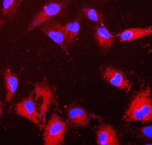
Two anti-PRDX6 antibodies have paraffin-section IHC images from human tissue; one also has an IF image from A549 cells (PB9350 and M01847-1 image captions).

Real IHC data IHC analysis of PRDX6 using anti-PRDX6 antibody (PB9350). PRDX6 was detected in paraffin-embedded section of Human Mammary Cancer tissues. Heat mediated antigen retrieval was performed in citrate buffer (pH6, epitope retrieval solution) for 20 mins. The tissue section was blocked with 10% goat serum. The tissue section was then incubated with 1μg/ml rabbit anti-PRDX6 Antibody (PB9350) overnight at 4°C. Biotinylated goat anti-rabbit IgG was used as secondary antibody and incubated for 30 minutes at 37°C. The tissue section was developed using Strepavidin-Biotin-Complex (SABC)(Catalog # SA1022) with DAB as the chromogen.
Anti-Peroxiredoxin 6/PRDX6 Antibody ®
Cat # PB9350
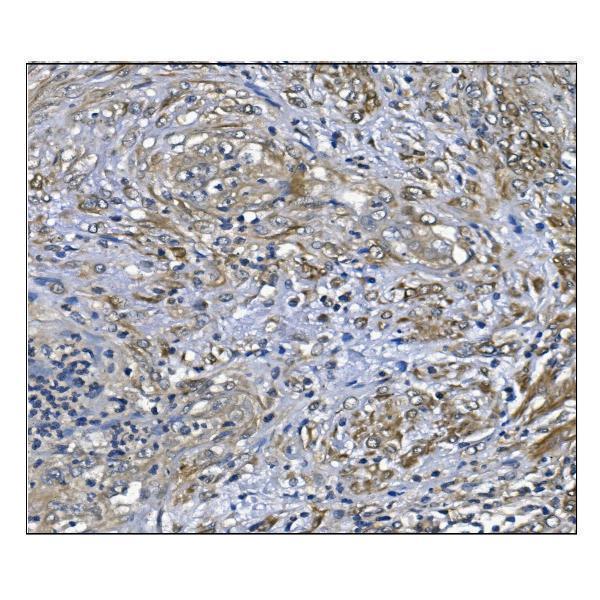
Real IHC data IHC analysis of PRDX6 using anti-PRDX6 antibody (M01847-1). PRDX6 was detected in paraffin-embedded section of human endometrial carcinoma tissue. Heat mediated antigen retrieval was performed in EDTA buffer (pH8.0, epitope retrieval solution). The tissue section was blocked with 10% goat serum. The tissue section was then incubated with 1μg/ml mouse anti-PRDX6 Antibody (M01847-1) overnight at 4°C. Biotinylated goat anti-mouse IgG was used as secondary antibody and incubated for 30 minutes at 37°C. The tissue section was developed using Strepavidin-Biotin-Complex (SABC) (Catalog # SA1021) with DAB as the chromogen.
Anti-PRDX6 Antibody ® (monoclonal, 6I8)
Cat # M01847-1

PB9350 has a human mammary cancer IHC image and an ICC image from PC-3 cells (PB9350 image captions). M01847-1 has a human endometrial carcinoma IHC image and an A549 IF image (M01847-1 image captions).

Which to pick: For tissue IHC, PB9350 has a shown paraffin-section result using citrate retrieval at pH 6 for 20 minutes; the fixative is unreported (PB9350 IHC caption). For IF/ICC, choose M01847-1: both applications are listed, and its A549 IF image uses Cy3 detection and DAPI counterstaining; its own paraffin-section IHC image uses EDTA retrieval at pH 8, with fixative unreported (M01847-1 catalog applications and IF/IHC captions). For cross-species planning, PB9350 lists Human, Mouse and Rat reactivity, while M01847-1 lists Human and Rat; both shown IHC images use human tissue (catalog reactivity; PB9350 and M01847-1 IHC captions).

Each figure is that product's own IHC / IF validation image from its datasheet.

References

  1. UniProt Consortium. UniProt entry P30041 (PRDX6_HUMAN, Peroxiredoxin-6).
  2. Human Protein Atlas. PRDX6 tissue IHC expression (reliability: Supported).
  3. Human Protein Atlas. PRDX6 subcellular location (ICC-IF): Mainly localized to the plasma membrane and cytosol..
  4. Human Protein Atlas. PRDX6 antibody validation summary (2 antibodies).
  5. PRDX6 knockout restrains the malignant progression of intrahepatic cholangiocarcinoma. Medical oncology (Northwood, London, England) 2022 — PMC9547796.
  6. Overexpression of Peroxiredoxin 6 (PRDX6) Promotes the Aggressive Phenotypes of Esophageal Squamous Cell Carcinoma. Journal of Cancer 2018 — PMC6218759.
  7. Effects of buyang huanwu decoction on ventricular remodeling and differential protein profile in a rat model of myocardial infarction. Evidence-based complementary and alternative medicine : eCAM 2012 — PMC3459299.
  8. Quantitative HDL Proteomics Identifies Peroxiredoxin-6 as a Biomarker of Human Abdominal Aortic Aneurysm. Scientific reports 2016 — PMC5146935.
  9. PubMed PMID:8999971 — UniProt-cited evidence.
  10. PubMed PMID:9050990 — UniProt-cited evidence.
  11. PubMed PMID:7788527 — UniProt-cited evidence.