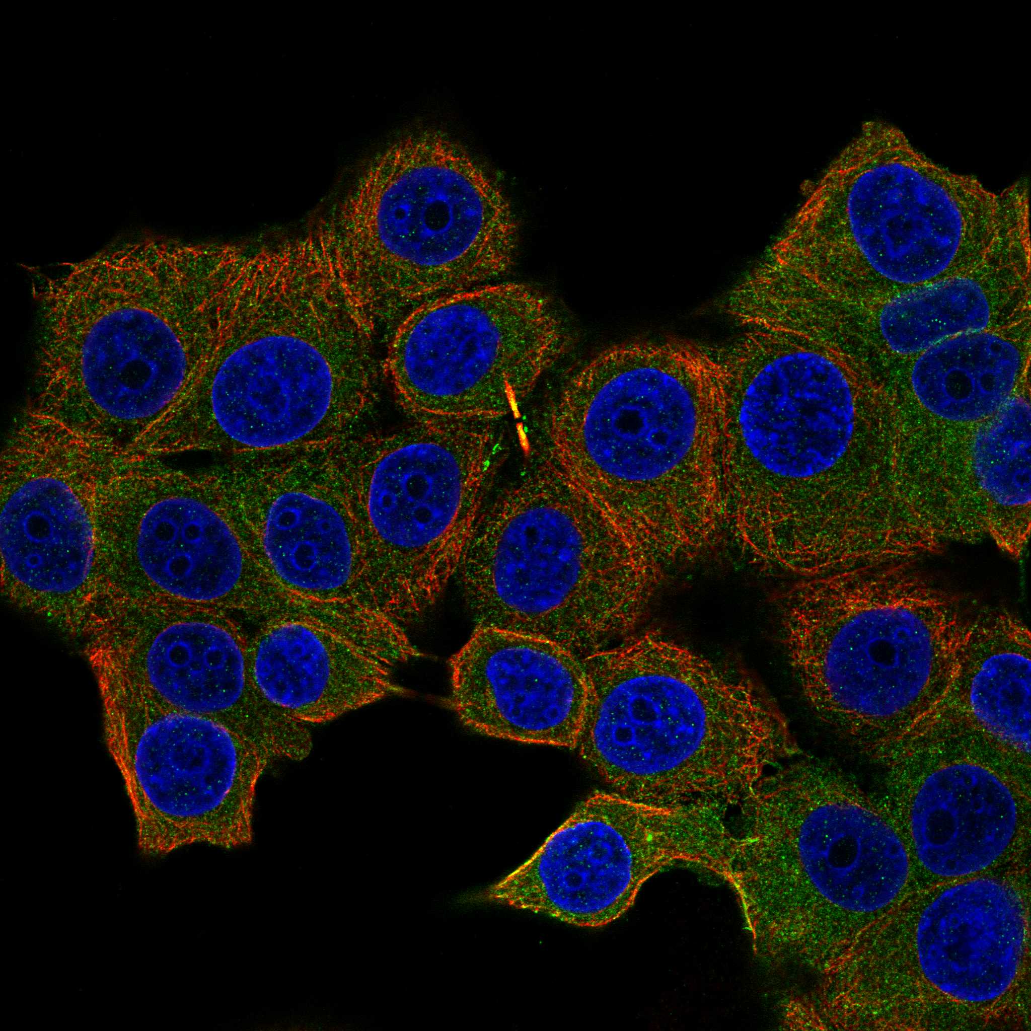
PRKACA / cAMP-dependent protein kinase catalytic subunit alpha · IHC design guide

Design Immunohistochemistry for PRKACA

Use the HPA cytoplasmic tissue pattern and high staining in cortical neurons and seminiferous duct cells as PRKACA IHC references (HPA tissue IHC). Compare the catalog antibodies’ specificity and IHC dilutions before scoring stained cell types and compartments (datasheets; standard IHC practice).

Evidence assembled Oct 2026 · For research use; verify linked source records and product datasheet before use
Immunohistochemistry protocol sheet for PRKACA (IHC for PRKACA): expected localisation Cytoplasmic tissue staining (HPA tissue IHC); nuclear translocation is possible (UniProt), antibody M00653-1, validated IHC image, and IHC protocol steps
Printable PRKACA IHC protocol sheet — expected localisation Cytoplasmic tissue staining (HPA tissue IHC); nuclear translocation is possible (UniProt), antibody M00653-1, controls and protocol steps. Open the full PRKACA IHC guide →

PRKACA Immunohistochemistry Experimental Design Guide

Expected localisation, validated protocols, controls and antibodies — the at-a-glance facts below, then the full design guide.

Must know before staining
Expected localisation Cytoplasmic tissue staining (HPA tissue IHC); nuclear translocation is possible (UniProt)
Staining pattern Widespread cytoplasmic staining across tissue cell types (HPA tissue IHC)
Antigen retrieval Tris-EDTA pH 9.0 HIER, 95–98 °C, 20 min (rule: nuclear antigen)
Positive control ⓘ Cerebral cortex+4 more · see all
Negative control ⓘ None in HPA (detected in all 45 tissues); use no-primary + isotype controls
Important caveats
Reasons your staining may differ from the expected pattern.
Fixation Keep fixation consistent across paraffin sections (standard IHC practice; not target-specific)
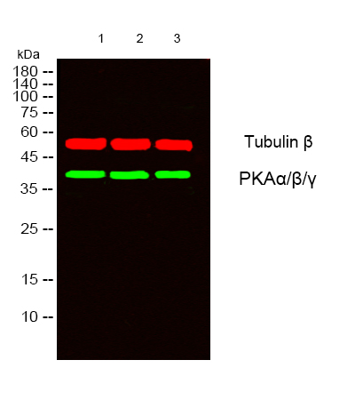
Caveat A00653 also targets PKA β/γ catalytic subunits (datasheet: A00653)
Regulation Isoform 2 enriched in pachytene cells (UniProt)
Isoform / epitope 2 isoforms; isoform 2 is sperm-specific; check epitope coverage (UniProt)
Section 1

Recommended PRKACA IHC & IF Protocols

Compare the catalog antibody’s IHC-P protocol with published PRKACA IHC methods for FFPE breast tumors and adrenal tissues (PMC4261061; PMC5427838).

Recommended immunohistochemistry (IHC-P) protocol parameters
SampleTissue sections; selected-image fixative not specified (standard IHC workflow)
FixationImage fixative and duration unreported (datasheet M00653-1); verify before use.
Sectioning4–5 µm sections on charged slides (standard)
DeparaffinisationXylene, graded ethanol series to water (standard)
Antigen retrievalHeat-induced epitope retrieval in Tris-EDTA buffer, pH 9.0, 20 min at 95–98 °C (standard rule: nuclear antigen)
Peroxidase block3% H2O2, 10 min, room temperature (standard)
Blocking10% normal serum of the secondary host, 30 min, room temperature (standard)
Primary antibodyRabbit anti-PRKACA, 1:2000-1:10000 (datasheet M00653-1)
Primary incubationOvernight at 4 °C (standard)
DetectionHRP-polymer secondary, DAB chromogen 5–10 min (standard)
CounterstainHematoxylin, blue, dehydrate and mount (standard)
Expected resultPRKACA-positive staining in neuronal cells of cerebral cortex (HPA tissue IHC: High). HPA tissue profile: Ubiquitous cytoplasmic expression. No signal in the no-primary control.
💡Decision noteStart with Tris-EDTA pH 9.0, 95–98 °C, 20 min (page retrieval); compare citrate pH 6.5, 13 min for adrenal sections (PMC5427838).
Section 2

What Is the Expected PRKACA Staining Pattern?

PRKACA should show predominantly cytoplasmic staining in paraffin sections, including neuronal cells in cerebral cortex and cells in testis seminiferous ducts (HPA: ubiquitous cytoplasmic expression; High in both cell groups). Nuclear or membrane-associated signal can also be biologically plausible (UniProt P17612: subcellular location). PRKACA has no transmembrane segment (UniProt P17612: topology). HPA rates its tissue IHC profile Approved, with medium consistency between staining and RNA data (HPA: tissue IHC reliability).

What am I looking at on my slide?
Clear cytoplasmic staining in cortical neuronal cells or cells in testis seminiferous ducts.This matches the strongest supplied tissue examples: HPA reports High staining in both cell groups (HPA: tissue IHC). Compare identifiable cells with their surrounding tissue and controls; a brown deposit alone does not establish cell identity (standard IHC practice).
Nuclear or membrane-adjacent staining accompanies a convincing cytoplasmic pattern.Do not reject it solely by compartment: free catalytic subunit can enter the nucleus, and PRKACA can associate with membranes (UniProt P17612: subcellular location). Judge whether the pattern is cellular and reproducible with the same staining workflow (standard IHC practice).
Staining is confined to luminal debris, extracellular material, or an implausible cell population.A deposit without a coherent cellular pattern warrants an artefact check (standard IHC practice). Because HPA describes ubiquitous cytoplasmic expression and supplies no negative tissue, staining in an unexpected cell population alone cannot prove cross-reactivity (HPA: tissue IHC).
Weak, widespread brown haze obscures cell borders and compartment assignment.Treat the slide as difficult to interpret until background is controlled (standard IHC practice). Compare a no-primary control and review blocking, antibody concentration, washes, and detection chemistry; haze cannot be scored as PRKACA localisation (standard IHC practice).
No cellular signal appears in a cortical neuronal or testis seminiferous-duct positive reference.This conflicts with HPA's High observations in those cell groups, but does not by itself show biological absence (HPA: tissue IHC). Check section quality and the staining workflow with appropriate controls before interpreting the test specimen (standard IHC practice).
💡Expected PRKACA appearanceA credible positive is distinct, mainly cytoplasmic cellular staining, expected to be High in cortical neuronal cells or cells in testis seminiferous ducts (HPA: tissue IHC); diffuse haze or deposits outside cells are suspect (standard IHC practice).
How each factor affects the staining
Tissue and cell choiceCerebral cortex neuronal cells and testis seminiferous-duct cells provide High examples; adipocytes and adrenal glandular cells are Medium (HPA: tissue IHC). HPA lists Low examples but no negative tissue, so a Low example is a poor absence control (HPA: tissue IHC).
CompartmentCytoplasm is the main tissue IHC expectation (HPA: ubiquitous cytoplasmic expression). UniProt also lists nucleus, membrane, mitochondrion, and cilium among locations; those annotations do not establish that each compartment will resolve in routine chromogenic sections (UniProt P17612: subcellular location; standard IHC practice).
Isoform coverageIsoform 1 is ubiquitous; isoform 2 is sperm-specific, enriched in pachytene spermatocytes and undetected in round spermatids (UniProt P17612: tissue specificity). The supplied record gives no antibody epitope, so a testis stain cannot be assigned to either isoform from this evidence alone (UniProt P17612: isoforms).
Antibody evidenceCAB010361 has Approved IHC status in the supplied HPA list (HPA: antibodies). The tissue profile is Approved with medium staining–RNA consistency, so it supports a pattern expectation without establishing that every positive deposit is specific (HPA: tissue IHC reliability; standard IHC practice).
IF/ICC: should it show the same location?Expect mainly cytosolic signal, but treat finer structures cautiously: HPA marks cytosol, microtubules, cytokinetic bridge, primary cilium, and basal body as uncertain and cautions that its subcellular summary uses antibodies targeting proteins from multiple genes (HPA: subcellular ICC-IF). HPA lists HPA071185 as ICC Approved (HPA: antibodies).
Why is my staining missing, weak or wrong?
SituationLikely causeNext action
Expected positive tissue has no stain.The result disagrees with HPA's High cortical neuronal and testis seminiferous-duct examples; an incomplete IHC workflow is one possible cause (HPA: tissue IHC; standard IHC practice).Confirm the intended cells are present, inspect a concurrently stained positive control, and review antigen retrieval, primary incubation, and detection steps (standard IHC practice). No PRKACA-specific retrieval condition is supplied.
All cells and empty spaces look uniformly brown.Diffuse background or endogenous detection activity may obscure cellular staining (standard IHC practice). This appearance does not establish ubiquitous PRKACA protein (HPA: tissue IHC; standard IHC practice).Inspect a no-primary control; review blocking, reagent concentration, washes, and the detection system, including its endogenous-activity controls where applicable (standard IHC practice).
Signal seems exclusively nuclear.Nuclear PRKACA is possible after catalytic-subunit translocation, but an exclusively nuclear chromogenic pattern is not the supplied HPA tissue profile (UniProt P17612: subcellular location; HPA: ubiquitous cytoplasmic expression).Verify nuclear and cytoplasmic boundaries with the counterstain and compare positive-reference and no-primary sections before assigning localisation (standard IHC practice).
A membrane or ciliary accent dominates the section.These locations are biologically plausible (UniProt P17612: subcellular location), while HPA's finer ICC-IF locations are uncertain and its tissue profile is cytoplasmic (HPA: subcellular ICC-IF; HPA: tissue IHC).Check whether the accent follows identifiable cells across fields and controls; report the observed compartment without treating it as the routine tissue pattern (standard IHC practice; HPA: tissue IHC).
Testis staining differs among seminiferous-duct cells.HPA reports High staining for cells in seminiferous ducts without resolving each cell stage (HPA: tissue IHC). UniProt reports stage-dependent isoform 2 expression, but antibody isoform coverage is unspecified (UniProt P17612: tissue specificity).Record the identifiable stained cell populations and intensity; avoid assigning isoform 2 or declaring a stage-specific negative from this antibody evidence alone (HPA: tissue IHC; UniProt P17612: isoforms).
A Low HPA example stains, or a Medium example looks weak.HPA levels describe reported observations, not a universal pass/fail threshold for every section (HPA: tissue IHC; standard IHC practice). HPA supplies no negative tissue in this payload (HPA: tissue IHC).Score cell identity, compartment, and control performance together; use the High cortical neuronal or testis seminiferous-duct examples to assess whether the run can detect the expected pattern (HPA: tissue IHC; standard IHC practice).

Sample controls for PRKACA IHC & IF

🧪Run cerebral cortex first and score neuronal cells, which have High PRKACA staining (HPA: cerebral cortex, neuronal cells, High). HPA reports PRKACA in all 45 scored tissues, so there is no validated negative tissue; use no-primary and isotype controls, and treat any unstained cells on the positive slide as an internal comparison without assuming a particular cell type is PRKACA-negative (HPA: no negative rows; detected in all 45 scored tissues).
Positive control tissue: Cerebral cortex (Neuronal cells, HPA High)
Negative control tissue: None in HPA: PRKACA is detected in all 45 scored tissues. Use a no-primary (secondary-only) and an isotype control instead.
ICC-IF cell lines (HPA subcellular resource): HPA ICC-IF images show PRKACA in MCF-7, PC-3, U2OS, ASC52telo, RPTEC/TERT1, hTERT-RPE1 (serum starved), Sperm, with annotated localisation: Cytosol (uncertain) (HPA subcellular).
Technical controls: Run a secondary-only slide and a host-species-, isotype-, and clonality-matched IgG control; where available, process PRKACA knockout material in parallel as a biological negative (standard IHC practice). For cerebral cortex chromogenic IHC, block endogenous peroxidase when using peroxidase detection and inspect tissue pigment before scoring neuronal staining (standard IHC practice).
⚠️Feasibility: A paraffin-section tissue stain is shown for M00653-1, but its fixative is unreported (selected-SKU tissue-IHC caption). A target-specific fixation window and antigen-retrieval dependency are unreported in the supplied evidence; the ICC-IF images do not establish that IF or frozen sections are easier (HPA: subcellular ICC-IF images). In cerebral cortex, tissue pigment can complicate chromogenic scoring, and lipofuscin autofluorescence can complicate IF (standard IHC/IF practice).

HPA tissue IHC evidence for PRKACA

Comprehensive Human Protein Atlas IHC scoring per tissue (reliability: Approved — Medium consistency between antibody staining and RNA expression data.). Rows are taken directly from the HPA tissue chart — click any row's HPA link to view the source.

Positive expression · recommended positive controls

TissueCell typeLevelEvidenceSource
Cerebral cortex Neuronal cells High Protein (IHC) HPA →
Testis Cells in seminiferous ducts High Protein (IHC) HPA →
Adipose tissue Adipocytes Medium Protein (IHC) HPA →
Adrenal gland Glandular cells Medium Protein (IHC) HPA →
Appendix Lymphoid tissue Medium Protein (IHC) HPA →

Undetected expression · recommended negative controls

TissueCell typeLevelEvidenceSource
None in HPA: PRKACA is detected in all 45 scored tissues. Use a no-primary (secondary-only) and an isotype control instead.
Section 3

Advanced PRKACA IHC Tips

Troubleshoot PRKACA staining in paraffin sections by checking retrieval, compartment patterns, antibody specificity and the controls used for scoring.

How should I adjust retrieval when PRKACA staining is weak in paraffin sections?
Start with Tris-EDTA pH 9.0 heat-induced epitope retrieval at 95–98 °C for 20 min (page retrieval setting). Keep section thickness, cooling and detection conditions consistent while comparing a weak run with a previously stained section; changing several variables together obscures the cause (standard IHC practice). If staining remains weak, test a shorter or longer heating interval on adjacent sections and inspect tissue preservation before increasing antibody concentration (standard IHC practice). The catalog antibody's tissue-IHC caption reports staining in human thyroid carcinoma but gives no retrieval conditions, so that image cannot establish an optimal retrieval time for this specimen (selected-SKU caption: M00653-1).
Could fixation explain weak or uneven PRKACA staining?
Record the fixative, time in fixative and processing history for each paraffin block before comparing PRKACA staining across specimens (standard IHC practice). Target-specific fixation sensitivity is unknown here: the catalog antibody's human thyroid carcinoma caption does not state a fixative, and the supplied record gives no fixation comparison (selected-SKU caption: M00653-1; UniProt P17612 record). Compare adjacent sections using the same retrieval setting, Tris-EDTA pH 9.0 at 95–98 °C for 20 min, and the same detection run (page retrieval setting; standard IHC practice). If staining varies with processing history, report that association without assigning a PRKACA-specific fixation mechanism (standard IHC practice).
Is nuclear PRKACA staining plausible alongside diffuse cytoplasmic staining?
Yes: PRKACA is reported in cytoplasm and nucleus, and free catalytic subunit can translocate into the nucleus after holoenzyme dissociation (UniProt P17612 subcellular location and subunit). Ubiquitous cytoplasmic tissue staining is the reported IHC pattern, with medium consistency between staining and RNA expression (HPA tissue IHC). Score nuclear and cytoplasmic staining separately within the same cell population, using a counterstain to define nuclei and matched exposure or chromogen development across sections (standard IHC practice). A membrane or ciliary focus is biologically possible, but require convincing cell outlines and controls before treating such a small structure as specific staining (UniProt P17612 subcellular location; standard IHC practice).
Can this IHC antibody distinguish PRKACA isoforms or phosphorylation states?
The supplied antibody caption identifies PRKACA staining but does not define its epitope or isoform selectivity, so treat the chromogenic signal as total detectable PRKACA until specificity is established (selected-SKU caption: M00653-1). Isoform 1 is ubiquitous, whereas isoform 2 is sperm-specific, enriched in pachytene spermatocytes and undetected in round spermatids (UniProt P17612 tissue specificity). PRKACA has reported modified residues, including phosphothreonine at position 196, but this antibody is not documented here as modification-specific (UniProt P17612 modified residues; selected-SKU caption: M00653-1). If isoform or modification identity matters, seek epitope documentation and validate with appropriate independently characterized controls before assigning a stained compartment to either species (standard IHC practice).
How can IF help assess the compartment pattern seen by chromogenic IHC?
On the separate IF/ICC workflow, pair PRKACA staining with a marker that identifies the expected cell population; neuronal cells and cells in seminiferous ducts show high tissue-IHC staining (HPA tissue IHC; standard IF practice). Choose spectrally separated fluorophores and consider a far-red channel when tissue autofluorescence interferes with shorter wavelengths, using unstained and single-channel controls to set interpretation thresholds (standard IF practice). PRKACA has no reported transmembrane segment, so assess whether permeabilisation provides access to the intracellular epitope, whose exact position is unspecified for this antibody (UniProt P17612 topology; selected-SKU caption: M00653-1; standard IF practice). Compare nuclear and cytoplasmic channels without assuming that an IF pattern automatically validates the chromogenic tissue signal (standard IF practice).
What should I check when PRKACA DAB staining appears diffuse or widespread?
Widespread cytoplasmic PRKACA staining can be expected, so evaluate whether cell boundaries and intensity gradients remain interpretable before calling the section background (HPA tissue IHC; standard IHC practice). Include a no-primary control and assess endogenous peroxidase blocking, secondary reagent background and excess DAB development within the same chromogenic workflow (standard IHC practice). Compare candidate staining with a lower-exposure or shorter-development section while holding Tris-EDTA pH 9.0 retrieval constant (page retrieval setting; standard IHC practice). Flag staining concentrated at folds, cut edges or damaged areas separately from intact-cell signal, and use the counterstain to check whether apparent pigment lies inside cells (standard IHC practice).
How should I quantify PRKACA IHC across sections with different cell composition? ⚠ ANSWER MARKED FOR VERIFICATION
Define the cell population and compartment before scoring: PRKACA has reported cytoplasmic tissue expression and can also enter nuclei (HPA tissue IHC; UniProt P17612 subcellular location). Within that population, record the percentage of positive cells and an intensity-based H-score from 0–300, keeping the intensity thresholds fixed across slides (standard IHC practice). Normalise positive-cell counts or stained area to the number of eligible cells or tissue area in mm², and exclude folds, necrosis and empty space using the same rules for every section (standard IHC practice). Report nuclear and cytoplasmic scores separately and retain the no-primary control when setting the positivity threshold (standard IHC practice).
How do I distinguish credible PRKACA staining from section artefact?
A credible result follows intact cell outlines and can include cytoplasmic staining, with nuclear staining assessed as a separate compartment (HPA tissue IHC; UniProt P17612 subcellular location; standard IHC practice). High staining in neuronal cells or cells in seminiferous ducts is consistent with the tissue-IHC profile, though that profile alone does not prove antibody specificity (HPA tissue IHC; standard IHC practice). Treat signal confined to section edges, folds or necrotic areas, or reproduced in a no-primary control, as suspect (standard IHC practice). Check endogenous peroxidase background when DAB appears outside intact cells, and avoid interpreting an isolated membrane or ciliary dot without corroborating morphology and controls (UniProt P17612 subcellular location; standard IHC practice).
Boster reagents

Best PRKACA / cAMP-dependent protein kinase catalytic subunit alpha IHC Antibodies

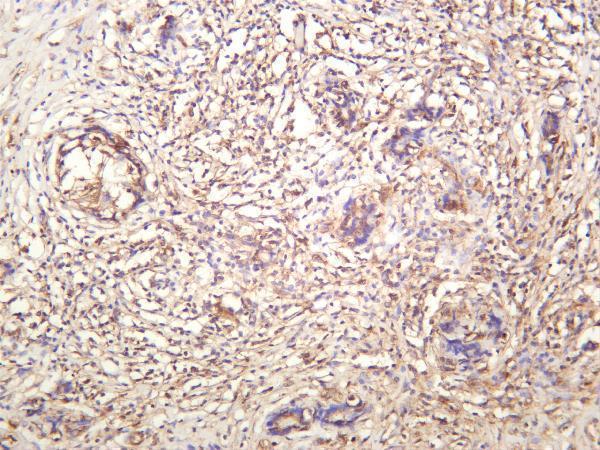
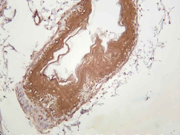
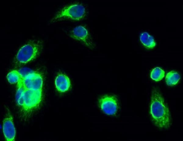
IHC images show PRKACA staining in human thyroid carcinoma and mouse and rat stomach (M00653-1 catalog images); IHC and IF images show human colon carcinoma and HeLa cells, respectively (A00653 catalog images).

Real IHC data Human thyroid carcinoma was stained with anti-PRKACA rabbit antibody
Anti-PRKACA Rabbit Monoclonal Antibody
Cat # M00653-1
Real IHC data Immunohistochemistry analysis of paraffin-embedded human colon carcinoma tissue, using PKA alpha/beta CAT Antibody. The picture on the right is blocked with the synthesized peptide.
Anti-PKA alpha/ beta/ gamma cat PRKACA Antibody
Cat # A00653

M00653-1 is listed for human, mouse and rat IHC and IF; its IHC images show human thyroid carcinoma and mouse and rat stomach (M00653-1 catalog). A00653 is listed for human, mouse, pig and rat IHC and IF; its images show paraffin-embedded human colon carcinoma by IHC and HeLa cells by IF (A00653 catalog).

Which to pick: For tissue IHC, choose the PRKACA monoclonal M00653-1 when its human thyroid carcinoma or mouse and rat stomach IHC examples match your sample (M00653-1 catalog images); tissue processing and fixative are unreported in those captions (M00653-1 catalog images). For IF, A00653 has a HeLa cell image, while its own IHC image supports paraffin sections with fixative unreported (A00653 catalog images). For the broadest listed species coverage among the rendered cards, choose A00653, which lists human, mouse, pig and rat reactivity (A00653 catalog); its title identifies alpha, beta and gamma catalytic subunits, so assess isoform specificity for a PRKACA-specific experiment (A00653 catalog title).

Each figure is that product's own IHC / IF validation image from its datasheet.

References

  1. UniProt Consortium. UniProt entry P17612 (KAPCA_HUMAN, cAMP-dependent protein kinase catalytic subunit alpha).
  2. Human Protein Atlas. PRKACA tissue IHC expression (reliability: Approved).
  3. Human Protein Atlas. PRKACA subcellular location (ICC-IF): Mainly localized to the cytosol. In addition localized to the microtubules, cytokinetic bridge, primary cilium and basal body. Caution: Based on antibodies targeting proteins from multiple genes..
  4. Human Protein Atlas. PRKACA antibody validation summary (2 antibodies).
  5. PRKACA mediates resistance to HER2-targeted therapy in breast cancer cells and restores anti-apoptotic signaling. Oncogene 2015 — PMC4261061.
  6. Differential expression of the protein kinase A subunits in normal adrenal glands and adrenocortical adenomas. Scientific reports 2017 — PMC5427838.
  7. The Association of Cholesterol Uptake and Synthesis with Histology and Genotype in Cortisol-Producing Adenoma (CPA). International journal of molecular sciences 2022 — PMC8875534.
  8. CRISPR/Cas9 Engineering of Adult Mouse Liver Demonstrates That the Dnajb1-Prkaca Gene Fusion Is Sufficient to Induce Tumors Resembling Fibrolamellar Hepatocellular Carcinoma. Gastroenterology 2017 — PMC5801691.
  9. PubMed PMID:2843813 — UniProt-cited evidence.
  10. PubMed PMID:14702039 — UniProt-cited evidence.
  11. PubMed PMID:15489334 — UniProt-cited evidence.