This website uses cookies to ensure you get the best experience on our website.


- Table of Contents
Plan PRKACB chromogenic IHC around the general cytoplasmic tissue pattern (HPA tissue IHC). Cerebral cortex neurons and appendix lymphoid tissue show high staining (HPA tissue IHC); titrate catalog antibody A05366-1 within 1:50–1:200 (datasheet).

Expected localisation, validated protocols, controls and antibodies — the at-a-glance facts below, then the full design guide.
| Expected localisation | General cytoplasmic staining (HPA tissue IHC) | |
| Staining pattern | Cytoplasmic staining across tissue cell types (HPA tissue IHC) | |
| Antigen retrieval | Tris-EDTA pH 9.0 HIER, 95–98 °C, 20 min (rule: nuclear antigen) | |
| Positive control | Appendix+4 more · see all | |
| Negative control | None in HPA (detected in all 45 tissues); use no-primary + isotype controls |
| Fixation | Keep fixation consistent across sections (standard IHC practice; not target-specific) | |
| Caveat | Splice or transcript discrepancy may affect staining (HPA tissue IHC) | |
| Regulation | Tissue-specific isoform expression (UniProt) | |
| Isoform / epitope | 10 isoforms; epitope coverage needs validation (UniProt) |
The catalog antibody’s IHC-P protocol is accompanied by published PRKACB IHC methods for gastric, osteosarcoma, testicular and esophageal tissue (PMC12006100; PMC10244582; PMC11559180; PMC12831332).
| Sample | Paraffin-embedded rat brain tissue; fixative not specified (datasheet A05366-1) |
| Fixation | Image fixative and duration unreported (datasheet A05366-1); verify before use. |
| Sectioning | 4–5 µm sections on charged slides (standard) |
| Deparaffinisation | Xylene, graded ethanol series to water (standard) |
| Antigen retrieval | Heat-induced epitope retrieval in Tris-EDTA buffer, pH 9.0, 20 min at 95–98 °C (standard rule: nuclear antigen) |
| Peroxidase block | 3% H2O2, 10 min, room temperature (standard) |
| Blocking | 10% normal serum of the secondary host, 30 min, room temperature (standard) |
| Primary antibody | Rabbit anti-PRKACB, 1:50-1:200 (datasheet A05366-1) |
| Primary incubation | Overnight at 4 °C (standard) |
| Detection | HRP-polymer secondary, DAB chromogen 5–10 min (standard) |
| Counterstain | Hematoxylin, blue, dehydrate and mount (standard) |
| Expected result | PRKACB-positive staining in lymphoid tissue of appendix (HPA tissue IHC: High). HPA tissue profile: General cytoplasmic expression. No signal in the no-primary control. |
PRKACB should appear mainly cytoplasmic in paraffin-section IHC, with high staining reported in cerebral-cortex neurons, pancreatic exocrine cells and selected lymphoid populations (HPA: general cytoplasmic expression; High in these cells). Nuclear localisation is biologically possible after catalytic-subunit translocation (UniProt P22694: subcellular location). PRKACB has no transmembrane segment, so a membrane-only pattern needs scrutiny (UniProt P22694: topology). HPA rates the tissue IHC profile Approved, with a splice/transcript-discrepancy caution (HPA: tissue reliability).
| Clear cytoplasmic stain in cerebral-cortex neurons, pancreatic exocrine cells or splenic white-pulp cells (HPA: High in these cells). | This fits the reported tissue pattern (HPA: tissue IHC). Compare the intended cell population with surrounding cells: a positive tissue need not stain uniformly because HPA reports intensity by cell population (HPA: tissue IHC). |
| Predominantly nuclear or sharply membrane-only stain, with little cytoplasmic signal (HPA: general cytoplasmic expression). | Nuclear signal can reflect catalytic-subunit translocation, but nuclear dominance alone does not establish pathway activation (UniProt P22694: subcellular location). A membrane-only pattern is less consistent with the reported cytoplasmic profile and no-transmembrane topology; check staining controls (HPA: tissue IHC; UniProt P22694: topology; general IHC practice). |
| Strong stain mainly in bronchial respiratory epithelium or liver cholangiocytes, while expected positive cells are weak (HPA: Low in these cells). | That reversal conflicts with the reported cell-level pattern (HPA: tissue IHC). Consider off-target binding or detection-system activity, then compare an appropriate negative control and a known-positive section (general IHC practice). Low means some signal may be genuine; it does not mean absent expression (HPA: Low). |
| Diffuse color over stroma, lumens and cells, obscuring cell boundaries (general IHC practice). | This is not the reported cell-associated cytoplasmic pattern (HPA: tissue IHC). Background may arise from nonspecific reagent binding, incomplete blocking or endogenous detection activity; use control sections to locate its source (general IHC practice). |
| No discernible stain in cerebral-cortex neurons or pancreatic exocrine cells (HPA: High in these cells). | Treat the run as unresolved before calling the specimen negative: verify tissue preservation, retrieval, antibody and detection steps against the catalog antibody’s IHC-P instructions and controls (general IHC practice). HPA's Approved status has a splice/transcript-discrepancy caution (HPA: tissue reliability). |
| Cell population and isoforms (HPA: tissue IHC; UniProt P22694: tissue specificity). | HPA reports high staining in several neuronal, glandular and lymphoid populations; UniProt reports tissue-dependent isoforms. A tissue name alone cannot predict every cell's signal (HPA: tissue IHC; UniProt P22694: tissue specificity). |
| Compartment and signaling state (UniProt P22694: subcellular location). | Inactive holoenzyme is cytoplasmic; free catalytic subunit can enter the nucleus. Interpret nuclear signal alongside the dominant cytoplasmic IHC pattern and controls (UniProt P22694: subcellular location; HPA: tissue IHC; general IHC practice). |
| Antibody validation scope (HPA: antibodies; tissue reliability). | HPA029754 and CAB010363 are IHC Approved, but the tissue profile carries a splice/transcript-discrepancy caution. Approved status does not identify which isoform an individual stain detects (HPA: antibodies; tissue reliability; UniProt P22694: isoforms). |
| Antigen retrieval and detection (general IHC practice). | For paraffin IHC, follow the catalog antibody’s IHC-P retrieval and detection instructions and include run controls. These sources give no PRKACB-specific fixation sensitivity or retrieval effect (general IHC practice; HPA: tissue IHC; UniProt P22694: record). |
| IF/ICC Q: Where should PRKACB appear? (HPA: subcellular ICC-IF). | A: Mainly cytosol; microtubules are an additional approved location. Cilium, basal-body and cytokinetic-bridge calls are uncertain, and HPA cautions that the summary draws on antibodies targeting multiple genes (HPA: subcellular ICC-IF). |
| Situation | Likely cause | Next action |
|---|---|---|
| Known-positive cells are unstained (HPA: High in cerebral-cortex neurons and pancreatic exocrine cells). | Possible failure in retrieval, primary-antibody or chromogenic detection steps (general IHC practice). | Repeat with a known-positive section and the catalog antibody’s IHC-P instructions; check each run control before interpreting the specimen (general IHC practice). |
| Strong signal appears in a no-primary control (general IHC practice). | Endogenous detection activity or nonspecific detection-reagent binding may contribute (general IHC practice). | Review the detection system and its appropriate blocking and control steps; reassess specimen signal only after control background is resolved (general IHC practice). |
| Color covers the section diffusely (general IHC practice). | Possible nonspecific binding, inadequate blocking or excessive detection background (general IHC practice). | Compare no-primary and known-positive controls; check reagent concentrations, wash steps and blocking against the established IHC workflow (general IHC practice). |
| Nuclei dominate while cytoplasm is faint (HPA: general cytoplasmic expression). | Nuclear entry is possible, but staining alone cannot establish activation; technical background is also possible (UniProt P22694: subcellular location; general IHC practice). | Check controls and whether nuclear staining is confined to relevant cells; report the observed compartment without inferring signaling state (general IHC practice; UniProt P22694: subcellular location). |
| HPA-low cells stain more intensely than expected high cells (HPA: tissue IHC). | Possible off-target staining, detection background or differences among cell populations; HPA Low is not absence (general IHC practice; HPA: Low). | Compare matched positive and negative controls, then score the named cell populations separately instead of labeling the whole tissue positive (general IHC practice; HPA: tissue IHC). |
| IHC and IF/ICC show different minor compartments (HPA: tissue IHC; subcellular ICC-IF). | The assays report different observations; HPA's ICC-IF summary cautions that antibodies target proteins from multiple genes (HPA: subcellular ICC-IF). | Prioritize the IHC cell-level cytoplasmic profile when scoring paraffin sections; describe any additional compartment with its assay and HPA confidence level (HPA: tissue IHC; subcellular ICC-IF). |
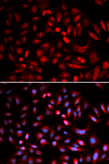
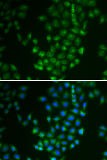


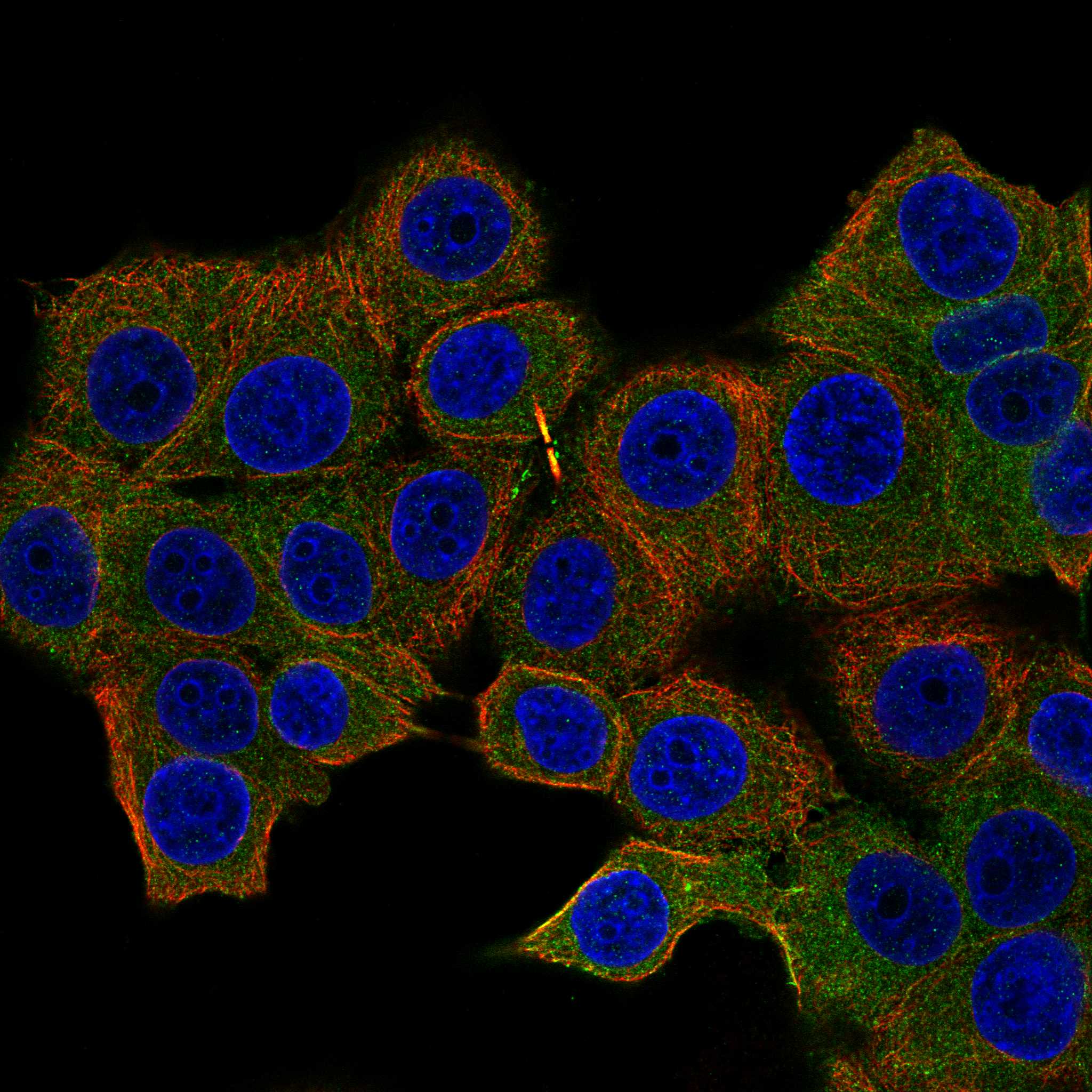
Comprehensive Human Protein Atlas IHC scoring per tissue (reliability: Approved — Caution, Splice and/or transcript discrepancy exists.). Rows are taken directly from the HPA tissue chart — click any row's HPA link to view the source.
| Tissue | Cell type | Level | Evidence | Source |
|---|---|---|---|---|
| Appendix | Lymphoid tissue | High | Protein (IHC) | HPA → |
| Cerebral cortex | Neuronal cells | High | Protein (IHC) | HPA → |
| Lymph node | Non-germinal center cells | High | Protein (IHC) | HPA → |
| Pancreas | Exocrine glandular cells | High | Protein (IHC) | HPA → |
| Spleen | Cells in white pulp | High | Protein (IHC) | HPA → |
| Tissue | Cell type | Level | Evidence | Source |
|---|---|---|---|---|
| None in HPA: PRKACB is detected in all 45 scored tissues. Use a no-primary (secondary-only) and an isotype control instead. | ||||
Troubleshoot PRKACB chromogenic IHC by checking retrieval, tissue handling, cellular localisation and scoring against the reported expression patterns.
The catalog lists PRKACB antibodies for IHC and IF/ICC in human, mouse and rat; IHC images show paraffin-embedded rat brain and human appendix (catalog: applications, reactivity and A05366-1 IHC captions).
A05366-1 lists IHC and IF/ICC for human, mouse and rat, with IHC images of paraffin-embedded rat brain and human appendix (catalog: A05366-1 applications, reactivity and IHC captions). A05366-3 lists IHC and IF/ICC for the same species, but has no IHC or IF image in the payload (catalog: A05366-3 applications, reactivity and image fields).
Which to pick: For tissue IHC, choose A05366-1: its own captions document paraffin-embedded rat brain and human appendix at 1:100 after microwave retrieval in 10 mM PBS, pH 7.2; the fixative is unreported (catalog: A05366-1 IHC captions). For IF/ICC, A05366-1 lists both applications at 1:50–1:200; A05366-3 is a listed polyclonal option at 1:50, with no IF image reported for either SKU (catalog: applications, dilution fields, A05366-3 dilution_raw and image fields). Both SKUs list human, mouse and rat reactivity, but the documented tissue IHC examples belong to A05366-1 (catalog: reactivity and A05366-1 IHC captions).