PRKCG / Protein kinase C gamma type · IHC design guide

Design Immunohistochemistry for PRKCG

Plan PRKCG IHC in paraffin sections using cerebellar Purkinje cells as a high-expression reference (HPA tissue IHC). This guide covers fixation consistency, expected cytoplasmic staining and interpretation of stimulus-dependent movement to synaptic membranes (HPA tissue IHC; UniProt).

Evidence assembled Oct 2026 · For research use; verify linked source records and product datasheet before use
Immunohistochemistry protocol sheet for PRKCG (IHC for PRKCG): expected localisation Cytoplasmic in Purkinje cells and neuropil (HPA tissue IHC), antibody A01890, validated IHC image, and IHC protocol steps
Printable PRKCG IHC protocol sheet — expected localisation Cytoplasmic in Purkinje cells and neuropil (HPA tissue IHC), antibody A01890, controls and protocol steps. Open the full PRKCG IHC guide →

PRKCG Immunohistochemistry Experimental Design Guide

Expected localisation, validated protocols, controls and antibodies — the at-a-glance facts below, then the full design guide.

Must know before staining
Expected localisation Cytoplasmic in Purkinje cells and neuropil (HPA tissue IHC)
Staining pattern Selective cytoplasmic Purkinje cell and neuropil staining (HPA tissue IHC)
Antigen retrieval Citrate pH 6 HIER, heat-mediated (datasheet A01890)
Positive control ⓘ Cerebellum+1 more · see all
Negative control ⓘ Adipose tissue+4 more · see all
Important caveats
Reasons your staining may differ from the expected pattern.
Fixation Keep fixation conditions consistent across sections. (standard IHC practice; not target-specific)
Caveat Stimulation may shift staining toward synaptic membranes (UniProt)
Regulation Stimulus-linked synaptic translocation (UniProt)
Isoform / epitope 2 isoforms; epitope differences are unspecified (UniProt)
Section 1

Recommended PRKCG IHC & IF Protocols

The catalog antibody protocol is accompanied by one published PRKCG IHC protocol using paraffin-embedded renal tissue (PMC8832145).

Recommended immunohistochemistry (IHC-P) protocol parameters
SampleParaffin-embedded human glioma tissues; fixative not specified (datasheet A01890)
FixationImage fixative and duration unreported (datasheet A01890); verify before use.
Sectioning4–5 µm sections on charged slides (standard)
DeparaffinisationXylene, graded ethanol series to water (standard)
Antigen retrievalHeat retrieval: Citrate pH 6, 20 min (datasheet A01890)
Peroxidase block3% H2O2, 10 min, room temperature (standard)
Blocking10% goat serum (datasheet A01890)
Primary antibodyRabbit anti-PRKCG, 1μg/ml (datasheet A01890)
Primary incubationOvernight at 4 °C (datasheet A01890)
DetectionStreptavidin-biotin complex (SABC), DAB chromogen (datasheet A01890)
CounterstainHematoxylin, blue, dehydrate and mount (standard)
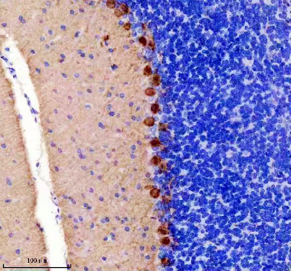
Expected resultPRKCG-positive staining in purkinje cells of cerebellum (HPA tissue IHC: High). HPA tissue profile: Selective cytoplasmic expression in Purkinje cells and neuropil. No signal in the no-primary control.
💡Decision noteStart with heat-mediated citrate pH 6 retrieval for the catalog antibody (datasheet: A01890); the published renal tissue excerpt does not specify retrieval (PMC8832145).
Section 2

What Is the Expected PRKCG Staining Pattern?

PRKCG staining should be strongest in cerebellar Purkinje cells and hippocampal neurons, with cytoplasmic signal and staining in neuropil (HPA: High in both cell populations; selective cytoplasmic expression in Purkinje cells and neuropil). Perinuclear, dendritic and synaptic or membrane-associated signal can fit its annotated distribution (UniProt P05129: subcellular location; no transmembrane segment). The tissue IHC pattern has Enhanced reliability, reflecting consistency with RNA expression (HPA: reliability description).

What am I looking at on my slide?
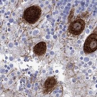
Strong cytoplasmic Purkinje-cell staining, with signal extending into nearby neuropil.This matches the clearest positive tissue pattern (HPA: High in cerebellar Purkinje cells; selective cytoplasmic expression in Purkinje cells and neuropil). Compare cell bodies and processes with the counterstain before scoring; neuropil signal alone gives less certainty about which cells contain PRKCG (general IHC interpretation).
Strong staining in hippocampal neuronal cells, or weaker neuropil staining in cerebral cortex.Both can be compatible with the tissue record, but they serve different purposes: hippocampal neurons provide another strong positive reference, whereas cerebral cortex neuropil is recorded at a low level (HPA: High in hippocampal neuronal cells; Low in cerebral cortex neuropil). Compare each slide with its own tissue context rather than applying one intensity threshold to every region (general IHC interpretation).
Predominantly nuclear staining, with little cytoplasmic or neuronal-process signal.An isolated nuclear pattern conflicts with the annotated cytoplasmic, perinuclear, dendritic and synaptic distribution (UniProt P05129: subcellular location). Treat it as suspect until controls and morphology support it; check whether the counterstain, precipitated chromogen or nonspecific staining is being mistaken for target signal (general IHC practice).
Strong staining across cell types recorded as negative, such as adipocytes or bone-marrow hematopoietic cells.That distribution is inconsistent with those HPA tissue observations (HPA: Not detected in adipose-tissue adipocytes and bone-marrow hematopoietic cells). Cross-reactivity, endogenous detection activity or background are possibilities, not established causes. Review a detection-only control and tissue morphology before calling the staining PRKCG (general IHC practice).
Diffuse staining over tissue and empty areas, or no staining in cerebellar Purkinje cells.Diffuse signal without cellular structure limits interpretation; absent Purkinje-cell signal fails a strong positive reference (HPA: High in cerebellar Purkinje cells). Neither observation identifies a PRKCG-specific cause. Inspect controls, section quality and detection performance before concluding that expression differs from the reference (general IHC practice).
💡Expected PRKCG appearanceCall positive when high cytoplasmic staining marks cerebellar Purkinje cells and associated neuropil, with hippocampal neuronal staining as another reference; widespread staining of HPA-negative cell types is suspect (HPA: tissue IHC profile and levels).
How each factor affects the staining
Location and activationCytoplasmic and perinuclear signal can coexist with dendritic, synaptic and membrane-associated signal; stimulation can move PRKCG toward synaptic membranes (UniProt P05129: subcellular location). A membrane-associated component alone therefore does not establish an artefact. Its extent cannot be predicted for an unstated specimen condition (UniProt P05129: stimulation-dependent translocation).
Positive and negative tissue referencesCerebellar Purkinje cells and hippocampal neurons are strong positive references; adipose-tissue adipocytes and bone-marrow hematopoietic cells are recorded as not detected (HPA: tissue IHC levels). These are cell-specific observations, so a negative entry should not be read as proof that every cell in that tissue is negative (HPA: cell-specific tissue entries).
Antibody evidence and molecular coverageThe listed rabbit polyclonal antibody HPA054560 has Enhanced IHC validation (HPA: antibody validation). PRKCG has two reported isoforms and multiple modified residues (UniProt P05129: isoforms; modified residues). The supplied records give no epitope or isoform-recognition data, so they cannot establish which molecular forms a particular IHC antibody detects.
IF/ICC Q&A: Can the IHC pattern be used as an IF/ICC result?No ICC-IF image-bearing cell lines or main subcellular location are supplied (HPA: subcellular record). UniProt annotates plausible compartments, but the IHC tissue pattern does not itself validate an IF/ICC staining result (UniProt P05129: subcellular location; HPA: tissue IHC profile).
Why is my staining missing, weak or wrong?
SituationLikely causeNext action
Purkinje cells are unstained in a cerebellar section.A technical failure is possible because these cells are a strong positive reference; the slide alone cannot identify the failed step (HPA: High in cerebellar Purkinje cells).Check the tissue control, antibody and detection run; review section morphology and the recorded retrieval conditions before interpreting a negative result (general IHC practice).
Signal appears mainly nuclear.The pattern does not match annotated PRKCG compartments (UniProt P05129: subcellular location). Counterstain or nonspecific deposit may complicate the readout (general IHC practice).Inspect a detection-only control and compare nuclear boundaries with cytoplasm and neuronal processes; withhold a PRKCG-positive call if the signal remains exclusively nuclear (general IHC practice).
Many HPA-negative cell types show strong chromogen.Cross-reactivity, endogenous enzyme activity or nonspecific detection are possible; the pattern differs from the cited negative cell entries (HPA: Not detected in adipocytes and bone-marrow hematopoietic cells).Compare those exact cell types with a detection-only control, check endogenous activity blocking where relevant, and score signal by cell type rather than by whole-tissue darkness (general IHC practice).
The section has broad haze or scattered chromogen deposits.Background or precipitate may obscure the selective neuronal pattern (HPA: selective cytoplasmic expression in Purkinje cells and neuropil; general IHC practice).Review washing, blocking and detection conditions, then reassess whether signal follows recognizable cells and neuropil; do not score diffuse haze as expression (general IHC practice).
Cortical neuropil is faint while cerebellar Purkinje cells are clear.The difference can agree with the reported tissue levels (HPA: Low in cerebral cortex neuropil; High in cerebellar Purkinje cells).Use the cerebellar positive reference to judge run performance and record cortical staining in its own context; avoid treating equal intensity across regions as a requirement (general IHC interpretation).
A membrane-associated signal varies between specimens.PRKCG can translocate to synaptic membranes on stimulation, but the supplied records do not establish stimulation status for these specimens (UniProt P05129: subcellular location).Document the compartment and specimen conditions, and compare the cytoplasmic neuronal pattern before attributing the difference to PRKCG redistribution (UniProt P05129: subcellular location; general IHC interpretation).

Sample controls for PRKCG IHC & IF

🧪Run cerebellum first and require staining in Purkinje cells (HPA: High in cerebellar Purkinje cells). Use adipose tissue as the negative tissue (HPA: Not detected in adipocytes); on the cerebellar slide, compare Purkinje cells with neighboring non-Purkinje cells, without assuming those cells are PRKCG-negative.
Positive control tissue: Cerebellum (Purkinje cells, HPA High)
Negative control tissue: Adipose tissue (HPA Not detected)
ICC-IF cell lines (HPA subcellular resource): HPA carries no ICC-IF cell line for PRKCG; derive a cell-line control from the positive tissue's cell type (Purkinje cells) and confirm it by RNA or western blot first.
Technical controls: Include a no-primary (secondary-only) slide, an isotype control matched to the primary antibody’s host and clonality, and PRKCG-knockout tissue as a biological negative. Block endogenous peroxidase and assess endogenous biotin background because the reported cerebellum-compatible chromogenic workflow uses a biotinylated secondary and DAB (selected-SKU caption: biotinylated goat anti-rabbit IgG, streptavidin–biotin complex, DAB).
⚠️Feasibility: A target-specific fixation window or fixation effect is unreported, and the selected-SKU paraffin-section caption does not state a fixative (selected-SKU caption: fixative unreported). The reported IHC procedure uses heat retrieval in citrate buffer at pH 6 for 20 minutes, but retrieval dependence has not been established (selected-SKU caption: citrate retrieval for 20 minutes). Frozen sections or IF cannot be judged easier from the supplied evidence; for IF, check cerebellar autofluorescence with an unstained section before interpreting neuronal signal.

HPA tissue IHC evidence for PRKCG

Comprehensive Human Protein Atlas IHC scoring per tissue (reliability: Enhanced — High consistency between antibody staining and RNA expression data.). Rows are taken directly from the HPA tissue chart — click any row's HPA link to view the source.

Positive expression · recommended positive controls

TissueCell typeLevelEvidenceSource
Cerebellum Purkinje cells High Protein (IHC) HPA →
Hippocampus Neuronal cells High Protein (IHC) HPA →

Undetected expression · recommended negative controls

TissueCell typeLevelEvidenceSource
Adipose tissue Adipocytes Not detected Protein (IHC) HPA →
Adrenal gland Glandular cells Not detected Protein (IHC) HPA →
Appendix Endocrine cells Not detected Protein (IHC) HPA →
Bone marrow Hematopoietic cells Not detected Protein (IHC) HPA →
Breast Adipocytes Not detected Protein (IHC) HPA →
Section 3

Advanced PRKCG IHC Tips

Troubleshoot PRKCG staining in paraffin sections by checking retrieval, compartment, cell type and controls against the reported IHC pattern (datasheet A01890; HPA tissue IHC).

What retrieval should I try first when PRKCG staining is weak in paraffin sections?
Start with heat-mediated retrieval in citrate buffer at pH 6 for 20 minutes (datasheet A01890). The catalog antibody produced chromogenic staining in paraffin-embedded human glioma sections under those conditions, although the caption does not state the fixative (datasheet A01890). Compare a cerebellar section with the test section in the same staining run, assessing Purkinje-cell cytoplasm and neuropil as expected sites (HPA tissue IHC). If signal remains weak, check section attachment, heating consistency and antibody incubation before treating retrieval as the cause (standard IHC practice). Record any retrieval change alongside background and tissue morphology so the comparison remains interpretable (standard IHC practice).
Could fixation explain absent or diffuse PRKCG staining?
Target-specific sensitivity to fixation is unknown; the selected paraffin-section caption does not report its fixative (datasheet A01890). Document the fixative and fixation duration for each specimen, then compare sections processed under matched conditions with the same citrate pH 6 retrieval (standard IHC practice; datasheet A01890). Include a cerebellar control and assess whether Purkinje-cell cytoplasm and neuropil remain recognizable before attributing a negative result to fixation (HPA tissue IHC; standard IHC practice). Excessive fixation or poor preservation can change antigen accessibility and morphology in IHC generally, but these data do not establish a PRKCG-specific fixation effect (standard IHC practice; datasheet A01890).
Which staining compartments should count as plausible PRKCG signal?
Prioritize cytoplasmic staining in Purkinje cells and neuropil when evaluating cerebellar sections (HPA tissue IHC). Perinuclear cytoplasm, cell membrane, synaptosomes and dendrites are also listed localizations, and PRKCG can move to synaptic membranes after stimulation (UniProt P05129 subcellular location). A membrane-associated pattern therefore needs anatomical context and appropriate controls; a uniformly nuclear pattern alone does not match the listed localizations (UniProt P05129 subcellular location; standard IHC practice). Score cellular compartments separately where morphology permits, and compare the same compartments in a positive control and a no-primary control (standard IHC practice). Do not equate uneven chromogen deposition with stimulus-dependent translocation (standard IHC practice; UniProt P05129 subcellular location).
How can epitope choice affect interpretation across PRKCG isoforms?
PRKCG has 2 listed isoforms, but the supplied antibody caption does not identify its epitope or establish isoform selectivity (UniProt P05129 isoforms; datasheet A01890). Ask for the immunogen sequence and map it against both isoforms before describing staining as isoform-specific (standard IHC practice; UniProt P05129 isoforms). PRKCG has a C2 domain at residues 157–275 and a kinase domain at 351–614; known phosphorylation sites include residues 320, 322, 326 and 328 (UniProt P05129 domains and modified residues). Without epitope mapping, staining changes cannot establish loss of an isoform or a phosphorylation-dependent epitope (standard IHC practice; datasheet A01890).
How should I adapt the PRKCG IHC question to a multiplex IF experiment?
On the separate IF/ICC workflow, pair PRKCG with a marker that identifies the expected Purkinje cells, then assess whether the signals occupy the expected cellular regions (HPA tissue IHC; standard IF practice). Choose fluorophores after inspecting unstained tissue for autofluorescence, and place the weaker channel away from the strongest autofluorescent range (standard IF practice). Because PRKCG has no transmembrane segment and is listed in cytoplasm and membrane-associated compartments, use permeabilisation appropriate to an intracellular epitope while checking that cellular morphology is retained (UniProt P05129 topology and subcellular location; standard IF practice). Establish IF fixation and antibody conditions independently; the supplied catalog caption documents chromogenic paraffin-section IHC only (datasheet A01890).
How do I reduce widespread brown staining without losing PRKCG signal?
Run a no-primary control beside a cerebellar positive control to separate detection-system background from the expected Purkinje-cell and neuropil pattern (standard IHC practice; HPA tissue IHC). The selected procedure used 10% goat serum blocking, 1 μg/mL primary antibody overnight at 4°C, and biotin-based detection with DAB (datasheet A01890). If background persists, check peroxidase blocking, wash stringency, secondary-antibody binding and DAB development time as general chromogenic IHC variables (standard IHC practice). Adjust one variable at a time while preserving the same retrieval and positive control, then compare signal against the no-primary section (standard IHC practice; datasheet A01890).
What should I measure when comparing PRKCG staining between sections? ⚠ ANSWER MARKED FOR VERIFICATION
Define the anatomical region and cell population before scoring, because Purkinje cells and neuropil have different expected staining patterns (HPA tissue IHC; standard IHC practice). For identifiable Purkinje cells, report the percentage positive and an intensity-weighted H-score; for neuropil, report chromogen-positive area or optical density within a fixed region (standard IHC practice). Normalize cell counts to the number of evaluable Purkinje cells and area measurements to the measured tissue area, with consistent thresholds and acquisition settings (standard IHC practice). Exclude folds, tears and necrotic regions by a prespecified rule, and compare batches using the same positive-control section where feasible (standard IHC practice).
When is an apparent PRKCG-positive region more likely to be artefact?
A convincing cerebellar result follows selective cytoplasmic staining in Purkinje cells and neuropil, supported by the reported tissue pattern (HPA tissue IHC). Treat dominant staining in an unexpected cell population or an isolated nuclear signal cautiously, since the listed PRKCG locations are cytoplasmic, perinuclear, membrane-associated, synaptosomal and dendritic (UniProt P05129 subcellular location). Check whether staining concentrates at section edges, folds or necrotic areas, and whether it appears in the no-primary control; those findings favor a staining artefact (standard IHC practice). Residual endogenous peroxidase can produce DAB signal, so inspect the blocked control before assigning brown deposits to PRKCG (standard IHC practice).
Boster reagents

Best PRKCG / Protein kinase C gamma type IHC Antibodies

Anti-PRKCG antibodies have IHC images from human glioma, mouse brain and cerebellum, and rat brain paraffin sections, plus IF images from SH-SY5Y cells and rat cerebellum (catalog image captions).

Real IHC data IHC analysis of PKC gamma using anti-PKC gamma antibody (A01890). PKC gamma was detected in paraffin-embedded section of human glioma tissues. Heat mediated antigen retrieval was performed in citrate buffer (pH6, epitope retrieval solution) for 20 mins. The tissue section was blocked with 10% goat serum. The tissue section was then incubated with 1μg/ml rabbit anti-PKC gamma Antibody (A01890) overnight at 4°C. Biotinylated goat anti-rabbit IgG was used as secondary antibody and incubated for 30 minutes at 37°C. The tissue section was developed using Strepavidin-Biotin-Complex (SABC)(Catalog # SA1022) with DAB as the chromogen.
Anti-PKC gamma/PRKCG Antibody ®
Cat # A01890
Real IHC data IHC analysis of PKC gamma using anti-PKC gamma antibody (M01890-2). PKC gamma was detected in a paraffin-embedded section of mouse cerebellum tissue. Heat mediated antigen retrieval was performed in EDTA buffer (pH 8.0, epitope retrieval solution). The tissue section was blocked with 10% goat serum. The tissue section was then incubated with 1:50 rabbit anti-PKC gamma Antibody (M01890-2) overnight at 4°C. Peroxidase Conjugated Goat Anti-rabbit IgG was used as secondary antibody and incubated for 30 minutes at 37°C. The tissue section was developed using HRP Conjugated Rabbit IgG Super Vision Assay Kit (Catalog # SV0002) with DAB as the chromogen.
Anti-PKC gamma Rabbit Monoclonal Antibody
Cat # M01890-2

A01890 has IHC images from human glioma, mouse brain and rat brain paraffin sections, and IF images from SH-SY5Y cells and rat cerebellum (A01890 image captions). M01890-2 has an IHC image from mouse cerebellum paraffin sections and lists IHC, IF and ICC applications for human, mouse and rat (M01890-2 image caption; catalog applications and reactivity).

Which to pick: For tissue IHC, choose A01890 when its documented human glioma, mouse brain or rat brain paraffin-section examples match the sample; choose M01890-2 for a rabbit monoclonal with a documented mouse cerebellum paraffin-section example (A01890 IHC captions; M01890-2 IHC caption and catalog clone). For IF/ICC, A01890 has documented IF images from SH-SY5Y cells and rat cerebellum; M01890-2 lists IF/ICC applications but has no IF image in this payload (A01890 IF captions; M01890-2 catalog applications and image captions). For cross-species IHC, A01890 has images from human, mouse and rat samples, while both SKUs list reactivity with those species; the fixative is unreported in their IHC captions (A01890 IHC captions; catalog reactivity; M01890-2 IHC caption).

Each figure is that product's own IHC / IF validation image from its datasheet.

References

  1. UniProt Consortium. UniProt entry P05129 (KPCG_HUMAN, Protein kinase C gamma type).
  2. Human Protein Atlas. PRKCG tissue IHC expression (reliability: Enhanced).
  3. Human Protein Atlas. PRKCG subcellular location (ICC-IF): Highest expression in OE19: 2.2 nTPM.
  4. Human Protein Atlas. PRKCG antibody validation summary (1 antibodies).
  5. Metastatic Immune-Related Genes for Affecting Prognosis and Immune Response in Renal Clear Cell Carcinoma. Frontiers in molecular biosciences 2021 — PMC8832145.
  6. Targeted Inhibition of O-Linked β-N-Acetylglucosamine Transferase as a Promising Therapeutic Strategy to Restore Chemosensitivity and Attenuate Aggressive Tumor Traits in Chemoresistant Urothelial Carcinoma of the Bladder. Biomedicines 2022 — PMC9138654.
  7. PubMed PMID:14702039 — UniProt-cited evidence.
  8. PubMed PMID:15057824 — UniProt-cited evidence.
  9. PubMed PMID:15489334 — UniProt-cited evidence.