This website uses cookies to ensure you get the best experience on our website.


- Table of Contents
Plan PRKDC IHC in paraffin sections around the expected nuclear staining pattern (HPA tissue IHC). Use consistently fixed sections and assess nuclear signal; bone marrow hematopoietic cells provide a high-staining reference (HPA tissue IHC).

Expected localisation, validated protocols, controls and antibodies — the at-a-glance facts below, then the full design guide.
| Expected localisation | Predominantly nuclear in tissue sections (HPA tissue IHC) | |
| Staining pattern | Widespread nuclear staining across cell types (HPA tissue IHC) | |
| Antigen retrieval | Tris-EDTA pH 9.0 HIER, 95–98 °C, 20 min (rule: nuclear antigen) | |
| Positive control | Adipose tissue+4 more · see all | |
| Negative control | None in HPA (detected in all 45 tissues); use no-primary + isotype controls |
| Fixation | Matched tissue-IHC evidence does not establish the fixation claim. Validate the specimen-specific method before use. (selected-SKU IHC image M00645) | |
| Caveat | Broad staining limits tissue-negative controls (HPA tissue IHC) | |
| Regulation | Expression regulation is unreported (UniProt) | |
| Isoform / epitope | 2 isoforms; epitope coverage is unspecified (UniProt) |
Compare the catalog antibody's IHC-P protocol (datasheet) with 2 published PRKDC IHC methods (PMC13128572; PMC11084440).
| Sample | Paraffin-embedded human breast tissue; fixative not specified (datasheet M00645) |
| Fixation | Image fixative and duration unreported (datasheet M00645); verify before use. |
| Sectioning | 4–5 µm sections on charged slides (standard) |
| Deparaffinisation | Xylene, graded ethanol series to water (standard) |
| Antigen retrieval | Heat-induced epitope retrieval in Tris-EDTA buffer, pH 9.0, 20 min at 95–98 °C (standard rule: nuclear antigen) |
| Peroxidase block | 3% H2O2, 10 min, room temperature (standard) |
| Blocking | 10% normal serum of the secondary host, 30 min, room temperature (standard) |
| Primary antibody | Rabbit monoclonal (clone EIA-16) anti-PRKDC, 1:50 (datasheet M00645) |
| Primary incubation | Overnight at 4 °C (standard) |
| Detection | HRP-polymer secondary, DAB chromogen 5–10 min (standard) |
| Counterstain | Hematoxylin, blue, dehydrate and mount (standard) |
| Expected result | PRKDC-positive staining in adipocytes of adipose tissue (HPA tissue IHC: High). HPA tissue profile: Ubiquitous nuclear expression. No signal in the no-primary control. |
PRKDC should show predominantly nuclear staining in paraffin sections: HPA describes a ubiquitous nuclear pattern with Supported IHC reliability and high staining in several listed cell populations (HPA tissue IHC). UniProt also places PRKDC in the nucleolus and cytosol and reports no transmembrane segment, so assess compartment and tissue context together (UniProt P78527).
| Distinct nuclear chromogen in adipocytes, glandular cells, hematopoietic cells, respiratory epithelium, or the listed brain cells (HPA tissue IHC). | This agrees with HPA's ubiquitous nuclear profile. Its listed high staining includes adipocytes, glandular cells, hematopoietic cells, respiratory epithelial cells, caudate glia, and cerebellar granular layer cells (HPA tissue IHC). Compare nuclei with the counterstain before scoring; HPA's Supported rating reflects agreement between antibody staining and RNA data, not a guarantee for every specimen (HPA tissue IHC). |
| Strong membrane or extracellular staining dominates while nuclei remain faint or clear (UniProt P78527 topology; HPA tissue IHC). | Treat this as a suspect compartment pattern: PRKDC has no transmembrane segment, and HPA reports ubiquitous nuclear expression (UniProt P78527; HPA tissue IHC). UniProt also lists cytosol and nucleolus, so limited staining there alone does not establish an artefact (UniProt P78527). Check whether the suspect signal persists without primary antibody before interpreting it as PRKDC (general IHC practice). |
| Signal is restricted to an unexpected cell population while nuclei of adjacent HPA-listed cells lack staining (HPA tissue IHC). | Check cell identity and tissue morphology first. HPA reports low tissue specificity and supplies no negative cell population, so staining outside its examples is not automatically cross-reactivity (HPA tissue IHC). If the signal is confined to a population with an implausible compartment pattern, investigate cross-reactivity or endogenous detection activity using appropriate controls (general IHC practice; HPA tissue IHC). |
| Brown deposit spreads across nuclei, cytoplasm, and empty areas, obscuring cell boundaries (general chromogenic IHC practice). | This diffuse background prevents a reliable nuclear call against HPA's reported pattern (HPA tissue IHC). Inspect a no-primary control and assess blocking, washing, chromogen development, and antibody concentration as general IHC variables (general IHC practice). A background-free, compartment-resolved signal is needed before assigning cell-level PRKDC staining (general IHC practice). |
| No nuclear signal appears in a section containing an HPA-listed high-staining cell population (HPA tissue IHC). | Target-specific fixation effects are not established by the supplied assay evidence. Verify with a matched IHC source before attributing a result to fixation. |
| Compartment: What should guide IHC scoring? (HPA tissue IHC; UniProt P78527) | Prioritize discernible nuclear staining: HPA calls the tissue pattern ubiquitous and nuclear, while UniProt also lists nucleolus and cytosol (HPA tissue IHC; UniProt P78527). A cytosolic component therefore needs context; it should not replace the expected nuclear reference pattern (HPA tissue IHC; UniProt P78527). |
| Reference cells and validation (HPA tissue IHC; HPA antibodies) | HPA lists High staining in eight supplied tissue and cell entries; it supplies no negative or low examples in this payload (HPA tissue IHC). Both listed antibodies have Supported IHC status, so use their observations as reference patterns while retaining antibody-specific controls (HPA antibodies; general IHC practice). |
| Isoforms and modifications (UniProt P78527) | UniProt lists two isoforms and multiple modified residues, but this payload does not identify the catalog antibody's epitope or establish isoform-specific staining (UniProt P78527). Do not infer that a staining difference reflects an isoform or modification without epitope and validation evidence (UniProt P78527; general IHC practice). |
| IF/ICC Q&A: Where should the signal appear? (HPA subcellular ICC-IF) | Expect enhanced nucleoplasmic localization in HPA's ICC-IF record; its image list includes A-431, Hep-G2, and U2OS, and antibody HPA035174 has Enhanced ICC status (HPA subcellular ICC-IF; HPA antibodies). That cell imaging evidence supports an IF localization check, not an IHC-P protocol choice (HPA subcellular ICC-IF; HPA antibodies). |
| Situation | Likely cause | Next action |
|---|---|---|
| Nuclei are blank in a tissue containing HPA-listed high-staining cells (HPA tissue IHC). | The expected cells may be absent from the evaluated field, or the staining run may have failed; the slide alone cannot distinguish these possibilities (HPA tissue IHC; general IHC practice). | Locate the listed cells morphologically, check a positive control, then review the catalog antibody's IHC-P retrieval, dilution, and detection instructions (HPA tissue IHC; general IHC practice). |
| Most signal lies at cell borders or outside cells (HPA tissue IHC; UniProt P78527 topology). | That distribution conflicts with HPA's nuclear pattern and UniProt's lack of a transmembrane segment; nonspecific deposition or detection background is possible (HPA tissue IHC; UniProt P78527; general IHC practice). | Compare a no-primary control, inspect the counterstained nuclei, and score PRKDC only where compartment-resolved signal supports the nuclear pattern (HPA tissue IHC; general IHC practice). |
| All compartments appear uniformly dark, obscuring nuclei (general chromogenic IHC practice). | Excess antibody, inadequate washing, or overdevelopment can produce diffuse chromogenic background in IHC generally; this record does not assign a PRKDC-specific cause (general IHC practice). | Check the no-primary control, then adjust dilution, washing, or development according to the catalog IHC-P method while retaining a positive control (general IHC practice). |
| A suspicious cell population stains, but expected nearby nuclei do not (HPA tissue IHC). | HPA's low tissue specificity and absence of listed negative cells limit any exclusion by cell identity alone; cross-reactivity or endogenous detection activity remains possible (HPA tissue IHC; general IHC practice). | Confirm morphology, run no-primary and detection-system controls as appropriate, and compare the suspect signal with the reported nuclear pattern (HPA tissue IHC; general IHC practice). |
| Cytoplasmic signal accompanies a weaker nuclear signal (UniProt P78527; HPA tissue IHC). | UniProt lists cytosol as a PRKDC location, while HPA's tissue IHC profile emphasizes nuclei; this combination alone cannot establish a failed assay (UniProt P78527; HPA tissue IHC). | Score nuclear and cytoplasmic compartments separately, inspect controls for background, and report the observed distribution without assigning an unsupported cause (UniProt P78527; HPA tissue IHC; general IHC practice). |




Comprehensive Human Protein Atlas IHC scoring per tissue (reliability: Supported — High consistency between antibody staining and RNA expression data.). Rows are taken directly from the HPA tissue chart — click any row's HPA link to view the source.
| Tissue | Cell type | Level | Evidence | Source |
|---|---|---|---|---|
| Adipose tissue | Adipocytes | High | Protein (IHC) | HPA → |
| Adrenal gland | Glandular cells | High | Protein (IHC) | HPA → |
| Appendix | Glandular cells | High | Protein (IHC) | HPA → |
| Bone marrow | Hematopoietic cells | High | Protein (IHC) | HPA → |
| Breast | Glandular cells | High | Protein (IHC) | HPA → |
| Tissue | Cell type | Level | Evidence | Source |
|---|---|---|---|---|
| None in HPA: PRKDC is detected in all 45 scored tissues. Use a no-primary (secondary-only) and an isotype control instead. | ||||
Troubleshoot PRKDC staining by checking nuclear signal against retrieval, specimen handling, antibody specificity, and tissue controls.
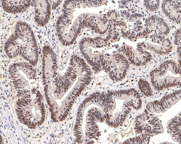
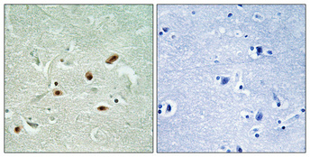
Three anti-PRKDC antibodies have IHC images from paraffin-embedded human breast, colon, and brain (IHC captions); all three list IF, with species coverage specified in the catalog (catalog applications/reactivity).
M00645 has a human breast IHC image and lists IF/ICC for human, mouse, and rat; P00645 has human colon and colon carcinoma IHC images and lists IF/ICC for human (IHC captions; catalog applications/reactivity). A00645S2612 has a human brain IHC image and lists IF for human, mouse, and rat (IHC caption; catalog applications/reactivity).
Which to pick: For paraffin-section IHC, choose M00645 to detect total PRKDC in human breast (M00645 title and IHC caption), P00645 to detect phospho-S2056 in human colon (P00645 title and IHC caption), or A00645S2612 to detect phospho-S2612 in human brain (A00645S2612 title and IHC caption). For IF/ICC and human, mouse, or rat samples, M00645 is the rabbit monoclonal with both applications listed; A00645S2612 lists IF and the same species, while P00645 lists IF/ICC for human only (catalog applications/reactivity/host/clone). The P00645 colon caption reports Tris-EDTA pH 9.0 retrieval for 20 minutes and primary antibody at 1:200, whereas its catalog IHC dilution is 1:50; the A00645S2612 brain caption reports Tris-EDTA pH 8.0 retrieval and 1:100 primary antibody, and fixative is unreported in all three captions (IHC captions; catalog IHC dilutions).