This website uses cookies to ensure you get the best experience on our website.
- Table of Contents
1 Citations 16 Q&As
Facts about Alternative prion protein.
Might be required for neuronal myelin sheath maintenance. May promote myelin homeostasis through acting as an agonist for ADGRG6 receptor.
Human | |
---|---|
Gene Name: | PRNP |
Uniprot: | F7VJQ1 |
Entrez: | 5621 |
Belongs to: |
---|
No superfamily |
CD230; CJD; fatal familial insomnia); GSS; prion protein (p27-30); prion protein PrP; prion protein; prion-related protein; PRIPMGC26679
Mass (kDA):
8.691 kDA
Human | |
---|---|
Location: | 20p13 |
Sequence: | 20; NC_000020.11 (4686456..4701588) |
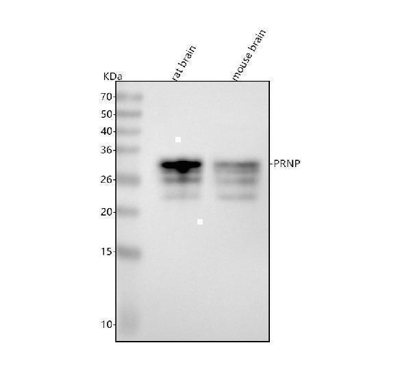
Detected in brain homogenate, primary neurons, and peripheral blood mononuclear cells (at protein level).
Mitochondrion outer membrane; Single-pass membrane protein.
In this article, we'll look at the Anti-Prion marker PRP/PRNP and its applications in neuroscience. We will also discuss validation and application of this marker to determine the neurotoxic properties of prion proteins and prion proteins. We will also discuss the current status of the PRNP marker in relation to neurodegenerative disorders. Boster Bio authors wrote this article.
The PRNP, or pron protein, is an antibody that detects prion disease. This protein is created by the PRNP gene. It can be found in a variety of tissues, including the nervous system. The antigen is highly conserved across all animal species and has more than 90% identity. There are, however, variations within and between species of this gene. The most well-known polymorphisms are found in sheep and goats, both of which have been linked to susceptibility for prion disease.
The antibodies recognize globular structures within the protein and extend from residues 128 to 233. The positions of the four main binding regions are illustrated in Figure S2. Despite their similarities, certain antibodies recognize an epitope conformational, while others have not been examined in Pepscan. The antibodies can be used to determine PrP that is recombinant.
This antibody binds with a specific prions protein found in mammals, but not in other types. This antibody is more specific than commercial prion antibodies. It is suitable to be used in diagnostics. Numerous studies have been conducted using the Boster Bio Anti-Prion Protein PrP/PRNP marker. However, the outcomes could differ.
Anti-Prion proteins are not infectious but can be neutralized for many illnesses. Antibodies will bind naturally occurring prion proteins in cell culture and in vivo, but without altering their properties. Antibodies against infectious prions also be able to bind to PrP Sc forms in high quantities, at least 50 percent. This makes them beneficial in the diagnosis and treatment of prions.
Monoclonal antibodies against prion proteins are valuable instruments to differentiate between various forms of the protein. They can be utilized for understanding the structure of the prions protein. They are not specific to humans and therefore , antibodies are needed to detect the disease. This is why there are few available anti-Prion antibody antibodies. The absence of a human-specific antibody in animal models has complicated the research process as well as the testing process.
The PRNP locus was used to confirm the PRNP marker. It has been linked to risk in multiple studies of the prion disease marker. Its polymorphic codon number of 129 was associated with risk of developing disease. In addition, the nearby SNP RARB was associated with an increased risk of vCJD. The association was not seen in patients with Iatrogenic CJD. However risk in cases of sporadic CJD was not associated to the PRNP locus.
The rs6116492T Allele increased risk through altered expression of PRNP. Other variants, including rs6794719 and rs1460163, posed risk due to altered expression of STMN2 or the RARB. The SNPs were selected from the stringency-filtered standard data and were evenly distributed with all autosomes. Despite their low frequency , they can be dangerous and should be considered as an indicator that could be used to guide further research.
The PRNP marker is an extremely conserved genetic marker in mammals. The gene is present in the majority of human tissues. It is also connected to XBP1 dependent transcription. There are numerous studies showing the increased expression of PRNP in cancer. PrPC levels that are higher than normal give proliferative, invasion, as well as migratory capabilities to cancer cells. These results suggest that the PRNP gene could play a role within cellular metabolism.
One use of the PRNP marker is the identification of genetic variations within the PRNP gene's code region. The PRNP gene consists of three exons, the third of which is the open reading frame. The scrapie phenotype is closely connected to polymorphisms that are found in this exon. Although susceptibility to scrapie can be affected by other genetic regions of PRNP gene artificial selection in the area of the ovine has eliminated codons that cause disease.
The PRNP gene codes for prion protein or PRP. While prions are known for their destructive properties, the PRNP gene is also implicated in Parkinson's and Alzheimer's diseases. Many other diseases are linked with the PRNP gene. This protein is found in a variety of tissues, including the brain. Research conducted by Puckett et al. In 1991, a high level of heterozygosity could be found in the PRNP gene.
The PRNP marker can also be used to discern phenotypic characteristics in healthy sheep. PRNP influences a variety of economic traits and growth performance. For instance, polymorphisms of the 3' UTR and indel-introns were identified in Asian sheep breeds. Although the research is limited to animals however, it could provide theories for future research.
As was previously demonstrated, as has been previously demonstrated, PRNP marker is expressed in isogenic iPSC derived neurons. PrP may play an important role in late-life neurodegenerative illnesses. It has been suggested that the treatment with PrP could be more effective in conditions that involve protein aggregation. We have identified the neurotoxic effects of PRP in this study. The neurotoxic effects of tau or a-synuclein are linked to PrP.
Previous studies have shown that PrP is a contributor to Alzheimer's disease. PrP might be able to recognize disease-associated proteins aggregates by binding to these proteins, thereby raising the possibility that PrP might be able to recognize these aggregates. Furthermore, PrPC is necessary for the spread of PrPSc in wild-type mice. The progression of the disease affected by the toxic effects of PrP. Prions-resistant mice lacking in prPC are also resistant.
The transformation of PrP to PrPSc is a significant biophysical and structural changes. The former is normally attached to the membrane of the cell via a GPI anchor. However, in prPSc, it changes to a b-sheet-like structure and is more resistant to proteases (proteinase K) and heat. PrPScs encompass several sized quasispecies. Therefore, it is important to analyze these properties before determining their neurotoxic effects.
We have previously observed in human instances that cells of PrPC have the capacity to express more than one PRNP copy. The cells have distinct conformers that encode phenotypes associated with disease. These changes could be related to the a–syn or Ab misfolding mechanism and we believe that this unifying understanding will allow us to better know the pathogenic mechanisms of folded proteins.
The aSynPRNP interactions highlight the potential for STAs to enhance the PRP binding. Ab-aSyn Oligomers are able to bind PrP, but their recombinant A-helical form is toxic to murine neuronal cell however, it is not toxic to fibroblasts. A-helical PrP binds in vitro to PrP and toxicities are caused via intracerebrospinal injected of the monomeric form in mice brain.
PMID: 21478263 by Vanderperre B., et al. An overlapping reading frame in the PRNP gene encodes a novel polypeptide distinct from the prion protein.
*More publications can be found for each product on its corresponding product page