PSEN2 · Western blot design guide

Design a Western Blot for PSEN2

Real validated PSEN2 Western blot protocols, expected-band and isoform facts, troubleshooting for weak or shifted signal, and recommended anti-PSEN2 WB antibodies. Everything you need to plan the experiment before you commit precious samples.
Last reviewed: May 2026 · Scientific review: Boster Bio technical team
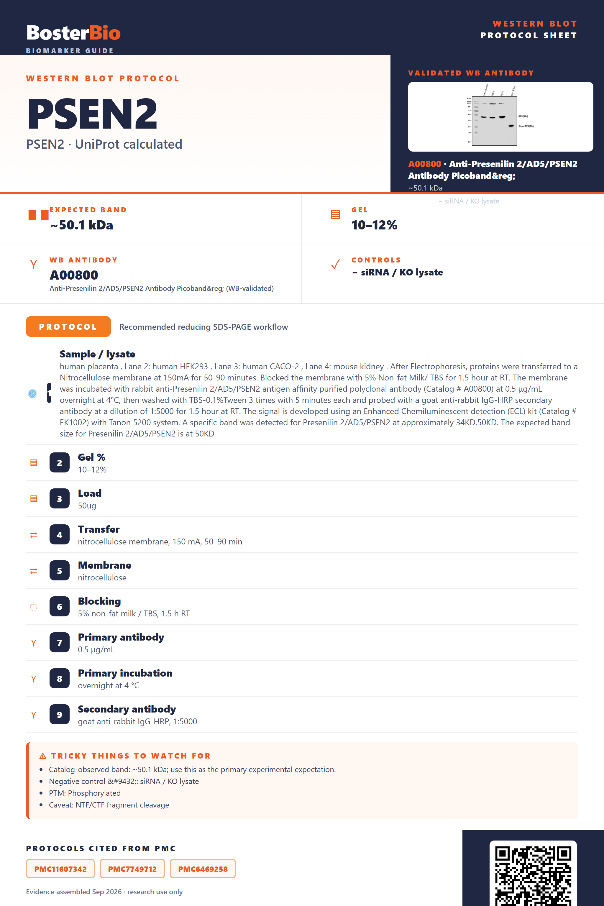
Western blot protocol sheet for PSEN2: expected band ~50.1 kDa, antibody A00800, and PMC-cited SDS-PAGE protocol steps
PSEN2 Western blot protocol sheet — expected band ~50.1 kDa, antibody A00800, controls and PMC citations. Open the full PSEN2 WB guide →

PSEN2 Western Blot Experimental Design Guide

Expected bands, validated protocols, controls and antibodies — the at-a-glance facts below, then the full design guide.

Must know before running
Expected band ~50.1 kDa
Observed band ~34 kDa and ~50 kDa
Gel 10–12%
Negative control ⓘ siRNA / KO lysate
Important caveats
Reasons your observed band may differ from the expected size.
PTM Phosphorylated
Caveat NTF/CTF fragment cleavage
Regulation Apoptosis
Isoform 3 isoform(s)
Section 1

Real Curated PSEN2 Western Blot Protocols

Literature-validated Western blot parameters for PSEN2 — gel percentage, transfer, blocking, antibody incubation and detection, extracted from published methods.

Recommended Western blot protocol parameters
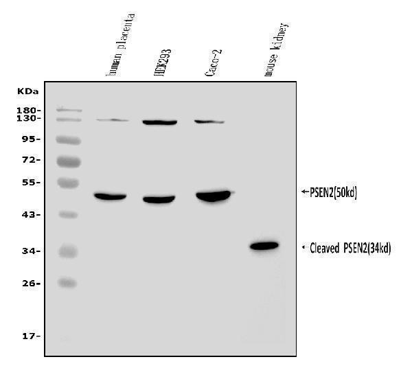
Sample / lysatehuman placenta , Lane 2: human HEK293 , Lane 3: human CACO-2 , Lane 4: mouse kidney . After Electrophoresis, proteins were transferred to a Nitrocellulose membrane at 150mA for 50-90 minutes. Blocked the membrane with 5% Non-fat Milk/ TBS for 1.5 hour at RT. The membrane was incubated with rabbit anti-Presenilin 2/AD5/PSEN2 antigen affinity purified polyclonal antibody (Catalog # A00800) at 0.5 μg/mL overnight at 4°C, then washed with TBS-0.1%Tween 3 times with 5 minutes each and probed with a goat anti-rabbit IgG-HRP secondary antibody at a dilution of 1:5000 for 1.5 hour at RT. The signal is developed using an Enhanced Chemiluminescent detection (ECL) kit (Catalog # EK1002) with Tanon 5200 system. A specific band was detected for Presenilin 2/AD5/PSEN2 at approximately 34KD,50KD. The expected band size for Presenilin 2/AD5/PSEN2 is at 50KD
Gel %10–12%
Load50ug
Transfernitrocellulose membrane, 150 mA, 50–90 min
Membranenitrocellulose
Blocking5% non-fat milk / TBS, 1.5 h RT
Primary antibody0.5 µg/mL
Primary incubationovernight at 4 °C
Secondary antibodygoat anti-rabbit IgG-HRP, 1:5000
WashTBS-0.1% Tween, 3 × 5 min
DetectionECL
Exposure / imagingTanon 5200
Observed band34 kDa, 50 kDa
Section 2

What Is the Expected PSEN2 Western Blot Band Size?

PSEN2 has a 50.1 kDa predicted full-length mass but is endoproteolytically processed, so blots typically show both a ~50 kDa full-length band and a ~34 kDa NTF fragment.

What am I looking at on my blot?
band at ~50 kDafull-length, uncleaved PSEN2 holoprotein matching the predicted monomer mass
band at ~34 kDaendoproteolytically cleaved N-terminal fragment (NTF) of presenilin-2, the predominant mature processed form
two bands present together (~50 kDa and ~34 kDa)simultaneous detection of the full-length precursor and its cleaved NTF fragment, reflecting normal partial endoproteolysis
band at roughly double the monomer mass under non-reducing conditionsincompletely dissociated PSEN2 homodimer or NTF/CTF heterodimer complex
several bands at different molecular weightsdetection of multiple PSEN2 splice isoforms (isoforms 1, 2, 3) rather than a single processing product
single sharp band near 50 kDa with no smearingunmodified full-length monomer, consistent with the absence of glycosylation sites on PSEN2
💡Expected PSEN2 appearanceExpect a band near 50 kDa for full-length PSEN2 and/or a ~34 kDa band for its endoproteolytically cleaved N-terminal fragment (NTF), the normal processed form of this multi-pass membrane protein.
How each factor affects band size
predicted mass (50.1 kDa, 448 aa)sets the baseline size for the full-length, unprocessed PSEN2 monomer
endoproteolytic cleavage into NTF/CTF fragmentsgenerates a smaller ~34 kDa mature fragment distinct from the 50 kDa full-length precursor
homodimer / NTF-CTF heterodimer formationcan produce a higher-molecular-weight species if the complex is not fully dissociated by reducing and denaturing conditions
alternative splicing (isoforms 1, 2, 3)can add extra bands of differing size relative to the canonical isoform, though exact isoform sizes are not defined
multi-pass membrane protein with no glycosylation sitesband position is not raised by glycosylation, so any upward shift is not attributable to glycan load
Why is my band missing or off?
SituationLikely causeNext action
Multiple bandsantibody detects both full-length PSEN2 (~50 kDa) and the cleaved NTF fragment (~34 kDa), and/or distinct splice isoformsmap each band against the expected full-length, NTF, and isoform sizes before calling any band nonspecific
Band lower than expectedsample is dominated by the mature, endoproteolytically processed NTF fragment rather than the full-length precursorcompare the lower band to the ~34 kDa NTF size and confirm identity with an N-terminal-directed antibody
Fragments below expected sizeadditional proteolytic degradation during lysis on top of normal NTF/CTF processingadd protease inhibitors and keep lysates cold throughout preparation to separate degradation from genuine processing
Band higher than expectedincompletely dissociated PSEN2 homodimer or NTF/CTF heterodimer complexincrease reducing agent and SDS concentration and extend boiling time before loading
Weak or no signallow PSEN2 expression in the tissue tested or poor extraction of this multi-pass membrane proteinuse tissue or cell lines with confirmed PSEN2 expression and optimize detergent-based membrane protein extraction

Sample controls for PSEN2 Western blot

🧪For positive controls for PSEN2 in Western blot, you can use a common mammalian cell line lysate such as HEK293 or HeLa, since PSEN2 is ubiquitously expressed as a core subunit of the gamma-secretase complex and no tissue-specific HPA expression data is available to point to a more specific source.
Positive control: HEK293 cell lysate
Negative control: ubiquitously expressed; use siRNA knockdown or KO line
Loading controls: Run GAPDH and β-actin as loading controls alongside a total-protein stain (e.g., stain-free gel, Ponceau S, or REVERT).
⚠️Feasibility: With no HPA expression data and PSEN2 broadly distributed across ER, Golgi, endosome, and lysosome membranes, no tissue is likely to serve as a clean negative, so siRNA knockdown or a knockout cell line is the more feasible negative control.

Comprehensive Human Protein Atlas IHC scoring per tissue. Rows are taken directly from the HPA tissue chart — click any row's HPA link to view the source.

Positive expression · recommended positive controls

TissueCell typeLevelEvidenceSource

Undetected expression · recommended negative controls

TissueCell typeLevelEvidenceSource
Section 3

Advanced PSEN2 Western Blot Tips

Deeper troubleshooting and optimisation questions for PSEN2, answered from its protein features.

Why does PSEN2 show bands at both ~34 kDa and ~50 kDa?
PSEN2 is synthesized as a ~50 kDa full-length protein but undergoes endoproteolytic cleavage into N-terminal (NTF) and C-terminal (CTF) fragments that predominantly assemble as a heterodimer. The ~34 kDa band likely represents the NTF cleavage product, while ~50 kDa reflects uncleaved or newly synthesized precursor. Detecting both is expected, and antibody epitope location (N- vs C-terminal) determines which band predominates.
Do PSEN2 isoforms affect the observed band pattern?
PSEN2 has three annotated isoforms (1, 2, 3) generated by alternative splicing. These may differ slightly in length and predicted mass, contributing minor additional bands or size heterogeneity beyond the NTF/CTF doublet. Confirm which isoform your antibody targets and whether your cell or tissue model expresses multiple isoforms before interpreting extra bands as nonspecific.
Is PSEN2 phosphorylation likely to shift its band?
PSEN2 is annotated as a phosphoprotein with four modified residues. Phosphorylation can subtly increase apparent molecular weight or produce closely spaced doublets, particularly on the full-length or CTF fragment. Phosphatase treatment of a parallel sample can confirm whether such shifts are phosphorylation-dependent rather than isoform- or degradation-related.
How should blocking be optimized for PSEN2 blots?
Because PSEN2 is a phosphoprotein, avoid milk-based blocking buffers, as casein phosphoproteins can cause high background with phospho-specific antibodies; use BSA instead. As a multi-pass ER/Golgi membrane protein, extended blocking (1 hour, room temperature) helps offset the low protein-to-lipid ratio typical of membrane-enriched lysates.
What transfer method to use for PSEN2 Western blot?
PSEN2 is a multi-pass transmembrane protein residing in ER, Golgi, endosome, and lysosome membranes, so it can transfer poorly with standard conditions. Use wet transfer with reduced methanol (10%) or a semi-dry system optimized for hydrophobic proteins, and PVDF membrane, which binds membrane proteins more efficiently than nitrocellulose.
Which PSEN2 band should be used for quantitation?
Because PSEN2 is detected as both full-length (~50 kDa) and cleaved NTF (~34 kDa) species, quantify only the band matching your antibody's validated epitope and consistently present across samples. Mixing signal from both species will distort normalization; select one band and confirm specificity with a knockdown or knockout control.
Why might extra high-molecular-weight PSEN2 bands appear?
PSEN2 forms homodimers and heterodimeric NTF/CTF complexes, and is a core component of the gamma-secretase complex with other subunits. Under incompletely denaturing or non-reducing conditions, these associations can persist as higher-molecular-weight bands. Ensure samples are fully denatured (SDS, reducing agent, heat) to resolve monomeric fragments.
Boster reagents

Best PSEN2 Western Blot Antibodies

BosterBio's PSEN2 antibodies are among the best-performing WB antibodies on the market — well cited, thoroughly validated, and orthogonally cross-validated against negative tissues and complementary methods.

Real WB data Western blot analysis of Presenilin 2/AD5/PSEN2 using anti-Presenilin 2/AD5/PSEN2 antibody (A00800). Electrophoresis was performed on a 5-20% SDS-PAGE gel at 70V (Stacking gel) / 90V (Resolving gel) for 2-3 hours. The sample well of each lane was loaded with 50ug of sample under reducing conditions. Lane 1: human placenta tissue lysates, Lane 2: human HEK293 whole cell lysates, Lane 3: human CACO-2 whole cell lysates, Lane 4: mouse kidney tissue lysates. After Electrophoresis, proteins were transferred to a Nitrocellulose membrane at 150mA for 50-90 minutes. Blocked the membrane with 5% Non-fat Milk/ TBS for 1.5 hour at RT. The membrane was incubated with rabbit anti-Presenilin 2/AD5/PSEN2 antigen affinity purified polyclonal antibody (Catalog # A00800) at 0.5 μg/mL overnight at 4°C, then washed with TBS-0.1%Tween 3 times with 5 minutes each and probed with a goat anti-rabbit IgG-HRP secondary antibody at a dilution of 1:5000 for 1.5 hour at RT. The signal is developed using an Enhanced Chemiluminescent detection (ECL) kit (Catalog # EK1002) with Tanon 5200 system. A specific band was detected for Presenilin 2/AD5/PSEN2 at approximately 34KD,50KD. The expected band size for Presenilin 2/AD5/PSEN2 is at 50KD.
Anti-Presenilin 2/AD5/PSEN2 Antibody Picoband®
Cat # A00800
Real WB data Anti-Presenilin 2 antibody, PA1358, Western blotting Lane 1: Rat Brain Tissue Lysate Lane 2: Rat Brain Tissue Lysate Lane 3: MCF-7 Cell Lysate Lane 4: HELA Cell Lysate Lane 5: SMMC Cell Lysate Lane 6: CEM Cell Lysate
Anti-Presenilin 2/PSEN2 Antibody Picoband®
Cat # PA1358

These anti-PSEN2 antibodies are top-performing, extensively cited reagents rigorously validated by western blot, with specificity confirmed through orthogonal testing against negative tissue controls and complementary detection methods, giving researchers reliable, reproducible results for presenilin 2 detection across sample types.

Which to pick: Both A00800 and PA1358 include real WB validation images. PA1358 shows broader validation across rat brain, MCF-7, HeLa, and SMMC lysates, making it a strong default; choose A00800 if you prefer its specific PSEN2 SDS-PAGE validation instead.

Source: BosterBio PSEN2 gene-info card — filtered to Western-blot-capable antibodies; each card shows that product's actual WB validation figure.

References

  1. UniProt Consortium. UniProt entry P49810.
  2. Human Protein Atlas. PSEN2 tissue expression.