PTGFRN / Prostaglandin F2 receptor negative regulator · IHC design guide

Design Immunohistochemistry for PTGFRN

Plan PTGFRN staining in paraffin sections around a membranous and cytoplasmic tissue pattern (HPA tissue IHC). Compare high-staining adrenal glandular cells with adipocytes where staining was not detected (HPA tissue IHC), and start with the catalog antibody at 1:100 (datasheet).

Evidence assembled Oct 2026 · For research use; verify linked source records and product datasheet before use
Immunohistochemistry protocol sheet for PTGFRN (IHC for PTGFRN): expected localisation Membranous and cytoplasmic staining in most tissues (HPA tissue IHC), antibody A09624, validated IHC image, and IHC protocol steps
Printable PTGFRN IHC protocol sheet — expected localisation Membranous and cytoplasmic staining in most tissues (HPA tissue IHC), antibody A09624, controls and protocol steps. Open the full PTGFRN IHC guide →

PTGFRN Immunohistochemistry Experimental Design Guide

Expected localisation, validated protocols, controls and antibodies — the at-a-glance facts below, then the full design guide.

Must know before staining
Expected localisation Membranous and cytoplasmic staining in most tissues (HPA tissue IHC)
Staining pattern Glandular and respiratory epithelial cells: membrane and cytoplasm (HPA tissue IHC)
Antigen retrieval Citrate pH 6.0 HIER, 95–98 °C, 20 min (rule: cytoplasmic / membrane antigen)
Positive control ⓘ Adrenal gland+4 more · see all
Negative control ⓘ Adipose tissue+4 more · see all
Important caveats
Reasons your staining may differ from the expected pattern.
Fixation Keep paraffin-section fixation consistent (standard IHC practice; not target-specific)
Caveat Low staining–RNA consistency; verify the pattern (HPA tissue IHC)
Regulation Expression regulation is not annotated (UniProt)
Isoform / epitope No isoforms annotated; extracellular versus cytoplasmic epitope matters (UniProt)
Section 1

Recommended PTGFRN IHC & IF Protocols

The catalog antibody’s IHC-P protocol is accompanied by published PTGFRN protocols for lung adenocarcinoma sections (PMC13049031) and patient-derived tissue microarrays (PMC12185247).

Recommended immunohistochemistry (IHC-P) protocol parameters
SampleParaffin-embedded human liver tissue; fixative not specified (datasheet A09624)
FixationImage fixative and duration unreported (datasheet A09624); verify before use.
Sectioning4–5 µm sections on charged slides (standard)
DeparaffinisationXylene, graded ethanol series to water (standard)
Antigen retrievalHeat-induced epitope retrieval in citrate buffer, pH 6.0, 20 min at 95–98 °C (standard rule: cytoplasmic / membrane antigen)
Peroxidase block3% H2O2, 10 min, room temperature (standard)
Blocking10% normal serum of the secondary host, 30 min, room temperature (standard)
Primary antibodyRabbit anti-PTGFRN, 1:100 recommended; image 1:200 (datasheet A09624)
Primary incubationOvernight at 4 °C (standard)
DetectionHRP-polymer secondary, DAB chromogen 5–10 min (standard)
CounterstainHematoxylin, blue, dehydrate and mount (standard)
Expected resultPTGFRN-positive staining in glandular cells of adrenal gland (HPA tissue IHC: High). HPA tissue profile: Membranous and cytoplasmic expression in most tissues. No signal in the no-primary control.
💡Decision noteStart with citrate pH 6.0 retrieval at 95–98 °C for 20 min (page retrieval); the 1B4 microarray protocol used no retrieval (PMC12185247).
Section 2

What Is the Expected PTGFRN Staining Pattern?

PTGFRN staining is expected at cell membranes and in cytoplasm across many tissues (HPA tissue IHC). Its extracellular region spans residues 26–832, followed by a transmembrane segment at 833–853; UniProt also places it on endoplasmic reticulum and trans-Golgi network membranes (UniProt Q9P2B2 topology and subcellular annotation). Interpret tissue patterns cautiously: HPA rates the IHC evidence Approved but reports low agreement with RNA data and pending external verification (HPA tissue IHC).

What am I looking at on my slide?
Membranous staining with some cytoplasmic signal in bronchial respiratory epithelium or placental trophoblasts.This fits the reported pattern. Both cell populations stain High in HPA tissue IHC; assess the named cells rather than assigning one score to the whole section (HPA tissue IHC).
Predominantly nuclear staining, especially without a convincing membrane or cytoplasmic pattern.Nuclei are outside the reported PTGFRN locations (HPA tissue IHC; UniProt Q9P2B2 subcellular annotation). Treat this as suspect staining and review controls before scoring it as PTGFRN (general IHC practice).
Strong signal in adipocytes, marrow hematopoietic cells, or skeletal myocytes.HPA reports PTGFRN as Not detected in these specific cell populations (HPA tissue IHC). Such signal merits checks for cross-reactivity or endogenous detection activity; it does not establish that the entire tissue is negative (general IHC practice).
Uniform chromogen across tissue, empty spaces, or the whole section with little cell-level contrast.This is less interpretable than the membranous and cytoplasmic cell pattern reported by HPA (HPA tissue IHC). Check reagent background and detection controls before calling it a broad PTGFRN distribution (general IHC practice).
No signal in an adequately sampled bronchus or placenta section.HPA reports High staining in bronchial respiratory epithelial cells and placental trophoblasts (HPA tissue IHC). Confirm that those cells are present, then review the IHC run and antibody performance (general IHC practice).
💡Expected PTGFRN appearanceA convincing positive shows High membranous with possible cytoplasmic staining in bronchial respiratory epithelial cells or placental trophoblasts (HPA tissue IHC); isolated nuclear or cell-free diffuse color is suspect (HPA tissue IHC; general IHC practice).
How each factor affects the staining
Subcellular contextPTGFRN has an extracellular domain, one transmembrane segment, and a short cytoplasmic tail (UniProt Q9P2B2 topology). HPA reports both membranous and cytoplasmic tissue staining, so cytoplasmic color alone is not automatically discordant (HPA tissue IHC).
Choice of comparison cellsAdrenal glandular cells, cervical and endometrial glandular cells, and nasopharyngeal respiratory epithelial cells are also High (HPA tissue IHC). HPA's Not detected calls apply to specified cells, such as adipocytes, rather than every cell in those organs (HPA tissue IHC).
Evidence strengthThe antibody's IHC status is Approved, not Enhanced (HPA antibody validation). HPA notes low consistency between antibody staining and RNA expression and says external verification is pending; use the pattern as a comparison, not independent proof of identity (HPA tissue IHC).
Antigen retrieval and fixationTarget-specific fixation effects are not established by the supplied assay evidence. Verify with a matched IHC source before attributing a result to fixation.
What does IF/ICC add?HPA's ICC-IF summary says Membrane but supplies no cell-line images or detailed main location (HPA subcellular). This supports only a limited membrane comparison; IF/ICC conditions and interpretation belong on its own guide page (HPA subcellular).
Why is my staining missing, weak or wrong?
SituationLikely causeNext action
A high-expression comparison section is blank.The sampled positive cell type may be absent, or the IHC run may have failed (HPA tissue IHC; general IHC practice).Locate bronchial respiratory epithelial cells or placental trophoblasts first; then check the detection controls and repeat the run if needed (HPA tissue IHC; general IHC practice).
Only nuclei stain strongly.That compartment conflicts with the reported membrane and cytoplasmic pattern (HPA tissue IHC; UniProt Q9P2B2 subcellular annotation).Inspect a positive comparison section and detection controls; avoid scoring nuclear-only color as PTGFRN without independent support (general IHC practice).
Expected negative comparison cells stain strongly.Cross-reactivity or endogenous detection activity is possible; HPA calls adipocytes and marrow hematopoietic cells Not detected (HPA tissue IHC; general IHC practice).Review cell identity and a control omitting primary antibody, then reassess background before interpreting the signal (general IHC practice).
Diffuse color obscures membrane detail.Background can make a cell-specific pattern hard to distinguish (general IHC practice).Review blocking, antibody dilution, washes, detection controls, and chromogen development within the established IHC workflow (general IHC practice).
Membranous staining appears in a cell type listed as low.HPA levels vary by cell type; lung macrophages and intestinal glandular cells are reported Low (HPA tissue IHC).Score intensity in the named cells against a positive comparison section; do not convert a low HPA call into an expected strong result (HPA tissue IHC; general IHC practice).
Cytoplasmic staining is present with weak membrane staining.HPA reports cytoplasmic as well as membranous tissue expression, while UniProt places PTGFRN on internal membranes (HPA tissue IHC; UniProt Q9P2B2 subcellular annotation).Compare cell type, intensity, and controls before rejecting or accepting the result; the records do not specify a required membrane-to-cytoplasm ratio (HPA tissue IHC; UniProt Q9P2B2).

Sample controls for PTGFRN IHC & IF

🧪Run adrenal gland first: glandular cells should stain (HPA: High in adrenal gland glandular cells). Run adipose tissue as the negative comparator (HPA: Not detected in adipocytes); on the adrenal slide, use adjacent unstained cells as an internal background reference, without assuming their PTGFRN status.
Positive control tissue: Adrenal gland (Glandular cells, HPA High)
Negative control tissue: Adipose tissue (HPA Not detected)
ICC-IF cell lines (HPA subcellular resource): HPA carries no ICC-IF cell line for PTGFRN; derive a cell-line control from the positive tissue's cell type (Glandular cells) and confirm it by RNA or western blot first.
Technical controls: Include a no-primary, secondary-only control and an isotype control matched to the primary antibody’s host species and clonality; use PTGFRN knockout tissue or a validated immunogen peptide block as a biological specificity control. Quench endogenous peroxidase on the adrenal section before chromogenic detection (standard IHC practice).
⚠️Feasibility: A PTGFRN-specific fixation window or fixation effect is unreported, and the selected A09624 paraffin-section caption does not state its fixative (caption: human liver, 1:200). Retrieval dependence is unreported; optimize antigen retrieval empirically for paraffin sections (standard IHC practice). The supplied evidence does not establish that frozen sections or IF are easier (HPA: no ICC-IF image cell lines), or identify a PTGFRN-specific adrenal artefact; assess endogenous pigment against the control sections (standard IHC practice).

HPA tissue IHC evidence for PTGFRN

Comprehensive Human Protein Atlas IHC scoring per tissue (reliability: Approved — Low consistency between antibody staining and RNA expression data. Pending external verification.). Rows are taken directly from the HPA tissue chart — click any row's HPA link to view the source.

Positive expression · recommended positive controls

TissueCell typeLevelEvidenceSource
Adrenal gland Glandular cells High Protein (IHC) HPA →
Bronchus Respiratory epithelial cells High Protein (IHC) HPA →
Cervix Glandular cells High Protein (IHC) HPA →
Endometrium Glandular cells High Protein (IHC) HPA →
Nasopharynx Respiratory epithelial cells High Protein (IHC) HPA →

Undetected expression · recommended negative controls

TissueCell typeLevelEvidenceSource
Adipose tissue Adipocytes Not detected Protein (IHC) HPA →
Bone marrow Hematopoietic cells Not detected Protein (IHC) HPA →
Cerebellum Cells in granular layer Not detected Protein (IHC) HPA →
Lymph node Germinal center cells Not detected Protein (IHC) HPA →
Ovary Ovarian stroma cells Not detected Protein (IHC) HPA →
Section 3

Advanced PTGFRN IHC Tips

Troubleshoot PTGFRN staining in paraffin section chromogenic IHC, using the catalog image and tissue expression data as context for controls.

How should I adjust retrieval when PTGFRN staining is weak?
Start with citrate pH 6.0 heat induced epitope retrieval at 95–98 °C for 20 min (page retrieval setting). Cool sections in the retrieval solution before washing, and keep section thickness and heating conditions consistent across slides (standard IHC practice). If staining remains weak, compare a modestly longer retrieval time on adjacent sections while checking whether tissue structure deteriorates (standard IHC practice). The catalog image shows PTGFRN staining in paraffin embedded human liver at 1:200, but its caption does not report retrieval conditions (catalog A09624 caption). Record both staining intensity and tissue preservation before selecting a revised condition (standard IHC practice).
Can I infer the best fixation for PTGFRN from the catalog image?
No target specific fixation sensitivity is established by the supplied evidence: the paraffin embedded human liver caption leaves the fixative unreported (catalog A09624 caption). When validating a local fixation workflow, compare sections processed with documented fixation conditions while holding retrieval, antibody dilution, and detection constant (standard IHC practice). Evaluate preservation of cell boundaries alongside signal intensity, because damaged morphology makes a membranous pattern difficult to score (standard IHC practice; HPA: membranous and cytoplasmic expression). Use the catalog image’s 1:200 dilution as a documented comparison point, without treating that image as evidence for any fixative (catalog A09624 caption).
Where should convincing PTGFRN staining appear in tissue sections?
Assess staining at cell boundaries and in cytoplasm, the two patterns reported across tissues by HPA (HPA: membranous and cytoplasmic expression in most tissues). PTGFRN has an extracellular region at residues 26–832, one transmembrane segment at 833–853, and a cytoplasmic tail at 854–879 (UniProt Q9P2B2 topology). Endoplasmic reticulum and trans Golgi network membrane annotations also support assessing intracellular signal in its cellular context (UniProt Q9P2B2 subcellular). Compare each compartment with a matched negative control and the same cell type across sections; diffuse stain over empty spaces or section edges is less persuasive (standard IHC practice).
Could epitope position explain different PTGFRN staining patterns?
First obtain the antibody’s immunogen or mapped epitope, because the supplied catalog caption reports a 1:200 dilution but no epitope (catalog A09624 caption). PTGFRN’s extracellular region spans residues 26–832, whereas its cytoplasmic tail spans 854–879; epitope location therefore matters when comparing staining accessibility (UniProt Q9P2B2 topology; standard IHC practice). Six Ig like domains and 9 annotated glycosylation sites occur in the extracellular region, but their effect on this antibody’s staining is untested here (UniProt Q9P2B2 domains and glycosylation). The supplied record lists 0 isoforms, so do not assign divergent staining to a particular splice isoform without separate evidence (supplied UniProt record: isoforms).
How can I compare PTGFRN IHC with a multiplex IF experiment?
Treat multiplex IF as a separate validation experiment: the supplied HPA subcellular record lists no cell lines with ICC/IF images (HPA subcellular record). Pair PTGFRN with an independently validated marker for the expected cell type, such as a respiratory epithelial marker when examining bronchus, where HPA reports high signal in respiratory epithelial cells (HPA: bronchus, respiratory epithelial cells, High; standard IF practice). Select spectrally separated fluorophores and inspect unstained tissue for autofluorescence before choosing channels (standard IF practice). If the antibody recognizes the extracellular region, minimize permeabilisation; if it recognizes the 854–879 cytoplasmic tail, compare gentle permeabilisation conditions after fixation (UniProt Q9P2B2 topology; standard IF practice).
What should I check when PTGFRN staining covers the whole section?
Run a no primary control through the same chromogenic detection steps to identify secondary reagent or substrate background (standard IHC practice). Include a peroxidase block before a peroxidase based DAB workflow, and compare a shorter development time if precipitate obscures cell boundaries (standard IHC practice). Titrate around the catalog image’s 1:200 antibody dilution while keeping retrieval and detection constant; that dilution documents one paraffin section image, not an optimum for every specimen (catalog A09624 caption; standard IHC practice). Compare the same cell type across sections and inspect edges and folds separately, because uneven reagent exposure can mimic widespread positive staining (standard IHC practice).
How should I score heterogeneous PTGFRN staining across specimens? ⚠ ANSWER MARKED FOR VERIFICATION
Define the cell population and compartment before scoring: HPA reports membranous and cytoplasmic expression in most tissues (HPA: tissue IHC profile). For epithelial samples, record the percentage of positive target cells and an intensity based H-score, reporting membrane and cytoplasmic scores separately when both are visible (standard IHC scoring practice). If counting scattered positive cells, report density per mm² of viable tissue and normalize to the measured viable area rather than total section area (standard IHC quantification practice). Apply one threshold, counterstain standard, and image acquisition method across specimens, and exclude folds, necrosis, and section edges before comparison (standard IHC practice).
How do I distinguish credible PTGFRN signal from staining artefact?
A credible result has interpretable membrane or cytoplasmic staining in intact cells and exceeds its matched negative control (HPA: tissue IHC profile; standard IHC practice). Check cell identity: HPA reports high signal in bronchial respiratory epithelial cells but no detected signal in adipocytes, so these annotated populations provide context without proving antibody specificity (HPA: bronchus, High; adipose tissue, Not detected). Reject apparent positives confined to folds, edges, necrotic regions, or pigment and endogenous enzyme deposits when the cellular pattern is absent (standard IHC practice). Interpret discordance cautiously because HPA calls its tissue IHC reliability Approved while noting low consistency with RNA expression and pending external verification (HPA: reliability description).
Boster reagents

Best PTGFRN / Prostaglandin F2 receptor negative regulator IHC Antibodies

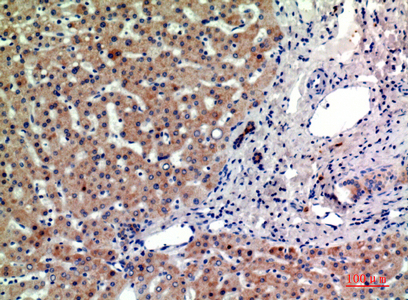
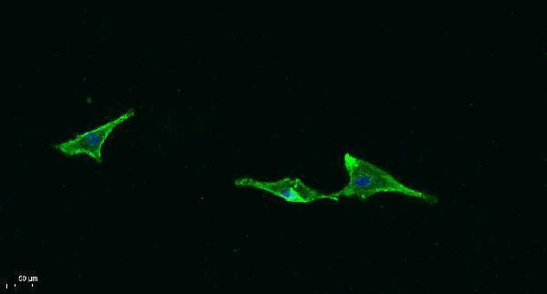
A09624 has IHC data from paraffin-embedded human liver and IF data from A549; catalog reactivity includes human, mouse, and rat (IHC/IF image captions; catalog reactivity).

Real IHC data Immunohistochemical analysis of paraffin-embedded human-liver, antibody was diluted at 1:200
Anti-CD315 PTGFRN Antibody
Cat # A09624

A09624 will render with an IHC image of paraffin-embedded human liver using 1:200 antibody dilution (IHC image caption). It is also listed for IF, with an A549 IF image using 1:200 primary dilution (catalog applications; IF image caption).

Which to pick: For tissue IHC, choose A09624: its own image documents paraffin-embedded human liver; the fixative is unreported (IHC image caption). For IF/ICC, A09624 has an A549 IF image, while ICC-specific validation is unreported (IF image caption; catalog applications). For mouse or rat samples, A09624 lists reactivity with both species and is rabbit polyclonal; the supplied IHC image documents human liver (catalog reactivity/host/dilution_raw; IHC image caption).

Each figure is that product's own IHC / IF validation image from its datasheet.

References

  1. UniProt Consortium. UniProt entry Q9P2B2 (FPRP_HUMAN, Prostaglandin F2 receptor negative regulator).
  2. Human Protein Atlas. PTGFRN tissue IHC expression (reliability: Approved).
  3. Human Protein Atlas. PTGFRN subcellular location (ICC-IF): Membrane.
  4. Human Protein Atlas. PTGFRN antibody validation summary (1 antibodies).
  5. Integrative analysis of cell adhesion molecules in glioblastoma identified prostaglandin F2 receptor inhibitor (PTGFRN) as an essential gene. BMC cancer 2022 — PMC9188228.
  6. Pan-cancer analysis reveals the oncogenic and immunomodulatory roles of PTGFRN across human cancers. Scientific reports 2026 — PMC13049031.
  7. 8C7: A Fully Human Anti-PTGFRN Monoclonal Antibody-Drug Conjugate Inhibiting Tumour Growth of Mesothelioma and Paediatric Medulloblastoma Cell Lines. Journal of cellular and molecular medicine 2025 — PMC12185247.
  8. Knockdown of long non‑coding RNA DLEU2 suppresses idiopathic pulmonary fibrosis by regulating the microRNA‑369‑3p/TRIM2 axis. International journal of molecular medicine 2021 — PMC7979258.
  9. PubMed PMID:10718198 — UniProt-cited evidence.
  10. PubMed PMID:14702039 — UniProt-cited evidence.
  11. PubMed PMID:16710414 — UniProt-cited evidence.