This website uses cookies to ensure you get the best experience on our website.


- Table of Contents
Plan PXDN staining in paraffin sections around fibroblast-associated and extracellular positivity (HPA tissue IHC). This guide covers fixation, antibody dilution and chromogenic detection using a documented 1 μg/ml IHC-P workflow (datasheet: DZ33984).

Expected localisation, validated protocols, controls and antibodies — the at-a-glance facts below, then the full design guide.
| Expected localisation | Extracellular tissue staining (HPA tissue IHC) | |
| Staining pattern | Fibroblast-associated and extracellular positivity (HPA tissue IHC) | |
| Antigen retrieval | EDTA pH 8.0 HIER, heat-mediated (datasheet DZ33984) | |
| Positive control | Bronchus+1 more · see all | |
| Negative control | Adipose tissue+4 more · see all |
| Fixation | Keep fixation consistent across sections. (standard IHC practice; not target-specific) | |
| Caveat | Only processed PXDN integrates into the matrix (UniProt) | |
| Regulation | Expression varies by tissue (UniProt) | |
| Isoform / epitope | 2 isoforms; a shorter form may lack the signal peptide, so check the epitope (UniProt) |
Compare the catalog antibody’s IHC-P protocol (datasheet DZ33984) with three published PXDN IHC protocols (PMC11603849; PMC6627806; PMC8858314).
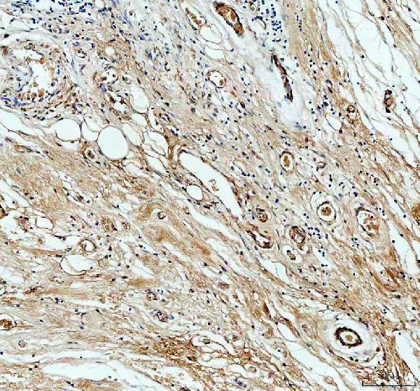
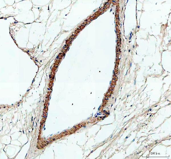
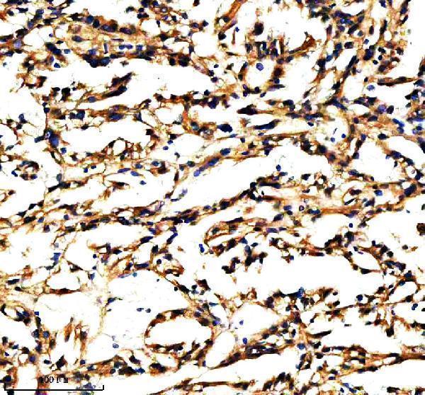
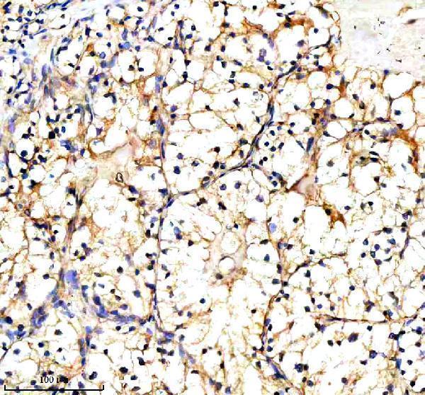
| Sample | Paraffin-embedded human pancrease cancer tissue; fixative not specified (datasheet DZ33984) |
| Fixation | Image fixative and duration unreported (datasheet DZ33984); verify before use. |
| Sectioning | 4–5 µm sections on charged slides (standard) |
| Deparaffinisation | Xylene, graded ethanol series to water (standard) |
| Antigen retrieval | Heat retrieval: EDTA pH 8.0 (datasheet DZ33984); 20 min, 95–100 °C (standard) |
| Peroxidase block | 3% H2O2, 10 min, room temperature (standard) |
| Blocking | 10% goat serum (datasheet DZ33984) |
| Primary antibody | Rabbit anti-PXDN, 1 μg/ml (datasheet DZ33984) |
| Primary incubation | Overnight at 4 °C (datasheet DZ33984) |
| Detection | Streptavidin-biotin complex (SABC), DAB chromogen (datasheet DZ33984) |
| Counterstain | Hematoxylin, blue, dehydrate and mount (standard) |
| Expected result | PXDN-positive staining in ciliated cells (cilia axoneme) of bronchus (HPA tissue IHC: Medium). HPA tissue profile: Selective expression in fibroblasts and extra cellular positivity. No signal in the no-primary control. |
PXDN is secreted into extracellular space and matrix, including basement membrane, and can also localise to the endoplasmic reticulum and cell surface; it has no transmembrane segment (UniProt Q92626). In paraffin-section IHC, expect selective fibroblast and extracellular staining, with medium staining reported in bronchial cilia axonemes and fallopian-tube cilia tips (HPA tissue IHC). HPA rates the tissue staining profile Approved (HPA tissue IHC).
| Selective staining around fibroblasts or in extracellular structures, with medium signal along bronchial cilia axonemes or at fallopian-tube cilia tips (HPA tissue IHC). | This matches the reported tissue profile and the ability of secreted PXDN to occupy extracellular matrix and basement membrane (HPA tissue IHC; UniProt Q92626). Assess the structure and its neighboring cells; extracellular signal need not trace a complete cell outline (UniProt Q92626 secretion). |
| Strong nuclear staining dominates, while extracellular structures and the reported ciliary sites are unstained. | A predominantly nuclear pattern is discordant with the listed secreted, matrix, cell-surface and endoplasmic-reticulum locations (UniProt Q92626). Treat it as suspect staining; compare the negative control and an HPA-reported positive structure before assigning it to PXDN (HPA tissue IHC). |
| Strong signal appears in adipocytes or adrenal glandular cells, despite little signal in the reported positive structures. | HPA reports PXDN as not detected in those specific cell populations, rather than declaring their whole tissues negative (HPA tissue IHC). Check for cross-reactivity or endogenous chromogenic detection activity, especially if the control without primary antibody also stains (standard IHC practice). |
| Chromogen coats most of the section, including stroma and cells, without a selective pattern. | Uniform colour obscures the selective fibroblast and extracellular profile reported by HPA (HPA tissue IHC). Review primary-antibody concentration, wash stringency and detection controls as general IHC background checks; the supplied sources do not establish a PXDN-specific cause for diffuse staining (standard IHC practice). |
| No convincing signal appears in bronchial cilia axonemes or fallopian-tube cilia tips. | Those structures have medium staining in HPA's tissue record, so an absent signal warrants a run-level check (HPA tissue IHC). Confirm that the sampled section contains the relevant ciliated cells, then review antibody, retrieval and detection conditions; a negative result alone does not prove PXDN is absent (standard IHC practice). |
| Secreted protein and compartment | PXDN occupies extracellular space, matrix and basement membrane, but is also listed at the cell surface and in the endoplasmic reticulum (UniProt Q92626). Score extracellular staining as well as cell-associated signal; tissue RNA and protein locations can differ for a secreted variant (HPA tissue IHC). |
| Proteolytic processing | Only proteolytically processed PXDN integrates into extracellular matrix (UniProt Q92626). A tissue section may therefore show a different balance of cellular and matrix staining; the supplied antibody record does not specify which processed forms its epitope detects (UniProt Q92626; HPA antibody record). |
| Isoforms and signal sequence | UniProt lists 2 isoforms and describes a shorter form, probably lacking the signal sequence, in testis and EB1 cells undergoing p53-dependent apoptosis (UniProt Q92626). Do not require every PXDN-positive context to show identical secretion; the supplied antibody record gives no isoform-specific staining validation (HPA antibody record). |
| Antibody validation | The supplied HPA antibody, HPA012375, is rated Approved for IHC; its ICC status is unreported, and no ICC-IF image cell lines are listed (HPA antibody record; HPA subcellular). The HPA subcellular summary says membrane and secreted, but supplies no main ICC-IF location, so it does not define an IF pattern (HPA subcellular). |
| Retrieval and detection settings | Antigen retrieval and primary-antibody concentration are routine paraffin-section IHC variables (standard IHC practice). Neither UniProt nor the supplied HPA record gives a PXDN-specific retrieval condition or fixation sensitivity; optimize these settings with controls and avoid treating a retrieval-dependent change as established PXDN biology (UniProt Q92626; HPA tissue IHC). |
| Situation | Likely cause | Next action |
|---|---|---|
| Reported ciliary or extracellular positives are blank. | The relevant structures may be absent from the section, or the IHC run may have insufficient usable signal; HPA reports medium staining at the specified ciliary sites (HPA tissue IHC). | Confirm ciliated cells are present and correctly oriented, then check the positive control, retrieval, primary-antibody incubation and detection steps in sequence (standard IHC practice). |
| Only a sharp nuclear pattern is visible. | Nuclear dominance conflicts with the supplied PXDN location record (UniProt Q92626). | Review the control without primary antibody and antibody specificity; require a reproducible, anatomically plausible extracellular or reported ciliary pattern before interpreting the signal as PXDN (HPA tissue IHC; standard IHC practice). |
| Adipocytes or adrenal glandular cells stain strongly. | Those cell types are listed as not detected by HPA; cross-reactivity or endogenous chromogenic activity is possible, but the pattern alone cannot distinguish them (HPA tissue IHC; standard IHC practice). | Compare a control without primary antibody and an HPA-reported positive structure in the same run; review blocking and detection if the control also develops colour (HPA tissue IHC; standard IHC practice). |
| Brown colour spreads across the section and masks tissue structure. | Excess antibody or incomplete removal of detection reagents can raise nonspecific background (standard IHC practice). | Review the antibody concentration, washes, peroxidase blocking and detection time against controls; score PXDN only where selective staining remains interpretable (standard IHC practice; HPA tissue IHC). |
| Extracellular signal is called negative because nearby cell bodies are pale. | That reading can miss secreted, matrix-associated PXDN; processed PXDN is the form that integrates into matrix (UniProt Q92626). | Inspect extracellular and basement-membrane regions alongside cell-associated staining, and record the compartment separately from the stained cell population (UniProt Q92626; standard IHC practice). |
| An IF image is used to set the paraffin-section IHC acceptance pattern. | The supplied HPA subcellular record has no ICC-IF image cell lines or main ICC-IF location, while the supplied antibody has an IHC Approved rating and no ICC rating (HPA subcellular; HPA antibody record). | Use the HPA tissue IHC observations to assess the chromogenic section. Treat IF localisation as a separate application requiring its own validation (HPA tissue IHC; HPA antibody record). |


Comprehensive Human Protein Atlas IHC scoring per tissue (reliability: Approved — At least one protein variant secreted, tissue location of RNA and protein might differ and correlation is complex. Antibody staining in cells/structures not annotated, view images.). Rows are taken directly from the HPA tissue chart — click any row's HPA link to view the source.
| Tissue | Cell type | Level | Evidence | Source |
|---|---|---|---|---|
| Bronchus | Ciliated cells (cilia axoneme) | Medium | Protein (IHC) | HPA → |
| Fallopian tube | Ciliated cells (tip of cilia) | Medium | Protein (IHC) | HPA → |
| Tissue | Cell type | Level | Evidence | Source |
|---|---|---|---|---|
| Adipose tissue | Adipocytes | Not detected | Protein (IHC) | HPA → |
| Adrenal gland | Glandular cells | Not detected | Protein (IHC) | HPA → |
| Appendix | Glandular cells | Not detected | Protein (IHC) | HPA → |
| Bone marrow | Hematopoietic cells | Not detected | Protein (IHC) | HPA → |
| Breast | Adipocytes | Not detected | Protein (IHC) | HPA → |
Troubleshoot PXDN staining in paraffin sections by checking retrieval, compartment, background, and the scoring unit against the supplied tissue evidence.
Two anti-PXDN antibodies have IHC images from human paraffin sections of renal or pancreatic cancer tissue; neither has IF images (catalog image captions; catalog IF image lists).
DZ33984 has IHC images from human pancreatic and renal cancer paraffin sections and lists Human, Mouse and Rat reactivity (DZ33984 image captions; catalog reactivity). A06456-1 has IHC images from human renal cancer paraffin sections and lists Human reactivity (A06456-1 image captions; catalog reactivity).
Which to pick: For human tissue IHC, choose either rabbit polyclonal antibody: DZ33984 was used at 1 μg/ml with EDTA pH 8 retrieval and biotin-based DAB detection, while A06456-1 was used at 2 μg/ml with EDTA pH 8 retrieval and peroxidase-based DAB detection (catalog clonality; each SKU’s IHC image captions). Neither SKU lists IF/ICC or provides an IF image, so there is no documented IF/ICC choice here (catalog applications; catalog IF image lists). DZ33984 is the cross-species candidate because its catalog lists Human, Mouse and Rat reactivity, but its shown IHC sections are human; both captions describe paraffin sections and leave the fixative unreported (catalog reactivity; each SKU’s IHC image captions).