This website uses cookies to ensure you get the best experience on our website.
- Table of Contents
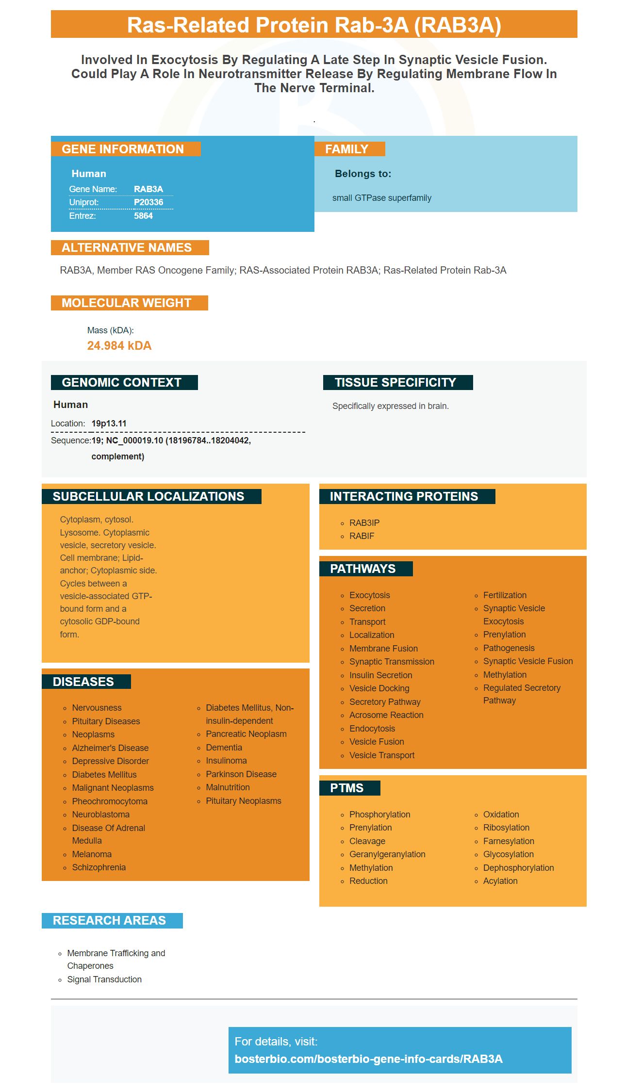
Facts about Ras-related protein Rab-3A.
.
Human | |
---|---|
Gene Name: | RAB3A |
Uniprot: | P20336 |
Entrez: | 5864 |
Belongs to: |
---|
small GTPase superfamily |
RAB3A, member RAS oncogene family; RAS-associated protein RAB3A; ras-related protein Rab-3A
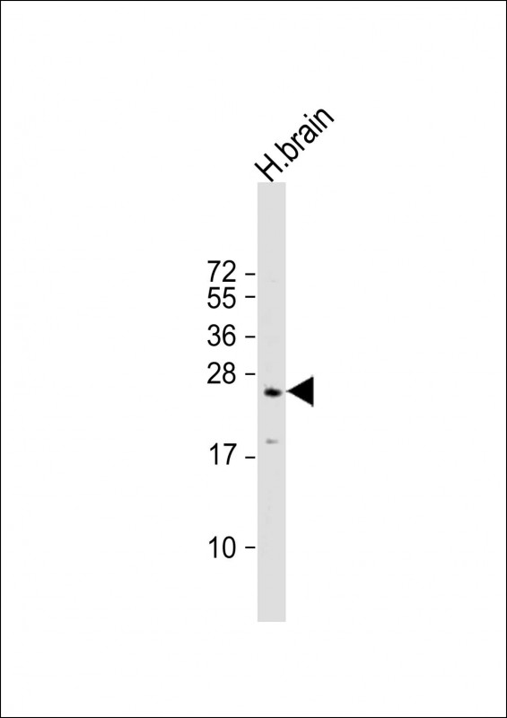
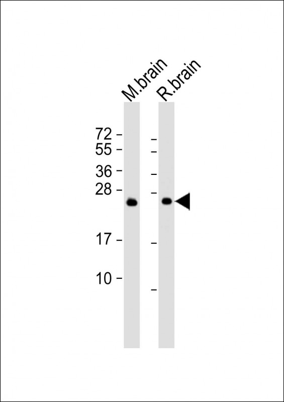
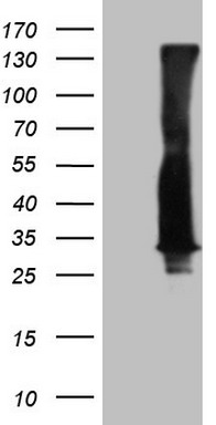
Mass (kDA):
24.984 kDA
Human | |
---|---|
Location: | 19p13.11 |
Sequence: | 19; NC_000019.10 (18196784..18204042, complement) |
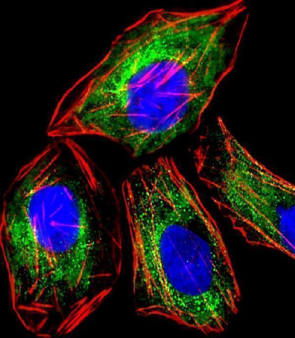
Specifically expressed in brain.
Cytoplasm, cytosol. Lysosome. Cytoplasmic vesicle, secretory vesicle. Cell membrane; Lipid-anchor; Cytoplasmic side. Cycles between a vesicle-associated GTP-bound form and a cytosolic GDP-bound form.
You've found the right place if you're looking for an anti-RAB3A marker. This article will give a brief overview about the Boster Bio Anti-RAB3A Marker. Its applications and validation. Read on to discover how this Rabbit polyclonal antibody can help you with your research. Here are some benefits of using Boster Bio Anti-RAB3A in your research.
Boster Bio Anti-RAB3A Antibody (Catalog number M03463) is a commercially available antibody that has been validated for use in WB and flow cytometry applications. It can keep at -20°C for up to one year. It reacts with human, mouse and rat samples. Boster Bio recommends using antibodies that have a greater length than 30 amino acid to achieve the best results.
This monoclonal antibody recognizes native or recombinant Rab3A proteins and is suitable for use in research studies involving Rab3A-mediated protein-protein interactions. The product can also be used with other commercially available antibodies. While it is only suitable for a limited number of applications, the versatility and usefulness of this antibody are remarkable. Boster Bio Anti–RAB3A marker will provide high-quality results for those who work on Rab3A-mediated proteins-protein interactions.
Boster Bio Rabbit Polyclonal Antibodies are sensitive, specific antibodies that are made by the rabbit immune. They can recognize minor epitope variations and cleaved forms of their target proteins. Their immune system has less immunodominance so they are more sensitive to minor epitope variations and can cleave target proteins with higher signal-to-noise than monoclonal mice antibodies.
Boster Bio Rabbit polyclonal antibody can be used in a variety of applications. The rabbit polyclonal protein has been developed and validated for use with different species. The production process involves rabbit immunization. Every two weeks, booster doses of the antigen will be administered. Ten days after the booster dose, the rabbits will be bled. The serum is then diluted twice in phosphate saline and incubated for 10 days with cyanogen bromide activated Sepharose. The Sepharose beads are able to elute the specific antibody.
The results were consistent and similar to previous studies. For both conditions, the same antibody was used. Recent research showed that RAB3A was colocalized to the dendritic marker, MAP2. To confirm these findings, we used a rabbit monoclonal antibody against MAP2 to visualize the mRNA from transfected neurons. The colocalization of Rab3A with 24-hydroxylase was limited or absent in the soma and processes.
Several different proteins are involved in synaptic vesicle fusion, including syntaxin 1 and synaptobrevins. This 32 kDa polyclonal antibody detects these proteins in membrane and cytosol fractions. It also recognizes SNAP-25/GS32 and SNAP-24. Syn16 (human-protein) is, however, highly expressed in the central nerve system.
The anti-GS32 antibody coimmunoprecipitates GS28 and syntaxin 6 in Golgi extracts. These proteins were not immunoprecipitated by the control rabbit IgG. These results suggest that antibodies against GS32 and GS28 preferentially interact with the syntaxin 6-containing complex. The antibodies used in this research are highly specific, and they can be used to analyze RAB3A's role in neuronal-cell differentiation.
There are many applications for the RAB3A indicator. It has been used in cytology to detect cancer cells. Although Rab3A exhibits a similar migration pattern to cortical granules, it disappears after exocytosis. The recombinant GSTRab3A is close to the target gene and triggers exocytosis.
The depolarization-evoked vesicle recruitment study involved the increased number of synaptic vesicles in the active zone. The total number of docked vessels was higher in wild-type nerve termins than in mutants that contain rab3A. RAB3A deficient nerve terminals did not increase the docked vesicles in the 100 ms following depolarization. The data were obtained using random sections of isolated nerve terminals to measure the number of docked Vesicles.
RAB3A was also found to play a critical role in synaptic recruit and transport. The protein is also involved with transporting synaptic and vesicles to the synapses. It is important to note that the RAB3A-deficient mice display a similar morphological phenotype to wild-type littermates.
These studies have shown that RAB3A can colocalise with secretogranin II(SGII), which is a key component of exocytosis. Rab3A also colocates with secretogranin (2) (SGII). The Rab proteins preferentially associate to newly synthesized and mature granules, while immature granules are associated with them after 20 minutes. The rate of exchange was not affected by inhibitions of the heat-shock proteins 90 (HSP90). During 300 mM ATP stimulation, Rab proteins were activated in live cells. During this time, ppANF-EGFP fluorescence decreased. Photobleaching also caused minimal changes in EGFP/Rab3A.
Bostern bio validated the RAB3A mark using two types tissue: cortex and hippocampus. The RAB3A marker was used to genotype the two tissues as (+/+), or Ebd/Ebd. Boster's validation protocol uses multiple independent methods to test antibodies for specificity or affinity. Boster gives product credits as a reward to the scientist who reviews a product.
HEK 293 cells received transfection with His-tagged Rab3A, and Myc–tagged rabphilin3A. The cells were lysed using lysis buffer containing 20 mm Tris-Cl, pH 7.5, 0.5 mm PMSF, and 1% Tween-20. Next, anti-HisG monoclonal antibodies was used to immunoprecipitate Rab3A.
The Rab3a allele was used to compare the effects of a Rab3a-/ allele on different aspects of brain function. Two-way analyses revealed that Rab3a/ mice displayed lower anxiety and a lower stress response during a zero-maze task. Rab3a/ mice also showed a reduced stress response during a forced swim test. The Rab3a-/ mice lacked the ability to perform cued fear conditioning.
The function of many organs is dependent on the Rab3a genes. The Rab3a gene is located on chromosome 8 and interacts with a number of effector proteins. It is also essential for the formation of cellular membranes. The social behavior and memory of Rab3a/ mice is different from wild-type mice.
Boster Bio's Rab3A anti-rabbit antibody is suitable both for flow cytometry and IF experiments. It is available in PBS containing only 0.09% sodium azide. It can be validated in various tissues with a variety if methods. The antibody is produced by a mouse immunized against recombinant RAB3A. If necessary, the immunogen length can also be altered.
It is important to note that Rab3A is a highly specific protein and is highly sensitive in detecting a range of diseases. Rab7L1 can be used as an adaptor for Rab3A. This allows LRRK2 to be transferred into enlarged lymphosomes. Boster Bio's Rab3A marker is also controlled by Rab7L1. We will continue to study Rab7L1 in order to improve our understanding of Lysosomes.
PMID: 2501306 by Zahraoui A., et al. The human Rab genes encode a family of GTP-binding proteins related to yeast YPT1 and SEC4 products involved in secretion.
PMID: 10574328 by Sullivan M., et al. Genomic organisation of the human cyclic AMP-specific phosphodiesterase PDE4C gene and its chromosomal localisation to 19p13.1, between RAB3A and JUND.