This website uses cookies to ensure you get the best experience on our website.


- Table of Contents
Plan RAB3B IHC-P around cytoplasmic staining in pancreatic islet cells, prostate glandular cells and gastrointestinal endocrine cells (HPA tissue IHC). The catalog antibody was used on formaldehyde-fixed paraffin sections at 1:25 (datasheet M09431 IHC-P); score staining by cell type.

Expected localisation, validated protocols, controls and antibodies — the at-a-glance facts below, then the full design guide.
| Expected localisation | Cytoplasmic staining in endocrine and glandular cells (HPA tissue IHC) | |
| Staining pattern | Cytoplasmic staining in islet, prostate and gut endocrine cells (HPA tissue IHC) | |
| Antigen retrieval | Citrate pH 6 HIER, heat-mediated (datasheet M09431) | |
| Positive control | Colon+4 more · see all | |
| Negative control | Adipose tissue+4 more · see all |
| Fixation | Formaldehyde-fixed paraffin sections were used (datasheet M09431 IHC-P) (selected-SKU IHC image M09431) | |
| Caveat | Staining varies by cell type within positive tissues (HPA tissue IHC) | |
| Regulation | Expression regulation not annotated (UniProt) | |
| Isoform / epitope | No isoforms annotated; epitope restriction unreported (UniProt) |
The catalog antibody’s IHC-P protocol is followed by four published RAB3B IHC protocols (PMC11151598; PMC12831361; PMC2951179; PMC13357932).
| Sample | Formaldehyde-fixed, paraffin-embedded tissue sections (datasheet M09431; sample unspecified) |
| Fixation | Image formalin-fixed; duration unreported (datasheet M09431); verify before use. |
| Sectioning | 4–5 µm sections on charged slides (standard) |
| Deparaffinisation | Xylene, graded ethanol series to water (standard) |
| Antigen retrieval | Heat retrieval: Citrate pH 6 (datasheet M09431); 20 min, 95–100 °C (standard) |
| Peroxidase block | 3% H2O2, 10 min, room temperature (standard) |
| Blocking | 10% normal serum of the secondary host, 30 min, room temperature (standard) |
| Primary antibody | Mouse monoclonal (clone 1543CT354.60.92) anti-RAB3B, 1:25 (datasheet M09431) |
| Primary incubation | 1 hours at 37°C (datasheet M09431) |
| Detection | HRP-polymer secondary, DAB chromogen 5–10 min (standard) |
| Counterstain | Hematoxylin, blue, dehydrate and mount (standard) |
| Expected result | RAB3B-positive staining in endocrine cells of colon (HPA tissue IHC: High). HPA tissue profile: Cytoplasmic expression in pancreatic islet cells, glandular cells in prostate and enteroendocrine cells of the gastrointestinal tract. No signal in the no-primary control. |
RAB3B is associated with the cell membrane and Golgi apparatus and has no transmembrane segment (UniProt P20337). In tissue IHC, expect cytoplasmic staining in pancreatic islet cells, prostate glandular cells, and gastrointestinal endocrine cells (HPA tissue IHC). HPA rates the tissue profile Enhanced: paired antibodies show similar staining, although staining and RNA expression have medium consistency (HPA tissue IHC).
| Distinct cytoplasmic staining in pancreatic islet cells, prostate glandular cells, or gastrointestinal endocrine cells. | This matches the reported tissue profile (HPA tissue IHC). Score the relevant cells separately from surrounding tissue; their distribution matters as much as staining strength (standard IHC practice). |
| Predominantly nuclear staining, with little or no cytoplasmic signal in the expected cells. | A nuclear-only pattern is inconsistent with the reported cytoplasmic IHC profile and UniProt membrane and Golgi locations (HPA tissue IHC; UniProt P20337). Check controls and staining conditions before interpreting it as RAB3B. |
| Strong staining throughout an unexpected cell population, including cells reported as undetected. | For example, adipocytes in adipose tissue are reported as Not detected (HPA tissue IHC). Such staining warrants checks for antibody cross-reactivity or endogenous detection activity; appearance alone cannot distinguish them (standard IHC practice). |
| Diffuse color across cells and surrounding tissue, without a discernible cell pattern. | This is background rather than a convincing match to the cell-restricted HPA profile (HPA tissue IHC). Evaluate blocking, washes, antibody concentration, and detection controls as general IHC checks (standard IHC practice). |
| No staining in pancreatic endocrine cells or another reported high-staining population. | These cells are reported as High, so an absent signal makes the run difficult to interpret (HPA tissue IHC). Review the positive control, retrieval, antibody and detection steps before assigning a biological negative result (standard IHC practice). |
| Which cells are suitable positive comparators? | Pancreatic endocrine cells, prostate glandular cells, and endocrine cells in colon, duodenum, rectum, and small intestine are rated High (HPA tissue IHC). Compare like cell populations within the section; an entire organ is not uniformly positive. |
| What does the antibody validation establish? | Two listed antibodies, HPA003159 and CAB023293, have Enhanced IHC status (HPA antibodies). This supports the reported profile, while HPA still describes only medium consistency with RNA expression (HPA tissue IHC). |
| How should subcellular location guide scoring? | HPA describes cytoplasmic tissue staining; UniProt lists cell membrane and Golgi locations without a transmembrane segment (HPA tissue IHC; UniProt P20337). The paraffin IHC result need not resolve those structures individually. |
| Does the IF/ICC image predict the paraffin IHC pattern? | No. HPA reports approved cell-junction localization by ICC-IF in U-251MG, while its tissue IHC profile is cytoplasmic in specified cell types (HPA subcellular; HPA tissue IHC). Interpret each assay against its own evidence. |
| Can processing or modification predict staining loss? | UniProt records no signal peptide, propeptide, or glycosylation sites, and lists several modified residues (UniProt P20337). Without an antibody epitope or target-specific fixation data, these annotations do not predict retrieval needs or staining sensitivity. |
| Situation | Likely cause | Next action |
|---|---|---|
| A reported High cell population is unstained. | The positive control or a general IHC step may have failed; the slide alone cannot identify which step (HPA tissue IHC; standard IHC practice). | Check control staining, antibody identity and dilution, retrieval conditions, and detection reagents against the validated IHC procedure (standard IHC practice). |
| All tissue compartments show a faint, even deposit. | Nonspecific reagent binding, incomplete washing, or excess detection signal can obscure a cell-restricted pattern (standard IHC practice). | Review blocking and washes, titrate primary antibody within the validated procedure, and compare with a primary-antibody omission control (standard IHC practice). |
| Nuclei dominate the staining pattern. | Nuclear-only staining conflicts with the cytoplasmic tissue profile and annotated membrane and Golgi locations (HPA tissue IHC; UniProt P20337). | Check the positive control and antibody identity; reassess retrieval and detection conditions before scoring nuclear signal as specific (standard IHC practice). |
| Reported negative cells stain strongly. | Cross-reactivity or endogenous detection activity is possible; adipocytes in adipose tissue are reported as Not detected (HPA tissue IHC; standard IHC practice). | Compare the expected positive cells and a primary-antibody omission control; review blocking and detection controls (standard IHC practice). |
| Endocrine cells stain in one gastrointestinal specimen but not another. | HPA rates endocrine cells High in colon, duodenum, rectum, and small intestine, but Not detected in appendix (HPA tissue IHC). | Verify the tissue and cell type before treating the difference as assay failure; use a reported High population as the positive comparator (HPA tissue IHC). |
| A weak result is called negative despite no positive control. | Without a working positive comparator, low signal cannot be separated confidently from a staining failure (standard IHC practice). | Repeat or review the run with a reported High cell population and score only the relevant cells; HPA's tissue profile has medium RNA-staining consistency (HPA tissue IHC; standard IHC practice). |


Comprehensive Human Protein Atlas IHC scoring per tissue (reliability: Enhanced — Medium consistency between antibody staining and RNA expression data. Paired antibodies with high similarity supports the protein profile.). Rows are taken directly from the HPA tissue chart — click any row's HPA link to view the source.
| Tissue | Cell type | Level | Evidence | Source |
|---|---|---|---|---|
| Colon | Endocrine cells | High | Protein (IHC) | HPA → |
| Duodenum | Endocrine cells | High | Protein (IHC) | HPA → |
| Pancreas | Pancreatic endocrine cells | High | Protein (IHC) | HPA → |
| Placenta | Syncytiotrophoblasts - cell body | High | Protein (IHC) | HPA → |
| Prostate | Glandular cells | High | Protein (IHC) | HPA → |
| Tissue | Cell type | Level | Evidence | Source |
|---|---|---|---|---|
| Adipose tissue | Adipocytes | Not detected | Protein (IHC) | HPA → |
| Appendix | Endocrine cells | Not detected | Protein (IHC) | HPA → |
| Bone marrow | Hematopoietic cells | Not detected | Protein (IHC) | HPA → |
| Breast | Adipocytes | Not detected | Protein (IHC) | HPA → |
| Bronchus | Respiratory epithelial cells | Not detected | Protein (IHC) | HPA → |
Troubleshoot chromogenic RAB3B IHC in paraffin sections by checking retrieval, cell identity, staining location and controls against the reported tissue pattern (HPA tissue IHC).
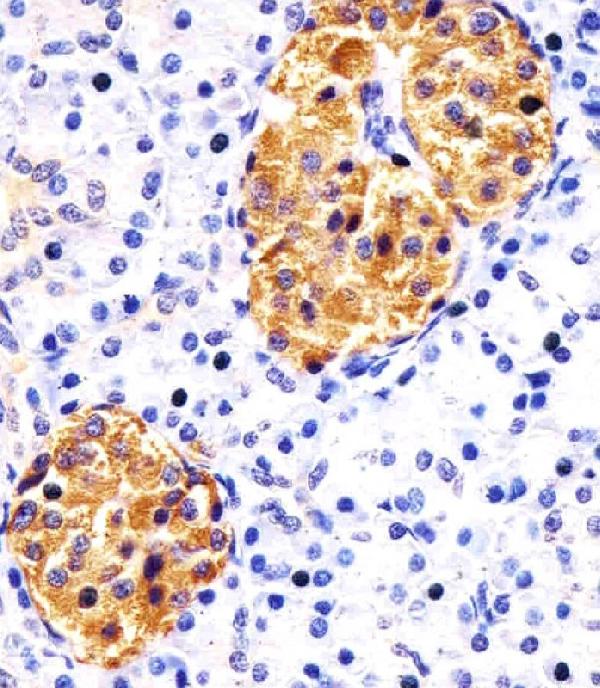
M09431 has real IHC-P data from human pancreas paraffin sections (M09431 IHC caption). Its listed reactivity covers human, mouse, and rat (catalog: reactivity); no IF data are supplied (catalog: applications, IF images).
M09431 is listed for IHC-P and shows staining in human pancreas paraffin sections (catalog: applications; M09431 IHC caption). It is listed as reactive with human, mouse, and rat, but the supplied IHC image documents human tissue only (catalog: reactivity; M09431 IHC caption).
Which to pick: For tissue IHC, choose M09431, a mouse monoclonal listed for IHC-P at 1:25 with an image from human pancreas paraffin sections (catalog: host, clone, applications, dilution; M09431 IHC caption). For IF/ICC, no listed SKU has an IF/ICC application or figure; M09431 would need independent validation (catalog: applications, IF images). For cross-species work, M09431 lists human, mouse, and rat reactivity, while its supplied IHC evidence covers only human pancreas sections; the caption reports paraformaldehyde-fixed paraffin sections and also describes formaldehyde fixation (catalog: reactivity; M09431 IHC caption).