This website uses cookies to ensure you get the best experience on our website.


- Table of Contents
Plan RAC2 staining in paraffin sections using cytoplasmic immune-cell staining as the expected pattern (HPA tissue IHC). Compare bone marrow, lymph node, spleen or tonsil positive cells with an appropriate negative control, and check for endogenous peroxidase signal in marrow (HPA tissue IHC; standard IHC practice).

Expected localisation, validated protocols, controls and antibodies — the at-a-glance facts below, then the full design guide.
| Expected localisation | Observed cytoplasm (HPA tissue IHC); membrane-associated when active (UniProt) | |
| Staining pattern | Cytoplasmic staining most abundant in immune cells (HPA tissue IHC) | |
| Antigen retrieval | Citrate pH 6.0 HIER, 95–98 °C, 20 min (rule: cytoplasmic / membrane antigen) | |
| Positive control | Bone marrow+3 more · see all | |
| Negative control | Cervix+4 more · see all |
| Fixation | Keep fixation consistent across paraffin sections (standard IHC practice; not target-specific); Matched tissue-IHC evidence does not establish the fixation claim. Validate the specimen-specific method before use. (selected-SKU IHC image A01714-1) | |
| Caveat | Marrow peroxidase can mimic chromogenic signal (HPA tissue IHC; standard IHC practice) | |
| Regulation | Hematopoietic-specific expression (UniProt) | |
| Isoform / epitope | No annotated isoforms; C-terminal processing may affect epitopes (UniProt) |
The catalog antibody’s IHC-P protocol is followed by 4 published RAC2 IHC protocols (PMC10750086; PMC7028469; PMC10198763; PMC6685597).
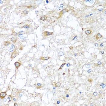
| Sample | Paraffin-embedded rat brain tissue; fixative not specified (datasheet A01714-1) |
| Fixation | Image fixative and duration unreported (datasheet A01714-1); verify before use. |
| Sectioning | 4–5 µm sections on charged slides (standard) |
| Deparaffinisation | Xylene, graded ethanol series to water (standard) |
| Antigen retrieval | Heat-induced epitope retrieval in citrate buffer, pH 6.0, 20 min at 95–98 °C (standard rule: cytoplasmic / membrane antigen) |
| Peroxidase block | 3% H2O2, 10 min, room temperature (standard) |
| Blocking | 10% normal serum of the secondary host, 30 min, room temperature (standard) |
| Primary antibody | Rabbit anti-RAC2, 1:50-1:100 (datasheet A01714-1) |
| Primary incubation | Overnight at 4 °C (standard) |
| Detection | HRP-polymer secondary, DAB chromogen 5–10 min (standard) |
| Counterstain | Hematoxylin, blue, dehydrate and mount (standard) |
| Expected result | RAC2-positive staining in hematopoietic cells of bone marrow (HPA tissue IHC: Medium). HPA tissue profile: Cytoplasmic expression in several tissues, most abundant in immune cells. No signal in the no-primary control. |
RAC2 staining should be mainly cytoplasmic in hematopoietic and other immune cells; membrane association can occur when RAC2 is activated (UniProt P15153). In tissue IHC, HPA reports cytoplasmic expression across several tissues, most abundant in immune cells, with medium staining in selected marrow and lymphoid compartments (HPA tissue IHC). HPA rates its tissue profile “Enhanced” but describes only medium consistency between staining and RNA data (HPA tissue IHC). RAC2 has no transmembrane segment (UniProt P15153 topology).
| Cytoplasmic staining in bone marrow hematopoietic cells and lymph node or tonsil non-germinal center cells. | This matches the reported cell and compartment pattern; HPA grades each listed cell population Medium, so compare signal within the identified cells rather than treating every cell in the section as positive (HPA tissue IHC). |
| A predominantly nuclear signal, or a crisp membrane-only outline without cytoplasmic staining. | Question the staining pattern and check controls: UniProt places RAC2 in the cytoplasm and notes membrane association when activated, while HPA reports cytoplasmic tissue expression (UniProt P15153; HPA tissue IHC). Compartment mismatch alone does not identify the artefact. |
| Strong staining in glandular or epithelial cells where the supplied HPA examples report no detection. | Investigate antibody cross-reactivity or endogenous chromogen-generating activity before assigning RAC2 positivity (general IHC practice). HPA reports no detection in cervix and duodenum glandular cells and esophageal squamous epithelial cells; that finding concerns those cells, not every cell in those tissues (HPA tissue IHC). |
| Diffuse color across tissue, extracellular space, or the negative control. | Interpret localization cautiously: broad background can obscure a cell-restricted cytoplasmic result (general IHC practice). HPA’s reported pattern is cytoplasmic and most abundant in immune cells, so diffuse color alone does not establish RAC2 staining (HPA tissue IHC). |
| No signal in the expected cell population of an otherwise evaluable bone marrow, lymph node, spleen, or tonsil section. | Treat the run as unresolved before calling the sample negative: HPA reports Medium staining in the specified hematopoietic, non-germinal center, or white-pulp cells (HPA tissue IHC). Check that those cells are present and that the positive and detection controls worked (general IHC practice). |
| Cell population and tissue context | HPA reports Medium staining in marrow hematopoietic cells, lymph node and tonsil non-germinal center cells, and spleen white-pulp cells; select and score the named cells rather than a whole-tissue average (HPA tissue IHC). |
| Negative comparison cells | HPA reports no detection in specific cells, including cervix glandular cells, colon endothelial cells, and liver cholangiocytes. These are cell-level observations, not proof that the entire tissue lacks RAC2-positive immune cells (HPA tissue IHC). |
| Localization and activation | RAC2 is cytoplasmic and can associate with the membrane when activated; it has no transmembrane segment. Membrane accentuation may be compatible with its biology, but this record supplies no activation-state IHC scoring rule (UniProt P15153). |
| Antibody evidence | HPA lists IHC status as Approved for HPA047820 and Enhanced for CAB022946. Those statuses support different validation strengths; neither establishes the performance of an unlisted antibody or a particular staining run (HPA antibodies). |
| Epitope, processing, and retrieval | UniProt annotates a chain spanning residues 1–189 and a 190–192 propeptide, but the supplied record gives no antibody epitope or RAC2-specific retrieval condition. Optimize retrieval against a known-positive control without claiming a target-specific fixation effect (UniProt P15153; general IHC practice). |
| IF/ICC: what pattern is supported? | A cytoplasmic signal is biologically plausible, with possible activation-associated membrane signal (UniProt P15153). HPA supplies no ICC-IF images or assigned subcellular location, and neither listed antibody has an ICC status here; this record cannot validate an IF/ICC pattern (HPA subcellular; HPA antibodies). |
| Situation | Likely cause | Next action |
|---|---|---|
| Known-positive immune cells show no chromogenic signal. | The target cells may be absent from the viewed area, or the IHC run may have failed; HPA reports Medium staining only in specified cells of its positive examples (HPA tissue IHC; general IHC practice). | Confirm cell identity on the counterstained section, then review the positive control, antibody conditions, retrieval, and detection reagents in that order (general IHC practice). Do not infer a RAC2-specific retrieval requirement from this record. |
| Only nuclei stain in a section containing identifiable immune cells. | The dominant compartment conflicts with cytoplasmic tissue expression and UniProt cytoplasmic localization (HPA tissue IHC; UniProt P15153). The pattern itself cannot distinguish non-specific binding from detection background (general IHC practice). | Compare with a matched negative control and an expected-positive cell population; review counterstain and detection background before assigning a RAC2 result (general IHC practice). |
| Glandular or squamous epithelial cells appear strongly positive. | HPA reports no detection in the specified cervix and duodenum glandular cells and esophageal squamous epithelial cells; cross-reactivity or endogenous detection activity is possible (HPA tissue IHC; general IHC practice). | Check cell identity and an appropriate negative control; assess endogenous enzyme blocking for the chromogenic detection system, then compare adjacent immune cells where present (general IHC practice). Do not label the whole tissue negative. |
| Color is widespread, including the negative control. | Diffuse background can arise from non-specific reagent binding or endogenous detection activity, preventing reliable cell-level interpretation (general IHC practice). It does not resemble the reported immune-cell-rich cytoplasmic profile (HPA tissue IHC). | Review blocking and washes, verify the negative control, and adjust detection conditions using the same positive and negative comparison sections (general IHC practice). |
| A membrane rim is seen alongside cytoplasmic staining. | Membrane association can occur when RAC2 is activated, although UniProt describes no transmembrane segment (UniProt P15153). A chromogenic rim does not by itself measure activation state. | Score the cytoplasmic signal and record the rim separately; compare its distribution with the negative control and avoid an activation claim without independent evidence (UniProt P15153; general IHC practice). |
| Two antibodies give different tissue patterns. | The supplied HPA entries have different IHC validation statuses: HPA047820 is Approved and CAB022946 is Enhanced. HPA’s tissue reliability description also notes medium staining–RNA consistency (HPA antibodies; HPA tissue IHC). | Compare the same named cell populations and controls across runs, document each antibody’s pattern, and treat discordant cells as unresolved rather than combining the stains into one positive call (HPA tissue IHC; general IHC practice). |


Comprehensive Human Protein Atlas IHC scoring per tissue (reliability: Enhanced — Medium consistency between antibody staining and RNA expression data. Antibody staining in cells/structures not annotated, view images.). Rows are taken directly from the HPA tissue chart — click any row's HPA link to view the source.
| Tissue | Cell type | Level | Evidence | Source |
|---|---|---|---|---|
| Bone marrow | Hematopoietic cells | Medium | Protein (IHC) | HPA → |
| Lymph node | Non-germinal center cells | Medium | Protein (IHC) | HPA → |
| Spleen | Cells in white pulp | Medium | Protein (IHC) | HPA → |
| Tonsil | Non-germinal center cells | Medium | Protein (IHC) | HPA → |
| Tissue | Cell type | Level | Evidence | Source |
|---|---|---|---|---|
| Cervix | Glandular cells | Not detected | Protein (IHC) | HPA → |
| Colon | Endothelial cells | Not detected | Protein (IHC) | HPA → |
| Duodenum | Glandular cells | Not detected | Protein (IHC) | HPA → |
| Endometrium | Cells in endometrial stroma | Not detected | Protein (IHC) | HPA → |
| Epididymis | Glandular cells | Not detected | Protein (IHC) | HPA → |
Troubleshoot RAC2 staining in paraffin sections by checking retrieval, cell identity and cytoplasmic localisation before interpreting chromogenic signal.
Anti-RAC2 options include a paraffin-section IHC image from rat brain and an IF image from mouse NIH/3T3 cells; catalog reactivity also includes human samples (catalog figures; catalog reactivity).
A01714-1 will render with its paraffin-section rat brain IHC figure, stained at 1:100 after microwave retrieval in 10 mM PBS, pH 7.2 (A01714-1 IHC caption). M01714 will render with its IF figure from paraformaldehyde-fixed, permeabilized NIH/3T3 cells, stained at 1:25 (M01714 IF caption).
Which to pick: Choose A01714-1 for paraffin-section tissue IHC: its own figure documents rat brain staining, while the fixative is unreported (A01714-1 IHC caption). Choose M01714 when a documented cell IF example matters; A01714-1 also lists ICC/IF, whereas M01714 lists IF but not ICC (M01714 IF caption; catalog applications). For work across human, mouse and rat, A01714-1 lists reactivity with all three, though its IHC figure documents rat only (catalog reactivity; A01714-1 IHC caption).