RAD50 / DNA repair protein RAD50 · IHC design guide

Design Immunohistochemistry for RAD50

Plan RAD50 IHC in paraffin sections using the widespread nuclear pattern as a reference (HPA tissue IHC). Start with the catalog antibody’s 2–5 μg/ml IHC range (datasheet A00347-2), and account for HPA’s caution that staining may reflect more than one gene (HPA tissue IHC).

Evidence assembled Oct 2026 · For research use; verify linked source records and product datasheet before use
Immunohistochemistry protocol sheet for RAD50 (IHC for RAD50): expected localisation Nuclear staining in tissue (HPA tissue IHC), antibody A00347-2, validated IHC image, and IHC protocol steps
Printable RAD50 IHC protocol sheet — expected localisation Nuclear staining in tissue (HPA tissue IHC), antibody A00347-2, controls and protocol steps. Open the full RAD50 IHC guide →

RAD50 Immunohistochemistry Experimental Design Guide

Expected localisation, validated protocols, controls and antibodies — the at-a-glance facts below, then the full design guide.

Must know before staining
Expected localisation Nuclear staining in tissue (HPA tissue IHC)
Staining pattern Nuclear staining across diverse cell types (HPA tissue IHC)
Antigen retrieval EDTA pH 8.0 HIER, heat-mediated (datasheet A00347-2)
Positive control ⓘ Appendix+4 more · see all
Negative control ⓘ None in HPA (detected in all 45 tissues); use no-primary + isotype controls
Important caveats
Reasons your staining may differ from the expected pattern.
Fixation Matched tissue-IHC evidence does not establish the fixation claim. Validate the specimen-specific method before use. (selected-SKU IHC image A00347-2)
Caveat Staining may reflect more than one gene (HPA tissue IHC)
Regulation Higher expression in testis (UniProt)
Isoform / epitope 3 isoforms; check epitope coverage (UniProt)
Section 1

Recommended RAD50 IHC & IF Protocols

Compare the catalog antibody’s IHC-P protocol with three published RAD50 chromogenic IHC protocols from ovarian, rectal, and renal tumor samples (PMC5223425; PMC5742811; PMC4155059).

Recommended immunohistochemistry (IHC-P) protocol parameters
SampleParaffin-embedded human testis cancer tissue; fixative not specified (datasheet A00347-2)
FixationImage fixative and duration unreported (datasheet A00347-2); verify before use.
Sectioning4–5 µm sections on charged slides (standard)
DeparaffinisationXylene, graded ethanol series to water (standard)
Antigen retrievalHeat retrieval: EDTA pH 8.0 (datasheet A00347-2); 20 min, 95–100 °C (standard)
Peroxidase block3% H2O2, 10 min, room temperature (standard)
Blocking10% goat serum (datasheet A00347-2)
Primary antibodyRabbit anti-RAD50, 2-5 μg/ml (datasheet A00347-2)
Primary incubationOvernight at 4 °C (datasheet A00347-2)
DetectionHRP-conjugated secondary, DAB chromogen (datasheet A00347-2)
CounterstainHematoxylin, blue, dehydrate and mount (standard)
Expected resultRAD50-positive staining in enterocytes of appendix (HPA tissue IHC: High). HPA tissue profile: Ubiquitous nuclear expression. No signal in the no-primary control.
💡Decision noteStart with the catalog antibody’s heat-mediated EDTA pH 8.0 retrieval (datasheet A00347-2); the published protocols used different retrieval conditions (PMC5742811; PMC4155059).
Section 2

What Is the Expected RAD50 Staining Pattern?

RAD50 should appear primarily in nuclei: UniProt places it in the nucleus and chromosomes and reports no transmembrane segment (UniProt Q92878). HPA describes ubiquitous nuclear tissue staining, with high staining in specified cell populations and low staining in others (HPA: tissue IHC). Its tissue IHC profile is Supported, with a caution that the antibodies target protein from more than one gene (HPA: tissue IHC reliability).

What am I looking at on my slide?
Nuclear chromogen in breast glandular cells or bone marrow hematopoietic cells (HPA: High).This matches the expected compartment and documented high staining in those cell populations (HPA: tissue IHC). Judge individual nuclei within the tissue, since the HPA levels describe cell populations rather than every cell in a section (HPA: tissue IHC).
Predominantly cytoplasmic or membrane staining, with little nuclear signal.This conflicts with RAD50's nuclear and chromosomal localization (UniProt Q92878) and HPA's ubiquitous nuclear IHC profile (HPA: tissue IHC). Treat it as suspect staining; check the detection controls and antibody conditions before assigning a biological meaning (general IHC practice).
Strong staining restricted to an unexpected cell population while expected nuclei are weak.Compare cells individually: HPA reports low staining in liver cholangiocytes, soft tissue fibroblasts and adipose tissue adipocytes, but does not call them negative (HPA: tissue IHC). Unexpected selective signal can reflect cross-reactivity or endogenous detection activity; controls are needed to distinguish them (general IHC practice).
Uniform chromogen across nuclei, cytoplasm and surrounding tissue.A diffuse distribution does not resolve the predominantly nuclear pattern (UniProt Q92878; HPA: tissue IHC). Evaluate reagent background, blocking and wash conditions with a no-primary control before scoring the section (general IHC practice).
No nuclear staining in appendix enterocytes or breast glandular cells (HPA: High).These are documented high-staining populations, so absent signal warrants a technical check (HPA: tissue IHC). Verify section quality, antigen retrieval, antibody dilution and detection with an appropriate positive control before interpreting the result as low RAD50 expression (general IHC practice).
💡Expected RAD50 appearanceCall a result positive when the expected cell population shows clear nuclear chromogen, especially a documented high-staining population; isolated cytoplasmic or tissue-wide diffuse color is suspect (HPA: tissue IHC; UniProt Q92878; general IHC practice).
How each factor affects the staining
Tissue and cell selectionHPA calls the tissue profile ubiquitous and nuclear, but lists High staining for specific populations, including appendix enterocytes and endometrial glandular cells, and Low staining for adipocytes (HPA: tissue IHC). Use the named cell population as the comparison unit.
Evidence and antibody specificityThe tissue pattern is Supported because antibody staining agrees with RNA expression, with an explicit warning that the antibodies target protein from more than one gene (HPA: tissue IHC reliability). A matching pattern supports interpretation but does not, by itself, establish RAD50-specific staining.
Expression evidenceUniProt reports very low expression in most tissues and higher expression in testis, whereas HPA reports low tissue specificity and several High IHC cell populations (UniProt Q92878; HPA: tissue IHC). Keep these source-specific observations distinct when choosing a positive control.
Protein forms and epitopeUniProt lists three isoforms, a single 1–1312 chain and modified residues, including phosphorylation and acetylation (UniProt Q92878). The payload gives no antibody epitope, so these features cannot predict which forms the IHC-validated antibody detects or how retrieval affects its signal.
IF/ICC Q: Where should RAD50 appear?A: Mainly in the nucleoplasm, with additional nuclear-body localization (HPA: subcellular ICC-IF). UniProt also reports discrete nuclear foci after genotoxic treatment (UniProt Q92878); that conditional finding is not a baseline requirement for untreated tissue IHC.
Why is my staining missing, weak or wrong?
SituationLikely causeNext action
Expected nuclei are unstained in a documented High population (HPA: tissue IHC).The cause is unresolved from the image alone; staining or detection steps may have failed (general IHC practice).Check a positive control and the IHC-validated antibody's stated IHC-P conditions; review retrieval, dilution and detection as general IHC steps (general IHC practice).
Color is concentrated outside nuclei.The distribution conflicts with RAD50's nuclear localization (UniProt Q92878; HPA: tissue IHC).Inspect a no-primary control for detection background, then review antibody dilution and washes before scoring nuclear signal (general IHC practice).
Broad diffuse color obscures cell boundaries.Nonspecific background or endogenous detection activity is possible; the pattern alone cannot identify which (general IHC practice).Run no-primary and detection controls, then adjust blocking and washing according to the chromogenic method (general IHC practice).
An unexpected population stains strongly while documented High cells do not (HPA: tissue IHC).Cross-reactivity is plausible because the tissue IHC record cautions that antibodies target protein from more than one gene (HPA: tissue IHC reliability).Compare with a separately validated IHC antibody and the expected cell-specific nuclear pattern, where available (HPA: antibody validation; HPA: tissue IHC).
A documented Low population shows weak nuclear color (HPA: tissue IHC).Low is not an absence call; HPA lists cholangiocytes, fibroblasts and adipocytes as Low (HPA: tissue IHC).Retain the observation as weak staining and compare its nuclear distribution with an on-run High population before drawing a specificity conclusion (HPA: tissue IHC; general IHC practice).
Punctate nuclear staining is interpreted as a required baseline pattern.UniProt describes discrete nuclear foci after genotoxic treatment, while HPA's tissue IHC summary describes ubiquitous nuclear expression (UniProt Q92878; HPA: tissue IHC).Record whether genotoxic treatment occurred; score routine sections by cell-specific nuclear staining without requiring foci (UniProt Q92878; HPA: tissue IHC).

Sample controls for RAD50 IHC & IF

🧪Run bone marrow first and expect nuclear staining in hematopoietic cells (HPA: High in bone marrow hematopoietic cells; UniProt Q92878: nuclear localization). HPA lists no negative tissue and detects RAD50 in all 45 scored tissues, so use no-primary and isotype controls; cells without detectable nuclear signal on the positive slide should show background staining, but should not be designated a known RAD50-negative cell type (HPA: no negative tissue rows).
Positive control tissue: Appendix (Enterocytes, HPA High)
Negative control tissue: None in HPA: RAD50 is detected in all 45 scored tissues. Use a no-primary (secondary-only) and an isotype control instead.
ICC-IF cell lines (HPA subcellular resource): HPA ICC-IF images show RAD50 in A-431, U-251MG, U2OS, A-549, THP-1, with annotated localisation: Nucleoplasm (supported) (HPA subcellular).
Technical controls: Include a no-primary, secondary-only control and a concentration-matched rabbit IgG isotype control, since the catalog IHC example uses a rabbit primary antibody (caption: A00347-2 rabbit anti-RAD50). For biological specificity, compare a RAD50 knockout sample or perform a validated immunizing-peptide block if the peptide is available; quench endogenous peroxidase in bone marrow before HRP/DAB detection and assess endogenous biotin if using biotin-based detection (standard IHC practice; HPA: bone marrow hematopoietic cells).
⚠️Feasibility: No target-specific fixation window or fixation effect is reported in the supplied evidence, and the selected paraffin-section caption does not state a fixative (caption: A00347-2). Heat retrieval in EDTA at pH 8.0 was used for that paraffin-section example, but its necessity across specimens is unreported; the supplied evidence does not establish whether frozen sections or IF are easier (caption: A00347-2; HPA: ICC-IF localization). Bone marrow can produce endogenous peroxidase background during HRP/DAB detection, so interpret nuclear staining against the controls (standard IHC practice; HPA: bone marrow hematopoietic cells).

HPA tissue IHC evidence for RAD50

Comprehensive Human Protein Atlas IHC scoring per tissue (reliability: Supported — High consistency between antibody staining and RNA expression data. Caution, targets protein from more than one gene.). Rows are taken directly from the HPA tissue chart — click any row's HPA link to view the source.

Positive expression · recommended positive controls

TissueCell typeLevelEvidenceSource
Appendix Enterocytes High Protein (IHC) HPA →
Bone marrow Hematopoietic cells High Protein (IHC) HPA →
Breast Glandular cells High Protein (IHC) HPA →
Bronchus Basal cells High Protein (IHC) HPA →
Cerebellum Purkinje cells High Protein (IHC) HPA →

Undetected expression · recommended negative controls

TissueCell typeLevelEvidenceSource
None in HPA: RAD50 is detected in all 45 scored tissues. Use a no-primary (secondary-only) and an isotype control instead.
Section 3

Advanced RAD50 IHC Tips

Use nuclear localisation and matched controls to troubleshoot RAD50 staining in paraffin sections; the catalog antibody has a documented chromogenic IHC workflow.

What should I change when nuclear RAD50 staining is weak after retrieval?
Start with heat-mediated antigen retrieval in EDTA at pH 8.0 for paraffin sections (datasheet A00347-2). The documented section was stained with 2 μg/ml primary antibody overnight at 4°C, so check those conditions before attributing weak signal to retrieval (datasheet A00347-2). Run a control section through the same heating and cooling cycle, then compare nuclear signal and tissue morphology (standard IHC practice). If staining remains weak, adjust retrieval duration in small steps while keeping EDTA at pH 8.0, recording both signal and structural damage (standard IHC practice). Judge improvement by nuclear staining because RAD50 is nuclear (UniProt Q92878 localisation).
Could fixation explain weak or patchy RAD50 staining in my sections?
Target-specific RAD50 sensitivity to fixation is unknown: the selected paraffin-section caption does not report a fixative (datasheet A00347-2). Record the fixative, fixation duration, tissue thickness and processing history for each section before comparing staining across cases (standard IHC practice). Uneven fixation can affect antigen accessibility and morphology in paraffin IHC, so compare well-preserved regions and repeat questionable sections with matched processing when possible (standard IHC practice). Keep retrieval at EDTA pH 8.0 and the documented 2 μg/ml primary concentration while examining that variable (datasheet A00347-2). Do not infer a RAD50-specific fixation effect from nuclear staining patterns or protein modifications (HPA tissue IHC; UniProt Q92878).
Where should RAD50 staining appear, and how should I assess cytoplasmic signal?
Assess convincing RAD50 signal in nuclei: tissue IHC describes ubiquitous nuclear expression, and cell imaging places it mainly in the nucleoplasm with additional nuclear bodies (HPA tissue IHC; HPA subcellular). RAD50 can form discrete nuclear foci after genotoxic treatment and localise to DNA double-strand breaks, but an untreated section need not show obvious foci (UniProt Q92878 localisation). Use a nuclear counterstain and compare staining within intact cells to the no-primary control before calling diffuse cytoplasmic colour positive (standard IHC practice). If cytoplasmic staining dominates, review retrieval, antibody concentration and peroxidase blocking alongside morphology (standard IHC practice). Score nuclear and cytoplasmic patterns separately so background cannot inflate nuclear positivity (standard IHC practice).
Could isoforms or modifications change what the antibody detects in tissue?
RAD50 has 3 annotated isoforms, while the supplied antibody caption does not define its epitope or isoform coverage (UniProt Q92878 isoforms; datasheet A00347-2). The protein includes a zinc-hook region at residues 635–734 and annotated modification sites at residues 635, 690 and 959 (UniProt Q92878 domains and modified residues). Those annotations alone cannot establish whether fixation, retrieval or modification changes binding by this antibody (UniProt Q92878; datasheet A00347-2). Compare sections processed together and verify an unusual pattern with an independently characterised antibody whose epitope is known (standard IHC practice). Report the antibody identity and retrieval conditions when comparing studies, without claiming isoform-specific detection (standard IHC practice).
How can I use IF to check the nuclear pattern seen by chromogenic IHC?
Treat IF as a separate assay: the selected antibody evidence describes paraffin-section chromogenic IHC, while cell imaging places RAD50 mainly in the nucleoplasm and also in nuclear bodies (datasheet A00347-2; HPA subcellular). Multiplex a nuclear counterstain with a validated marker for the cell population being assessed, then check whether RAD50 signal occupies those cells' nuclei (standard IF practice). Choose a far-red fluorophore when shorter-wavelength tissue autofluorescence interferes, and include single-colour and no-primary controls (standard IF practice). Because RAD50 is nuclear and has no transmembrane segment, assess permeabilisation sufficient for antibody access to the nuclear epitope, without assuming the epitope is known (UniProt Q92878 topology; standard IF practice).
How do I reduce diffuse brown staining without losing nuclear RAD50 signal?
First compare the stained section with a no-primary control and inspect whether brown colour follows intact nuclei or tissue edges and damaged areas (standard IHC practice). The documented workflow used 10% goat serum blocking, 2 μg/ml primary antibody overnight at 4°C, and a peroxidase-linked secondary for 30 minutes at 37°C (datasheet A00347-2). Confirm an effective endogenous peroxidase block and control DAB development time, since both steps affect chromogenic background (standard IHC practice). If diffuse staining persists, adjust antibody exposure or blocking systematically while retaining a matched positive control (standard IHC practice). Accept a change only if interpretable nuclear staining remains, consistent with RAD50 localisation (UniProt Q92878 localisation).
How should I score RAD50 when nuclear staining varies between cells? ⚠ ANSWER MARKED FOR VERIFICATION
Define the cell population and viable tissue area before scoring, and exclude folds, necrosis and poorly preserved regions using consistent criteria (standard IHC practice). Record the percentage of nuclei at each intensity level and calculate a nuclear H-score from 0–300, or report percentage of positive nuclei with a prespecified threshold (standard IHC practice). For spatial analyses, report positive-nucleus density per mm² of viable tissue and normalise to the number of evaluable nuclei or tissue area as appropriate (standard IHC practice). Keep acquisition, DAB development and scoring thresholds consistent across sections (standard IHC practice). Report nuclear measures separately from cytoplasmic colour because RAD50's supported location is nuclear (HPA tissue IHC; HPA subcellular).
When is apparent RAD50 positivity more likely to be an artefact?
Give greatest weight to staining inside intact nuclei, consistent with the nuclear tissue profile and mainly nucleoplasmic cell localisation of RAD50 (HPA tissue IHC; HPA subcellular). High staining is reported in several cell populations, including bone-marrow hematopoietic cells, whereas low staining is reported in liver cholangiocytes, soft-tissue fibroblasts and adipocytes; identify the cells before comparing cases (HPA tissue IHC). Treat colour concentrated at section edges, necrotic areas or in the no-primary control as suspect, and check endogenous peroxidase blocking (standard IHC practice). Predominantly cytoplasmic colour also warrants review because the supported RAD50 compartment is nuclear (UniProt Q92878 localisation; HPA subcellular). Interpret foci cautiously unless the experimental context includes genotoxic treatment (UniProt Q92878 localisation).
Boster reagents

Best RAD50 / DNA repair protein RAD50 IHC Antibodies

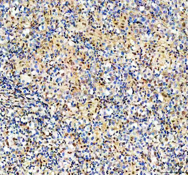
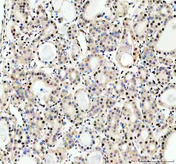
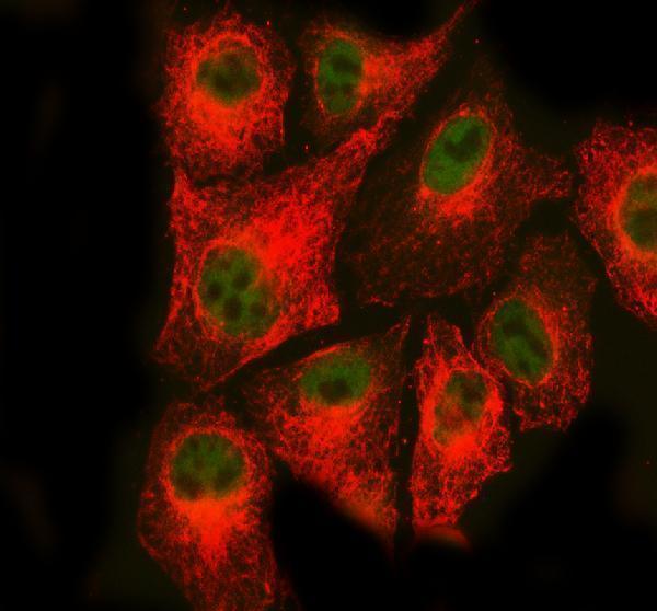
Cataloged anti-RAD50 antibodies cover human, mouse, and rat reactivity (catalog: both SKUs); IHC figures show human testis and thyroid cancer paraffin sections, while an IF figure shows A549 cells (A00347-2 captions).

Real IHC data IHC analysis of HRAD50 using anti-RAD50 antibody (A00347-2). RAD50 was detected in a paraffin-embedded section of human testis cancer tissue. Heat mediated antigen retrieval was performed in EDTA buffer (pH 8.0, epitope retrieval solution). The tissue section was blocked with 10% goat serum. The tissue section was then incubated with 2 μg/ml rabbit anti-RAD50 Antibody (A00347-2) overnight at 4°C. Peroxidase Conjugated Goat Anti-rabbit IgG was used as secondary antibody and incubated for 30 minutes at 37°C. The tissue section was developed using HRP Conjugated Rabbit IgG Super Vision Assay Kit (Catalog # SV0002) with DAB as the chromogen.
Anti-RAD50 Antibody ®
Cat # A00347-2
Real IF data Immunofluorescent analysis using the Antibody at 1:150 dilution.
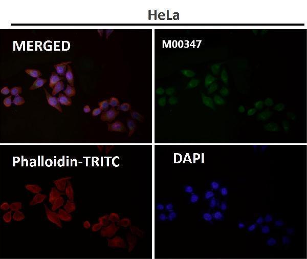
Anti-Rad50 Monoclonal Antibody
Cat # M00347

A00347-2 is listed for IHC and IF/ICC; its figures show human testis and thyroid cancer paraffin sections by IHC and A549 cells by IF (catalog: applications; A00347-2 captions). M00347 is listed for IF/ICC in human, mouse, and rat, with an IF figure whose specimen is unreported (catalog: applications/reactivity; M00347 IF caption).

Which to pick: Choose A00347-2 for tissue IHC: its own captions document staining of human paraffin sections, but do not report the fixative (A00347-2 IHC captions). For IF/ICC, M00347 offers a monoclonal option, while A00347-2 has an A549 IF example (catalog: M00347 clone/applications; A00347-2 IF caption). Both list human, mouse, and rat reactivity, but the documented IHC sections are human (catalog: reactivity; A00347-2 IHC captions).

Each figure is that product's own IHC / IF validation image from its datasheet.

References

  1. UniProt Consortium. UniProt entry Q92878 (RAD50_HUMAN, DNA repair protein RAD50).
  2. Human Protein Atlas. RAD50 tissue IHC expression (reliability: Supported).
  3. Human Protein Atlas. RAD50 subcellular location (ICC-IF): Mainly localized to the nucleoplasm. In addition localized to the nuclear bodies..
  4. Human Protein Atlas. RAD50 antibody validation summary (4 antibodies).
  5. Lack of MRE11-RAD50-NBS1 (MRN) complex detection occurs frequently in low-grade epithelial ovarian cancer. BMC cancer 2017 — PMC5223425.
  6. Early Postoperative Low Expression of RAD50 in Rectal Cancer Patients Associates with Disease-Free Survival. Cancers 2017 — PMC5742811.
  7. Synthetic lethality in ATM-deficient RAD50-mutant tumors underlies outlier response to cancer therapy. Cancer discovery 2014 — PMC4155059.
  8. RAD50 deficiency is a predictor of platinum sensitivity in sporadic epithelial ovarian cancers. Molecular biomedicine 2020 — PMC8607373.
  9. PubMed PMID:8756642 — UniProt-cited evidence.
  10. PubMed PMID:10415333 — UniProt-cited evidence.
  11. PubMed PMID:15372022 — UniProt-cited evidence.