This website uses cookies to ensure you get the best experience on our website.


- Table of Contents
Plan RAD51B staining in paraffin sections using its general nuclear tissue profile (HPA tissue IHC). This guide covers fixation, detection and interpretation, with an IHC-validated antibody diluted 1:100–1:300 (datasheet: A20019).

Expected localisation, validated protocols, controls and antibodies — the at-a-glance facts below, then the full design guide.
| Expected localisation | General nuclear staining (HPA tissue IHC) | |
| Staining pattern | Nuclei of glandular, hematopoietic and neuronal cells (HPA tissue IHC) | |
| Antigen retrieval | Tris-EDTA pH 9.0 HIER, 95–98 °C, 20 min (rule: nuclear antigen) | |
| Positive control | Adrenal gland+4 more · see all | |
| Negative control | None in HPA (detected in all 44 tissues); use no-primary + isotype controls |
| Fixation | Keep fixation consistent across paraffin sections. (standard IHC practice; not target-specific) | |
| Caveat | Antibody staining has medium consistency with RNA data (HPA tissue IHC) | |
| Regulation | DNA damage role; expression response unknown (UniProt) | |
| Isoform / epitope | 5 isoforms; check epitope coverage; no membrane topology (UniProt) |
The catalog antibody IHC-P protocol is accompanied by published RAD51B paraffin-section workflows (PMC4016303; PMC12827460).
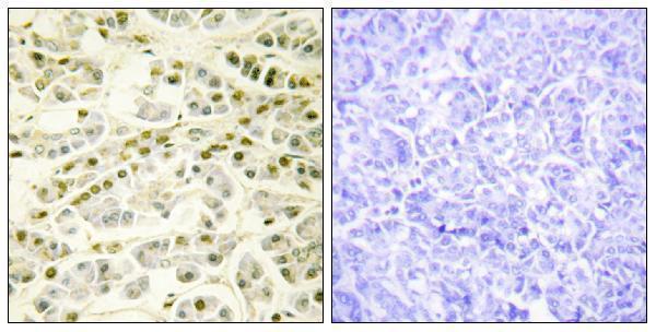
| Sample | Paraffin-embedded human pancreas tissue; fixative not specified (datasheet A20019) |
| Fixation | Image fixative and duration unreported (datasheet A20019); verify before use. |
| Sectioning | 4–5 µm sections on charged slides (standard) |
| Deparaffinisation | Xylene, graded ethanol series to water (standard) |
| Antigen retrieval | Heat-induced epitope retrieval in Tris-EDTA buffer, pH 9.0, 20 min at 95–98 °C (standard rule: nuclear antigen) |
| Peroxidase block | 3% H2O2, 10 min, room temperature (standard) |
| Blocking | 10% normal serum of the secondary host, 30 min, room temperature (standard) |
| Primary antibody | Rabbit anti-RAD51B, 1:100 - 1:300 (datasheet A20019) |
| Primary incubation | Overnight at 4 °C (standard) |
| Detection | HRP-polymer secondary, DAB chromogen 5–10 min (standard) |
| Counterstain | Hematoxylin, blue, dehydrate and mount (standard) |
| Expected result | RAD51B-positive staining in glandular cells of adrenal gland (HPA tissue IHC: High). HPA tissue profile: General nuclear expression. No signal in the no-primary control. |
RAD51B is a nuclear DNA repair protein with no annotated transmembrane segment (UniProt O15315: subcellular location and topology). In paraffin sections, expect predominantly nuclear staining across several cell types: HPA describes general nuclear expression and reports high staining in selected glandular, hematopoietic, respiratory epithelial, and neuronal cells (HPA: tissue IHC). HPA rates its tissue IHC profile Enhanced, while describing only medium consistency between antibody staining and RNA expression (HPA: tissue IHC reliability).
| Nuclear signal in expected cells, with limited staining outside nuclei. | This agrees with RAD51B nuclear localization (UniProt O15315) and HPA's general nuclear IHC profile (HPA: tissue IHC). High staining is reported in breast and appendix glandular cells, bone marrow hematopoietic cells, bronchial respiratory epithelial cells, and cerebral cortex neurons (HPA: tissue IHC). Compare cells within the same section before judging intensity (standard IHC practice). |
| Predominantly cytoplasmic, membranous, or extracellular stain. | These compartments disagree with the expected nuclear pattern (UniProt O15315: nucleus; HPA: tissue IHC). Review morphology and the negative reagent control before accepting the signal as RAD51B; nonspecific binding or detection background can produce misplaced chromogen (standard IHC practice). A small apparent rim around crowded nuclei needs careful microscopic review (standard IHC practice). |
| Strong stain in a cell population while the expected cells in that section lack nuclear signal. | Consider cross-reactivity or endogenous detection activity, especially if chromogen appears outside nuclei (standard IHC practice). RAD51B is broadly expressed, and HPA supplies no verified negative tissue in this payload, so an unlisted cell type alone cannot establish a false positive (UniProt O15315: tissue specificity; HPA: tissue IHC). |
| Diffuse chromogen over nuclei, cytoplasm, and surrounding tissue. | Widespread haze obscures the nuclear pattern and weakens interpretation (HPA: general nuclear IHC profile; standard IHC practice). Compare a no-primary control for detection background, then review blocking, washing, antibody concentration, and chromogen development as general IHC variables (standard IHC practice). |
| No nuclear signal in a reported high-staining tissue. | A blank result in bone marrow hematopoietic cells or breast glandular cells conflicts with HPA's reported high staining, but does not by itself prove target absence (HPA: tissue IHC). Check control staining, tissue preservation, retrieval, reagent performance, and detection settings using the assay's validated workflow (standard IHC practice). |
| Cell population and tissue | HPA reports High staining in adrenal, appendix, breast, and cervical glandular cells; bone marrow hematopoietic cells; bronchial respiratory epithelial cells; and caudate and cerebral cortex neurons (HPA: tissue IHC). These are reference observations, not guaranteed intensity in every specimen (standard IHC practice). |
| Antibody evidence | CAB016191 is mouse monoclonal and IHC Enhanced; HPA051869 is rabbit polyclonal with ICC Approved status and no IHC status in this payload (HPA: antibody validation). Keep assay-specific validation separate when comparing images or selecting an IHC antibody (standard IHC practice). |
| Isoforms and antigen identity | UniProt lists 5 RAD51B isoforms, but this payload gives no antibody epitope or isoform coverage (UniProt O15315; HPA: antibody validation). An unexpected pattern cannot be assigned to a particular isoform from these records alone. |
| Cell structure and processing | RAD51B is annotated as nuclear, with no transmembrane segment, signal peptide, or propeptide and one chain spanning residues 1–384 (UniProt O15315). These annotations support a nuclear readout; they provide no target-specific evidence about fixation or retrieval sensitivity. |
| IF/ICC appearance? | HPA ICC-IF places RAD51B mainly in nuclear bodies, with additional nucleoplasmic localization; images are listed for A-431, MCF-7, and U2OS (HPA: subcellular). This IF observation helps interpret localization, but does not define chromogenic IHC spot counts or intensity (standard assay interpretation). |
| Situation | Likely cause | Next action |
|---|---|---|
| Nuclei are blank in a reported high-staining reference tissue. | Weak assay signal, tissue quality, or a missed detection step is possible; the record does not identify a RAD51B-specific failure mechanism (HPA: tissue IHC; standard IHC practice). | Confirm the reference cell population, inspect section integrity, and review the validated IHC-P workflow, reagent controls, and detection steps (HPA: tissue IHC; standard IHC practice). |
| Chromogen is strongest outside nuclei. | The distribution conflicts with nuclear RAD51B and may reflect nonspecific antibody binding or detection activity (UniProt O15315; HPA: tissue IHC; standard IHC practice). | Compare the no-primary control, inspect morphology, and optimize blocking and washing within the established IHC workflow (standard IHC practice). |
| All cell types and extracellular spaces show a brown haze. | General background can obscure compartment-specific interpretation (standard IHC practice). | Review antibody concentration, wash steps, and chromogen development against the validated protocol; retain a no-primary control for comparison (standard IHC practice). |
| A bright signal appears only in an unlisted cell type. | HPA reports broad nuclear expression and provides no negative tissue here; the isolated signal remains uncertain (HPA: tissue IHC; UniProt O15315: tissue specificity). | Check whether the signal is nuclear, compare expected cells in the same section, and assess reagent controls before calling cross-reactivity (HPA: tissue IHC; standard IHC practice). |
| Two antibodies yield different distributions. | Their listed evidence covers different applications: CAB016191 has IHC Enhanced status, while HPA051869 has ICC Approved status only (HPA: antibody validation). | Interpret each in its validated application; for paraffin IHC, prioritize IHC validation and compare nuclear localization with tissue controls (HPA: antibody validation and tissue IHC; standard IHC practice). |
| IF/ICC shows punctate nuclei while IHC looks more evenly nuclear. | HPA localizes RAD51B mainly to nuclear bodies and additionally to nucleoplasm by ICC-IF; tissue IHC is summarized as general nuclear expression (HPA: subcellular; HPA: tissue IHC). | Score each application against its reported pattern; do not require discrete IF-like puncta in chromogenic paraffin sections (HPA: subcellular and tissue IHC; standard assay interpretation). |
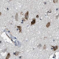


Comprehensive Human Protein Atlas IHC scoring per tissue (reliability: Enhanced — Medium consistency between antibody staining and RNA expression data.). Rows are taken directly from the HPA tissue chart — click any row's HPA link to view the source.
| Tissue | Cell type | Level | Evidence | Source |
|---|---|---|---|---|
| Adrenal gland | Glandular cells | High | Protein (IHC) | HPA → |
| Appendix | Glandular cells | High | Protein (IHC) | HPA → |
| Bone marrow | Hematopoietic cells | High | Protein (IHC) | HPA → |
| Breast | Glandular cells | High | Protein (IHC) | HPA → |
| Bronchus | Respiratory epithelial cells | High | Protein (IHC) | HPA → |
| Tissue | Cell type | Level | Evidence | Source |
|---|---|---|---|---|
| None in HPA: RAD51B is detected in all 44 scored tissues. Use a no-primary (secondary-only) and an isotype control instead. | ||||
Troubleshoot RAD51B staining by checking nuclear localisation, section processing, antibody specificity and cell-based scoring.
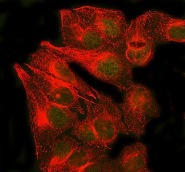
A20019 has IHC data from paraffin-embedded human pancreas (A20019 figure) and lists human, monkey and mouse reactivity (catalog); A02658-2 has IF data from human U2OS cells (A02658-2 figure).
A20019 will render with a paraffin-embedded human pancreas IHC image that includes a peptide-blocked comparison (A20019 figure). A02658-2 will render with an IF image of U2OS cells and lists human reactivity (A02658-2 figure; catalog).
Which to pick: Choose A20019 for tissue IHC: it is listed for IHC, is polyclonal, and has a paraffin-embedded human pancreas image (catalog; A20019 figure); its fixative is unreported (A20019 figure). Choose A02658-2 for human IF/ICC because both applications are listed and its U2OS IF image documents cell staining (catalog; A02658-2 figure). For cross-species work, A20019 lists human, monkey and mouse reactivity, while A02658-2 lists human only (catalog); A20019’s displayed IHC evidence is from human tissue (A20019 figure).