Real validated RAN Western blot protocols, expected-band and isoform facts, troubleshooting for weak or shifted signal, and recommended anti-RAN WB antibodies. Everything you need to plan the experiment before you commit precious samples.
Last reviewed: May 2026 · Scientific review: Boster Bio technical team
RAN Western blot protocol sheet — expected band ~24.4 kDa, antibody A00204-1, controls and PMC citations. Open the full RAN WB guide →
Expected bands, validated protocols, controls and antibodies — the at-a-glance facts below, then the full design guide.
Must know before running
Expected band
~24.4 kDa
Observed band
24 kDa
Gel
12–15%
Negative control ⓘ
siRNA / KO lysate
Important caveats
Reasons your observed band may differ from the expected size.
PTM
Phosphorylated + Acetylated
Caveat
PTM-induced mobility shift
Regulation
MYC up
Isoform
1 isoform(s)
Section 1
Real Curated RAN Western Blot Protocols
Literature-validated Western blot parameters for RAN — gel percentage, transfer, blocking, antibody incubation and detection, extracted from published methods.
Recommended Western blot protocol parameters
Sample / lysate
human PC-3, human RT4, human A549
Gel %
12–15%
Load
30 ug
Transfer
nitrocellulose membrane, 150 mA, 50–90 min
Membrane
nitrocellulose
Blocking
5% non-fat milk / TBS, 1.5 h RT
Primary antibody
0.5 µg/mL
Primary incubation
overnight at 4 °C
Secondary antibody
goat anti-rabbit IgG-HRP, 1:5000
Wash
TBS-0.1% Tween, 3 × 5 min
Detection
ECL
Exposure / imaging
Tanon 5200
Observed band
24 kDa
Section 2
What Is the Expected RAN Western Blot Band Size?
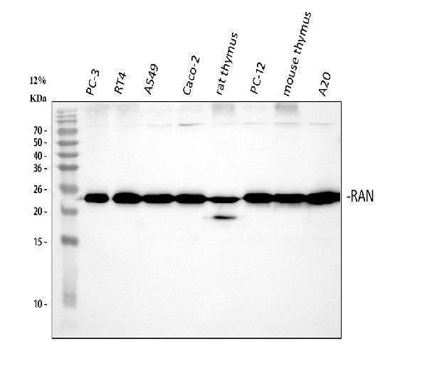
RAN's 24.4 kDa predicted monomeric mass closely matches its observed 24 kDa band, since it lacks glycosylation, disulfide dimerization, and cleavage that would otherwise shift its size.
What am I looking at on my blot?
single sharp band at ~24 kDa
matches the predicted 24.4 kDa mass of unmodified monomeric RAN
no doublet or extra bands near the main band
RAN has only one isoform, so no alternative splice product is expected
band position unchanged under reducing vs non-reducing conditions
RAN has no inter-chain disulfide bonds, so it does not dimerize on the gel
clear band present in standard whole-cell lysate
RAN is a nuclear/cytoplasmic protein, not secreted, so it is retained and detectable in lysate
no diffuse smear above the main band
RAN has no glycosylation sites, so there is no carbohydrate-driven heterogeneity to broaden the band
no higher-order band at roughly double the monomer mass
RAN functions as a monomer with no covalent dimerization to shift apparent size
💡Expected RAN appearanceRAN typically resolves as a single sharp band at 24 kDa, closely matching its predicted 24.4 kDa monomeric mass, since it has no signal peptide, glycosylation, or disulfide-linked dimerization to alter its apparent size.
How each factor affects band size
predicted mass from UniProt (24.4 kDa)
sets the baseline expected band position, consistent with the observed 24 kDa band
monomer, no inter-chain disulfide bonds
band position stays the same under reducing and non-reducing conditions with no dimer band appearing
single isoform (no additional splice variants)
only one species should be seen, not multiple isoform-driven bands
absence of glycosylation sites
no upward mass shift or smearing from carbohydrate addition
no signal peptide or propeptide
the full-length translated protein is the mature form, so no smaller cleaved fragment is generated
N-terminal and lysine acetylation, threonine phosphorylation
these small covalent modifications add negligible mass and do not measurably shift the band on SDS-PAGE
Why is my band missing or off?
Situation
Likely cause
Next action
Band higher than expected
incomplete denaturation leaves RAN in complex with binding partners like RANGAP1 or KPNB1, causing an apparent shift to higher mass
boil samples fully in fresh reducing sample buffer with adequate SDS before loading
Band lower than expected
proteolytic degradation during lysis clips the small 24 kDa protein into faster-migrating fragments
prepare lysates on ice with protease inhibitors and load samples promptly after preparation
Multiple bands
cross-reactivity with other small GTPases of similar mass, since RAN itself has only one isoform
confirm specificity with a RAN knockdown or knockout lysate control and titrate antibody concentration
Weak or no signal
standard cytoplasmic lysis buffers under-extract the nuclear pool of RAN
use a lysis buffer effective for nuclear proteins, such as RIPA with nuclease treatment, to fully release RAN
Broad smear instead of sharp band
overloading of lysate or poor transfer produces smearing even though RAN itself has no glycosylation to cause true heterogeneity
reduce total protein loaded per lane and confirm even transfer with a Ponceau stain
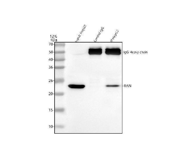
Real WB data behind the band-size expectations in this section.
Sample controls for RAN Western blot
🧪For positive controls for RAN in Western blot, you can use HeLa cell lysate, since RAN is a ubiquitously expressed nuclear protein required for nucleocytoplasmic transport in all proliferating cells.
Positive control: HeLa cells
Negative control: ubiquitously expressed; use siRNA knockdown or KO line
Loading controls: Run GAPDH and β-actin alongside a total-protein stain (e.g., stain-free gel, Ponceau S, or REVERT) to confirm equal loading.
⚠️Feasibility: As a ubiquitously expressed, essential nuclear GTPase, RAN has no tissue with a clean absence of signal, so specificity is best confirmed with siRNA knockdown or a CRISPR knockout line rather than a negative tissue.
Comprehensive Human Protein Atlas IHC scoring per tissue. Rows are taken directly from the HPA tissue chart — click any row's HPA link to view the source.
Deeper troubleshooting and optimisation questions for RAN, answered from its protein features.
Why does RAN run close to its predicted 24 kDa mass?
RAN has no signal peptide, propeptide, glycosylation, or disulfide bonds, so its unmodified sequence closely matches the observed 24 kDa band, with minimal denaturation-related shift under reducing SDS-PAGE conditions.
Should multiple RAN isoforms be expected on blots?
UniProt lists only one RAN isoform, so a single band near 24 kDa is expected; extra bands likely reflect degradation, nonspecific binding, or post-translationally modified forms rather than isoform diversity.
Do RAN modifications alter its apparent molecular weight?
RAN carries nine modified residues, including acetylation, phosphorylation, and isopeptide-linked ubiquitin-like conjugation. These modifications are typically too small to shift SDS-PAGE mobility much, but SUMOylated or ubiquitinated RAN can appear as faint higher-molecular-weight bands during mitosis.
How should blocking be optimized for RAN detection?
RAN lacks glycosylation and disulfide bonds, so standard 5% milk or BSA blocking works well; use BSA instead of milk only if probing phosphorylated RAN residues, since milk phosphoproteins can increase background on phospho-specific blots.
What transfer method to use for RAN Western blot?
RAN's small 24 kDa size and monomeric, non-glycosylated structure transfer efficiently onto PVDF or nitrocellulose using standard wet or semi-dry transfer at lower voltage and time; over-transfer risks blotting the protein through the membrane, so monitor transfer time closely.
Is RAN suitable as a loading control?
RAN is a monomeric, single-isoform, ubiquitously expressed nuclear-cytoplasmic shuttling protein with a stable predicted mass, making it a reasonably consistent reference band, though its cell-cycle-linked regulation means expression can vary in synchronized or mitotic samples.
What explains higher-molecular-weight RAN bands?
Isopeptide-linked ubiquitin-like conjugation (Ubl/SUMO) or interaction complexes with RANGAP1 or KPNB1 can produce higher-molecular-weight bands under non-reducing or incompletely denatured conditions; these are distinct from isoform variation since RAN has only one isoform.
Boster reagents
Best RAN Western Blot Antibodies
BosterBio's RAN antibodies are among the best-performing WB antibodies on the market — well cited, thoroughly validated, and orthogonally cross-validated against negative tissues and complementary methods.
Our recommended anti-RAN antibodies for Western blot are top-performing, extensively cited reagents validated across multiple systems, including orthogonal confirmation with negative-tissue controls and complementary detection methods, ensuring specificity and reproducible results for confident RAN detection.
Which to pick: Both A00204-1 and M00204-1 have genuine Western blot validation images against Ran, so either is a reliable pick; choose based on host species, clonality, or lot availability, since the catalog lists no reactivity differences between them.
Source: BosterBio RAN gene-info card — filtered to Western-blot-capable antibodies; each card shows that product's actual WB validation figure.