Click image to see more details
Product Info Summary
| SKU: | M00204-1 |
|---|---|
| Size: | 100 μg/vial |
| Reactive Species: | Human, Mouse, Rat |
| Host: | Mouse |
| Application: | Flow Cytometry, IF, ICC, WB |
Customers Who Bought This Also Bought

Product info
Product Name
Anti-Ran Antibody Picoband® (monoclonal, 5D5)
SKU/Catalog Number
M00204-1
Size
100 μg/vial
Form
Lyophilized
Description
Boster Bio Anti-Ran Antibody Picoband® (monoclonal, 5D5) catalog # M00204-1. Tested in Flow Cytometry, IF, ICC, WB applications. This antibody reacts with Human, Mouse, Rat. The brand Picoband indicates this is a premium antibody that guarantees superior quality, high affinity, and strong signals with minimal background in Western blot applications. Only our best-performing antibodies are designated as Picoband, ensuring unmatched performance.
Storage & Handling
Store at -20˚C for one year from date of receipt. After reconstitution, at 4˚C for one month. It can also be aliquotted and stored frozen at -20˚C for six months. Avoid repeated freeze-thaw cycles.
Cite This Product
Anti-Ran Antibody Picoband® (monoclonal, 5D5) (Boster Biological Technology, Pleasanton CA, USA, Catalog # M00204-1)
Host
Mouse
Contents
Each vial contains 4mg Trehalose, 0.9mg NaCl, 0.2mg Na2HPO4, 0.05mg NaN3.
Clonality
Monoclonal
Clone Number
5D5
Isotype
Mouse IgG2b
Immunogen
E. coli-derived human Ran recombinant protein (Position: A2-L216). Human Ran shares 100% amino acid (aa) sequence identity with both mouse and rat Ran.
Cross-reactivity
No cross-reactivity with other proteins.
Reactive Species
M00204-1 is reactive to RAN in Human, Mouse, Rat
Observed Molecular Weight
24 kDa
Calculated molecular weight
24.4 kDa
Background of RAN
RAN (ras-related nuclear protein) is a small GTP binding protein belonging to the RAS superfamily that is essential for the translocation of RNA and proteins through the nuclear pore complex. The RAN protein is also involved in control of DNA synthesis and cell cycle progression. Nuclear localization of RAN requires the presence of regulator of chromosome condensation 1 (RCC1). Mutations in RAN disrupt DNA synthesis. Because of its many functions, it is likely that RAN interacts with several other proteins. RAN regulates formation and organization of the microtubule network independently of its role in the nucleus-cytosol exchange of macromolecules. RAN could be a key signaling molecule regulating microtubule polymerization during mitosis. RCC1 generates a high local concentration of RAN-GTP around chromatin which, in turn, induces the local nucleation of microtubules. RAN is an androgen receptor (AR) coactivator that binds differentially with different lengths of polyglutamine within the androgen receptor. Polyglutamine repeat expansion in the AR is linked to Kennedy's disease (X-linked spinal and bulbar muscular atrophy). RAN coactivation of the AR diminishes with polyglutamine expansion within the AR, and this weak coactivation may lead to partial androgen insensitivity during the development of Kennedy's disease.
Antibody Validation
Boster validates all antibodies on WB, IHC, ICC, Immunofluorescence, and ELISA with known positive control and negative samples to ensure specificity and high affinity, including thorough antibody incubations.
Application & Images
Applications
M00204-1 is guaranteed for Flow Cytometry, IF, ICC, WB Boster Guarantee
Assay Dilutions Recommendation
The recommendations below provide a starting point for assay optimization. The actual working concentration varies and should be decided by the user.
Western blot, 0.1-0.5μg/ml
Immunocytochemistry/Immunofluorescence, 2μg/ml
Flow Cytometry (Fixed), 1-3μg/1x106 cells
Positive Control
WB: human 293T whole cell, human A375 whole cell, human Hela whole cel, human Jurkat whole cel, rat testis tissue, rat PC-12 whole cell, mouse testis tissue
ICC/IF: U20S cell
FCM: PC-3 cell
Validation Images & Assay Conditions
Click image to see more details
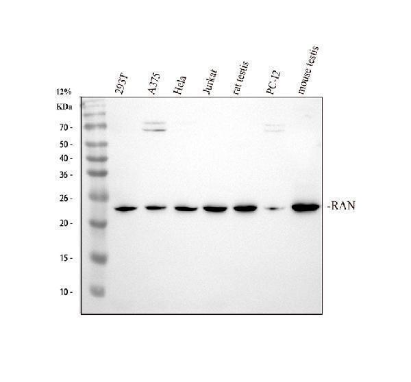
Western blot analysis of Ran using anti-Ran antibody (M00204-1).
Electrophoresis was performed on a 12% SDS-PAGE gel at 80V (Stacking gel) / 120V (Resolving gel) for 2 hours. The sample well of each lane was loaded with 30 ug of sample under reducing conditions.
Lane 1: human 293T whole cell lysates,
Lane 2: human A375 whole cell lysates,
Lane 3: human Hela whole cell lysates.
Lane 4: human Jurkat whole cell lysates,
Lane 5: rat testis tissue lysates,
Lane 6: rat PC-12 whole cell lysates,
Lane 7: mouse testis tissue lysates.
After Electrophoresis, proteins were transferred to a Nitrocellulose membrane at 150mA for 50-90 minutes. Blocked the membrane with 5% Non-fat Milk/ TBS for 1.5 hour at RT. The membrane was incubated with mouse anti-Ran antigen affinity purified monoclonal antibody (Catalog # M00204-1) at 0.5 μg/mL overnight at 4°C, then washed with TBS-0.1%Tween 3 times with 5 minutes each and probed with a goat anti-mouse IgG-HRP secondary antibody at a dilution of 1:10000 for 1.5 hour at RT. The signal is developed using an ECL Plus Western Blotting Substrate (Catalog # AR1196-200) with Tanon 5200 system. A specific band was detected for Ran at approximately 24 kDa. The expected band size for Ran is at 24 kDa.

Click image to see more details
Flow Cytometry analysis of PC-3 cells using anti-Ran antibody (M00204-1).
Overlay histogram showing PC-3 cells stained with M00204-1 (Blue line). To facilitate intracellular staining, cells were fixed with 4% paraformaldehyde and permeabilized with permeabilization buffer. The cells were blocked with 10% normal goat serum. And then incubated with mouse anti-Ran Antibody (M00204-1,1μg/1x106 cells) for 30 min at 20°C. DyLight®488 conjugated goat anti-mouse IgG (BA1126, 5-10μg/1x106 cells) was used as secondary antibody for 30 minutes at 20°C. Isotype control antibody (Green line) was mouse IgG (1μg/1x106) used under the same conditions. Unlabelled sample without incubation with primary antibody and secondary antibody (Red line) was used as a blank control.

Click image to see more details
IF analysis of Ran using anti-Ran antibody (M00204-1).
Ran was detected in immunocytochemical section of U20S cell. Enzyme antigen retrieval was performed using IHC enzyme antigen retrieval reagent (AR0022) for 15 mins. The cells were blocked with 10% goat serum. And then incubated with 2μg/mL mouse anti-Ran Antibody (M00204-1) overnight at 4°C. DyLight®488 Conjugated Goat Anti-Mouse IgG (BA1126) was used as secondary antibody at 1:100 dilution and incubated for 30 minutes at 37°C. Visualize using a fluorescence microscope and filter sets appropriate for the label used.
Specific Publications For Anti-Ran Antibody Picoband® (monoclonal, 5D5) (M00204-1)
Loading publications
Recommended Resources
Here are featured tools and databases that you might find useful.
- Boster's Pathways Library
- Protein Databases
- Bioscience Research Protocol Resources
- Data Processing & Analysis Software
- Photo Editing Software
- Scientific Literature Resources
- Research Paper Management Tools
- Molecular Biology Software
- Primer Design Tools
- Bioinformatics Tools
- Phylogenetic Tree Analysis
Customer Reviews
Have you used Anti-Ran Antibody Picoband® (monoclonal, 5D5)?
Share your experimental results or join a short interview to earn up to $1,000 in product credits or other rewards.
0 Reviews For Anti-Ran Antibody Picoband® (monoclonal, 5D5)
Customer Q&As
Have a question?
Find answers in Q&As, reviews.
Can't find your answer?
Submit your question
6 Customer Q&As for Anti-Ran Antibody Picoband® (monoclonal, 5D5)
Question
you antibody using your anti-Ran antibody (monoclonal, 5D5) for pre-mirna export from nucleus studies. Has this antibody been tested with western blotting on mouse lung tissue? We would like to see some validation images before ordering.
Verified Customer
Verified customer
Asked: 2019-08-07
Answer
Thank you for your inquiry. This M00204-1 anti-Ran antibody (monoclonal, 5D5) is tested on human a549, a549 whole cell lysates, u2os whole cell lysates, hepg2 whole cell lysates, sw620 whole cell lysates, mouse lung tissue, rat testis tissue, kidney tissue. It is guaranteed to work for Flow Cytometry, IF, ICC, WB in human, mouse, rat. Our Boster guarantee will cover your intended experiment even if the sample type has not been be directly tested.
Boster Scientific Support
Answered: 2019-08-07
Question
Do you have related data for WB, IF and IHC used by M00204-1?
Verified customer
Asked: 2019-04-23
Answer
For the Anti-Ran Antibody Picoband™ (Monoclonal, 5D5) (M00204-1), Please see the images on the product page. https://www.bosterbio.com/anti-ran-picoband-trade-antibody-monoclonal-m00204-1-boster.html
Boster Scientific Support
Answered: 2019-04-23
Question
We purchased anti-Ran antibody (monoclonal, 5D5) for Flow Cytometry on lymphoblast a few months ago. I am using rat, and We are going to use the antibody for ICC next. We are interested in examining lymphoblast as well as umbilical cord blood in our next experiment. Do you have any suggestion on which antibody would work the best for ICC?
Verified Customer
Verified customer
Asked: 2018-10-03
Answer
I took a look at the website and datasheets of our anti-Ran antibody (monoclonal, 5D5) and I see that M00204-1 has been validated on rat in both Flow Cytometry and ICC. Thus M00204-1 should work for your application. Our Boster satisfaction guarantee will cover this product for ICC in rat even if the specific tissue type has not been validated. We do have a comprehensive range of products for ICC detection and you can check out our website bosterbio.com to find out more information about them.
Boster Scientific Support
Answered: 2018-10-03
Question
We are currently using anti-Ran antibody (monoclonal, 5D5) M00204-1 for mouse tissue, and we are well pleased with the WB results. The species of reactivity given in the datasheet says human, mouse, rat. Is it true that the antibody can work on primate tissues as well?
G. Huang
Verified customer
Asked: 2014-08-29
Answer
The anti-Ran antibody (monoclonal, 5D5) (M00204-1) has not been validated for cross reactivity specifically with primate tissues, but there is a good chance of cross reactivity. We have an innovator award program that if you test this antibody and show it works in primate you can get your next antibody for free. Please contact me if I can help you with anything.
Boster Scientific Support
Answered: 2014-08-29
Question
We have been able to see staining in human umbilical cord blood. Do you have any suggestions? Is anti-Ran antibody (monoclonal, 5D5) supposed to stain umbilical cord blood positively?
D. Roberts
Verified customer
Asked: 2014-02-24
Answer
From what I have seen in literature umbilical cord blood does express RAN. From what I have seen in Uniprot.org, RAN is expressed in testis, teratocarcinoma, brain, umbilical cord blood, hippocampus, lymph, ovary, skin uterus, platelet, brain cajal-retzius cell, colon adenocarcinoma, cervix carcinoma, lymphoblast, melanoma, erythroleukemia, liver, among other tissues. Regarding which tissues have RAN expression, here are a few articles citing expression in various tissues:
Brain, Pubmed ID: 8421051, 10400640
Cervix carcinoma, Pubmed ID: 1961752
Erythroleukemia, Pubmed ID: 23186163
Hippocampus, Pubmed ID: 14702039
Liver, Pubmed ID: 24275569
Lymph, Ovary, Skin, and Uterus, Pubmed ID: 15489334
Lymphoblast, Pubmed ID: 14654843
Melanoma, Pubmed ID: 17081065
Teratocarcinoma, Pubmed ID: 2108320
Umbilical cord blood, Pubmed ID: 11042152
Boster Scientific Support
Answered: 2014-02-24
Question
Our lab were satisfied with the WB result of your anti-Ran antibody (monoclonal, 5D5). However we have observed positive staining in platelet nucleus using this antibody. Is that expected? Could you tell me where is RAN supposed to be expressed?
C. Lewis
Verified customer
Asked: 2014-02-20
Answer
From literature, platelet does express RAN. Generally RAN expresses in nucleus. Regarding which tissues have RAN expression, here are a few articles citing expression in various tissues:
Brain, Pubmed ID: 8421051, 10400640
Cervix carcinoma, Pubmed ID: 1961752
Erythroleukemia, Pubmed ID: 23186163
Hippocampus, Pubmed ID: 14702039
Liver, Pubmed ID: 24275569
Lymph, Ovary, Skin, and Uterus, Pubmed ID: 15489334
Lymphoblast, Pubmed ID: 14654843
Melanoma, Pubmed ID: 17081065
Teratocarcinoma, Pubmed ID: 2108320
Umbilical cord blood, Pubmed ID: 11042152
Boster Scientific Support
Answered: 2014-02-20
