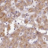
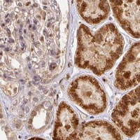
RANBP9 / Ran-binding protein 9 · IHC design guide

Design Immunohistochemistry for RANBP9

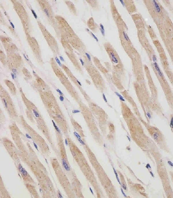
Plan RANBP9 staining in paraffin sections with the IHC-validated antibody at 1:25 (datasheet M03448-1). Compare nuclear and cytoplasmic staining in heart cardiomyocytes or cerebellar Purkinje cells, both reported as high (HPA tissue IHC).

Evidence assembled Oct 2026 · For research use; verify linked source records and product datasheet before use
Immunohistochemistry protocol sheet for RANBP9 (IHC for RANBP9): expected localisation General nuclear and cytoplasmic tissue staining (HPA tissue IHC), antibody M03448-1, validated IHC image, and IHC protocol steps
Printable RANBP9 IHC protocol sheet — expected localisation General nuclear and cytoplasmic tissue staining (HPA tissue IHC), antibody M03448-1, controls and protocol steps. Open the full RANBP9 IHC guide →

RANBP9 Immunohistochemistry Experimental Design Guide

Expected localisation, validated protocols, controls and antibodies — the at-a-glance facts below, then the full design guide.

Must know before staining
Expected localisation General nuclear and cytoplasmic tissue staining (HPA tissue IHC)
Staining pattern Nuclear and cytoplasmic staining in diverse cell types (HPA tissue IHC)
Antigen retrieval Citrate pH 6 HIER, heat-mediated (datasheet M03448-1)
Positive control ⓘ Adrenal gland+4 more · see all
Negative control ⓘ Adipose tissue+1 more · see all
Important caveats
Reasons your staining may differ from the expected pattern.
Fixation Keep fixation consistent (standard IHC practice; not target-specific); formaldehyde-fixed sections were used (selected-SKU IHC image M03448-1)
Caveat Antibody staining shows low consistency with RNA levels (HPA tissue IHC)
Regulation Broad expression; lowest in lung (UniProt)
Isoform / epitope 3 isoforms; check epitope coverage (UniProt)
Section 1

Recommended RANBP9 IHC & IF Protocols

The catalog antibody protocol uses citrate pH 6 retrieval (datasheet: M03448-1). The published IHC protocols below cover human colorectal cancer tissue (PMC8797687) and mouse tissues (PMC7188826).

Recommended immunohistochemistry (IHC-P) protocol parameters
SampleFormaldehyde-fixed, paraffin-embedded human heart tissue (datasheet M03448-1)
FixationImage formalin-fixed; duration unreported (datasheet M03448-1); verify before use.
Sectioning4–5 µm sections on charged slides (standard)
DeparaffinisationXylene, graded ethanol series to water (standard)
Antigen retrievalHeat retrieval: Citrate pH 6 (datasheet M03448-1); 20 min, 95–100 °C (standard)
Peroxidase block3% H2O2, 10 min, room temperature (standard)
Blocking10% normal serum of the secondary host, 30 min, room temperature (standard)
Primary antibodyMouse monoclonal (clone 1815CT202.22.8.37) anti-RANBP9, 1:25 (datasheet M03448-1)
Primary incubation1 hours at 37°C (datasheet M03448-1)
DetectionHRP-polymer secondary, DAB chromogen 5–10 min (standard)
CounterstainHematoxylin, blue, dehydrate and mount (standard)
Expected resultRANBP9-positive staining in glandular cells of adrenal gland (HPA tissue IHC: High). HPA tissue profile: General nuclear and cytoplasmic expression. No signal in the no-primary control.
💡Decision noteStart with heat-mediated citrate pH 6 retrieval (datasheet: M03448-1); the published mouse tissue protocol also specifies this buffer (PMC7188826).
Section 2

What Is the Expected RANBP9 Staining Pattern?

RANBP9 staining should appear mainly in nuclei and cytoplasm, including strongly stained Purkinje cells, cortical neurons, cardiomyocytes, and kidney tubule cells (HPA tissue IHC). A membrane-associated pool is possible for phosphorylated RANBP9, although the protein has no transmembrane segment (UniProt Q96S59). Interpret tissue staining cautiously: HPA rates its IHC profile Approved but reports low consistency with RNA expression data (HPA tissue IHC).

What am I looking at on my slide?
Nuclear and cytoplasmic staining in Purkinje cells, cortical neurons, cardiomyocytes, or kidney tubules.This matches the reported compartments and High staining in these cell types (HPA tissue IHC). Compare cells within the section; HPA intensity describes observed staining, not a required score for every specimen (HPA tissue IHC).
Predominantly extracellular or luminal staining, without convincing nuclear or cytoplasmic signal.Treat this as a possible artefact: RANBP9 has no signal peptide or transmembrane segment (UniProt Q96S59 topology). Do not automatically reject limited cell membrane staining, which can reflect the phosphorylated form (UniProt Q96S59 subcellular location).
Strong signal restricted to adipocytes or ovarian stromal cells while expected positive cells are unstained.HPA reports RANBP9 as Not detected in those cell populations (HPA tissue IHC). This discordance raises cross-reactivity or endogenous detection activity as possibilities (general IHC practice); check controls before assigning a target-specific signal.
Uniform color across cells, stroma, and blank areas, with little compartment definition.Diffuse staining does not reproduce the reported nuclear and cytoplasmic pattern (HPA tissue IHC). Nonspecific binding, residual detection reagent, or endogenous detection activity can produce background (general IHC practice).
No interpretable signal in a section containing Purkinje cells or kidney tubules.These are reported High cell populations (HPA tissue IHC). A blank result may reflect an assay failure or specimen variation (general IHC practice); it cannot, by itself, establish absence of RANBP9, particularly given HPA's reported staining–RNA inconsistency (HPA tissue IHC).
💡Expected RANBP9 appearanceCall the result positive when discrete nuclear and cytoplasmic staining identifies expected cells, such as High-staining Purkinje cells or kidney tubules (HPA tissue IHC); broad extracellular color without cell-associated signal is suspect (UniProt Q96S59 topology; general IHC practice).
How each factor affects the staining
Cell population and tissueHPA reports High staining in selected neuronal, epithelial, glandular, and cardiac cells, but Low staining in lung macrophages and bone marrow hematopoietic cells (HPA tissue IHC). Choose a reported High population for a positive control; weak staining in a Low population is less decisive.
Compartment and phosphorylationHPA tissue IHC describes general nuclear and cytoplasmic expression (HPA tissue IHC). UniProt places unphosphorylated RANBP9 predominantly in cytoplasm and associates a phosphorylated form with the plasma membrane (UniProt Q96S59); membrane staining needs cell-associated context.
Topology and processingRANBP9 has no transmembrane segment, signal peptide, or propeptide, and its annotated chain spans residues 1–729 (UniProt Q96S59). The record therefore gives no basis for expecting a routinely secreted or shed staining pattern; epitope position is not supplied.
Isoforms and antibody interpretationUniProt lists three isoforms (UniProt Q96S59). The payload does not identify which isoforms the IHC antibodies recognize, so differences between specimens cannot be assigned to a particular isoform without antibody-specific evidence.
IHC evidence strengthHPA lists HPA050007, HPA076784, and CAB033767 as IHC Approved (HPA antibody validation). Its tissue profile also reports low consistency between staining and RNA expression (HPA tissue IHC); interpret an unexpected distribution alongside controls.
IF/ICC Q: Where should fluorescence appear?A: HPA reports mainly nucleoplasmic signal, with supported nuclear body and cytosolic localization in ICC-IF (HPA subcellular). This is context for the separate IF/ICC guide, not an IHC protocol or a requirement that chromogenic sections resolve nuclear bodies.
Why is my staining missing, weak or wrong?
SituationLikely causeNext action
Reported High control tissue shows no staining.The section may have a failed staining or detection step (general IHC practice); High denotes an HPA observation, not a guarantee for every section (HPA tissue IHC).Check the run's positive and negative controls, reagent sequence, and chromogen development (general IHC practice). Reassess whether the relevant Purkinje cells, neurons, cardiomyocytes, or tubule cells are present (HPA tissue IHC).
Signal appears only outside cells or along empty spaces.That distribution conflicts with the reported nuclear and cytoplasmic pattern (HPA tissue IHC); RANBP9 lacks a signal peptide and transmembrane segment (UniProt Q96S59).Inspect a no-primary control and cell boundaries, then review washing and detection steps for background (general IHC practice). Interpret genuine cell-associated membrane signal separately because UniProt associates it with phosphorylated RANBP9 (UniProt Q96S59).
Adipocytes or ovarian stromal cells stain more strongly than the chosen positive cells.HPA reports RANBP9 as Not detected in those populations (HPA tissue IHC). Cross-reactivity or endogenous detection activity is possible (general IHC practice).Compare a no-primary control and an independently validated IHC antibody where available (general IHC practice; HPA antibody validation). Avoid assigning the unexpected signal to RANBP9 solely from its color or intensity.
Diffuse brown background obscures nuclei and cytoplasm.Nonspecific reagent binding or endogenous detection activity can reduce contrast (general IHC practice). HPA's expected tissue pattern requires interpretable cellular localization (HPA tissue IHC).Review blocking, washes, antibody concentration, and detection controls (general IHC practice). Adjust the workflow against the positive control until cell boundaries and compartments can be assessed.
Only weak signal appears in lung macrophages or bone marrow hematopoietic cells.HPA records Low staining in those cell populations (HPA tissue IHC). Weak signal there is less informative than staining in a reported High population.Assess a reported High control on the same run, such as kidney tubule cells or Purkinje cells (HPA tissue IHC). Score the weak population only after background and cellular localization are clear (general IHC practice).
Membrane-edge color appears alongside nuclear or cytoplasmic staining.A phosphorylated RANBP9 form can associate with the plasma membrane (UniProt Q96S59), while HPA describes general nuclear and cytoplasmic tissue expression (HPA tissue IHC).Check that the edge signal follows intact cells and exceeds control background (general IHC practice). Report membrane staining as a qualified observation; its appearance alone does not establish phosphorylation state (UniProt Q96S59).

Sample controls for RANBP9 IHC & IF

🧪Run cerebellum first and expect Purkinje cells to stain (HPA: High in Purkinje cells). Use adipose tissue as the negative tissue, where adipocytes should lack specific staining (HPA: Not detected in adipocytes); on the cerebellum slide, cells without specific signal should show counterstain only, but their target-negative status is unconfirmed.
Positive control tissue: Adrenal gland (Glandular cells, HPA High)
Negative control tissue: Adipose tissue (HPA Not detected)
ICC-IF cell lines (HPA subcellular resource): HPA ICC-IF images show RANBP9 in EFO-21, SK-MEL-30, U-251MG, A-431, U2OS, with annotated localisation: Nucleoplasm (supported) (HPA subcellular).
Technical controls: Include a no-primary, secondary-only control; an isotype control matched to the primary antibody’s host species, immunoglobulin subclass and concentration; and RANBP9-knockout material as a biological specificity control (standard IHC practice). For chromogenic cerebellum IHC, quench endogenous peroxidase and check for endogenous biotin if using the biotinylated secondary described in the selected-SKU caption; for IF, assess tissue autofluorescence (M03448-1 caption; standard IHC/IF practice).
⚠️Feasibility: A target-specific fixation window or fixation effect is unreported, and the selected-SKU fixative is unreported (M03448-1 caption). The selected-SKU paraffin-section caption uses heat-mediated citrate retrieval at pH 6, but retrieval dependence is unreported (M03448-1 caption). The supplied evidence does not establish that frozen sections or tissue IF are easier; cerebellar autofluorescence should be checked if IF is attempted (HPA: ICC-IF images; standard IF practice).

HPA tissue IHC evidence for RANBP9

Comprehensive Human Protein Atlas IHC scoring per tissue (reliability: Approved — Low consistency between antibody staining and RNA expression data.). Rows are taken directly from the HPA tissue chart — click any row's HPA link to view the source.

Positive expression · recommended positive controls

TissueCell typeLevelEvidenceSource
Adrenal gland Glandular cells High Protein (IHC) HPA →
Cerebellum Purkinje cells High Protein (IHC) HPA →
Cerebral cortex Neuronal cells High Protein (IHC) HPA →
Esophagus Squamous epithelial cells High Protein (IHC) HPA →
Gallbladder Glandular cells High Protein (IHC) HPA →

Undetected expression · recommended negative controls

TissueCell typeLevelEvidenceSource
Adipose tissue Adipocytes Not detected Protein (IHC) HPA →
Ovary Ovarian stroma cells Not detected Protein (IHC) HPA →
Section 3

Advanced RANBP9 IHC Tips

Troubleshoot RANBP9 staining in paraffin sections using the catalog antibody’s IHC evidence, protein localisation, and appropriate controls (datasheet M03448-1; UniProt Q96S59).

What retrieval should I try when RANBP9 staining is weak in paraffin sections?
Begin with heat-mediated citrate retrieval at pH 6 for RANBP9 IHC-P (datasheet M03448-1). Keep section thickness, heating duration, and cooling conditions consistent across cases, then compare signal in heart cardiomyocytes with a no-primary control (standard IHC practice; HPA: High in heart muscle cardiomyocytes). The selected antibody’s tissue image used 1:25 primary antibody for 1 hour at 37°C, providing a documented starting condition for this reagent (caption M03448-1). If staining remains weak, adjust retrieval time in a controlled series before changing buffer chemistry, and reject conditions that increase diffuse background or damage morphology (standard IHC practice).
Could fixation explain inconsistent RANBP9 staining between sections?
RANBP9-specific fixation sensitivity is unknown because no target-specific fixation comparison is supplied (evidence payload: no fixation comparison). Record fixative, fixation duration, tissue thickness, and processing history for each paraffin block before comparing staining intensity across specimens (standard IHC practice). When troubleshooting, stain matched sections from differently processed blocks in one run and hold citrate retrieval at pH 6 constant (standard IHC practice; datasheet M03448-1). Compare preserved cellular morphology alongside nuclear and cytoplasmic signal, since damaged or poorly processed regions can distort compartment scoring (standard IHC practice; HPA tissue IHC: general nuclear and cytoplasmic expression). Treat any apparent fixation effect as a result requiring confirmation in matched material (standard IHC practice).
Should RANBP9 appear in nuclei, cytoplasm, or at cell borders?
Score nuclear and cytoplasmic staining separately because tissue IHC reports both compartments for RANBP9 (HPA tissue IHC: general nuclear and cytoplasmic expression). Nucleoplasmic localisation is supported by subcellular imaging, with nuclear bodies and cytosol also supported (HPA subcellular: supported locations). UniProt additionally lists cell membrane localisation and associates a phosphorylated form with the plasma membrane, while describing the unphosphorylated form as predominantly cytoplasmic (UniProt Q96S59 localisation). For chromogenic sections, assess whether any border accentuation is reproducible within intact cells rather than assuming it identifies a phosphorylation state (standard IHC practice; UniProt Q96S59 localisation). Use a nuclear counterstain to distinguish nuclear signal from adjacent cytoplasm (standard IHC practice).
How can epitope choice affect interpretation of RANBP9 IHC staining?
RANBP9 has 3 listed isoforms, so determine which sequences the antibody immunogen and epitope cover before calling staining pan-isoform (UniProt Q96S59 isoforms; standard antibody validation practice). The protein has B30.2/SPRY, LisH, and CTLH regions, making epitope position relevant when comparing reagents (UniProt Q96S59 domains). Its listed modified residues include acetyllysine 405 and phosphoserines 477 and 487; proximity to an epitope would require experimental assessment, not an assumed staining effect (UniProt Q96S59 modified residues; standard antibody validation practice). Document the mapped epitope where available, then compare matched positive and negative controls under identical citrate pH 6 retrieval (standard IHC practice; datasheet M03448-1).
How should I investigate RANBP9 localisation by IF alongside a cell-type marker?
For the separate IF/ICC workflow, multiplex RANBP9 with a validated marker for the cell population being examined, such as cardiomyocytes in heart sections (HPA tissue IHC: High in heart muscle cardiomyocytes; standard IF practice). Choose spectrally separated fluorophores and inspect an unstained section in every channel to identify tissue autofluorescence before interpreting colocalisation (standard IF practice). RANBP9 lacks a transmembrane segment and has nuclear and cytosolic locations, so assess permeabilisation for access to intracellular epitopes after fixation (UniProt Q96S59 topology; HPA subcellular: supported nucleoplasm and cytosol; standard IF practice). Include single-label controls and evaluate each channel separately before overlaying images (standard IF practice).
What should I check when RANBP9 IHC shows widespread brown background?
First compare the stained section with a no-primary control to separate detection-system background from primary-antibody-dependent staining (standard IHC practice). The selected heart-tissue image used 3% BSA blocking for 0.5 hour and a biotinylated secondary, so review blocking and biotin-related background when reproducing that workflow (caption M03448-1; standard IHC practice). For a peroxidase and DAB workflow, check endogenous peroxidase quenching, reagent exposure, and wash quality as general IHC controls (standard IHC practice). Reassess primary concentration around the documented 1:25 condition using matched sections, while preserving morphology and the expected nuclear or cytoplasmic pattern (caption M03448-1; HPA tissue IHC: general nuclear and cytoplasmic expression; standard IHC practice).
How should I quantify RANBP9 staining across tissue samples? ⚠ ANSWER MARKED FOR VERIFICATION
Define the cell population and compartment before scoring, because RANBP9 tissue staining can be nuclear and cytoplasmic (HPA tissue IHC: general nuclear and cytoplasmic expression). For a chromogenic assay, report the percentage of positive cells and an H-score calculated from intensity categories, or count positive cells per mm² within a predefined region (standard IHC quantification practice). Normalise positive-cell counts to the number of eligible cells or analysed tissue area, and exclude folds, necrosis, and section edges using the same rules for every case (standard IHC quantification practice). Keep retrieval, detection, counterstain, imaging, and thresholds consistent, then report nuclear and cytoplasmic scores separately (standard IHC quantification practice; datasheet M03448-1: citrate pH 6 retrieval).
How can I distinguish credible RANBP9 staining from artefact?
Seek reproducible staining in intact cells with nuclear or cytoplasmic distribution, consistent with the reported tissue pattern (HPA tissue IHC: general nuclear and cytoplasmic expression; standard IHC practice). Heart cardiomyocytes and cerebellar Purkinje cells provide reported high-staining cell populations, whereas adipocytes are reported as not detected (HPA tissue IHC: cell-level observations). Treat signal concentrated at section edges, necrotic areas, or endogenous enzyme-rich regions as suspect until controls resolve it (standard IHC practice). A no-primary control tests detection background, while matched sections and independent antibody validation help assess unexpected compartments or cell populations (standard IHC practice). Interpret discordant staining cautiously because the HPA tissue profile is Approved but has low consistency with RNA expression (HPA tissue IHC: reliability description).
Boster reagents

Best RANBP9 / Ran-binding protein 9 IHC Antibodies

The catalog includes RANBP9 IHC data from human paraffin-embedded heart sections and IF data from human A549 cells; another ICC/IF listing covers human, mouse, and rat (catalog: image captions, applications, reactivity).

Real IHC data M03448-1 staining RANBP9 in human heart tissue sections by Immunohistochemistry (IHC-P -paraformaldehyde-fixed, paraffin-embedded sections). Tissue was fixed with formaldehyde and blocked with 3% BSA for 0. 5 hour at room temperature; antigen retrieval was by heat mediation with a citrate buffer (pH6). Samples were incubated with primary antibody (1/25) for 1 hours at 37°C. A undiluted biotinylated goat polyvalent antibody was used as the secondary antibody.
Anti-RANBP9 Antibody
Cat # M03448-1
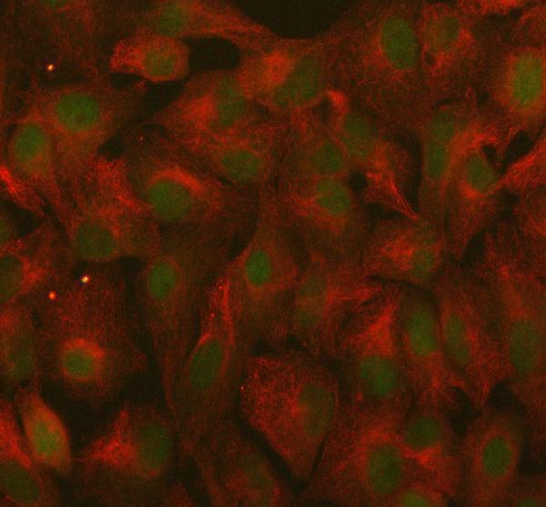
Real IF data IF analysis of RANBP9 using anti-RANBP9 antibody (A03448-1) and anti-Beta Tubulin antibody (M01857-3). RANBP9 was detected in immunocytochemical section of A549 cell. Enzyme antigen retrieval was performed using IHC enzyme antigen retrieval reagent (AR0022) for 15 mins. The cells were blocked with 10% goat serum. And then incubated with 5 μg/mL rabbit anti-RANBP9 Antibody (A03448-1) and mouse anti-Beta Tubulin antibody (M01857-3) overnight at 4°C. DyLight®488 Conjugated Goat Anti-Rabbit IgG (BA1127) and Cy3 Conjugated Goat Anti-Mouse IgG (BA1031) were used as secondary antibody at 1:500 dilution and incubated for 30 minutes at 37°C. The section was counterstained with DAPI. Visualize using a fluorescence microscope and filter sets appropriate for the label used.
Anti-RANBP9 Antibody ®
Cat # A03448-1

M03448-1 has human IHC-P data from paraffin-embedded heart sections (M03448-1 image caption; catalog: IHC-P, Human). A03448-1 has human ICC/IF data from A549 cells (A03448-1 image caption; catalog: ICC/IF, Human).

Which to pick: Choose M03448-1 for human tissue IHC-P: this mouse monoclonal was used at 1:25 on paraffin-embedded heart sections with citrate pH 6 heat retrieval; its caption describes fixation as both paraformaldehyde and formaldehyde (catalog: host, applications, reactivity; M03448-1 image caption). Choose A03448-1 for human IF/ICC: this rabbit antibody has A549-cell IF data at 5 μg/ml (catalog: host, applications, reactivity; A03448-1 image caption). For mouse or rat ICC/IF, M03448-2 lists both species and that application, but has no figure in the supplied catalog (catalog: applications, reactivity, image alts).

Each figure is that product's own IHC / IF validation image from its datasheet.

References

  1. UniProt Consortium. UniProt entry Q96S59 (RANB9_HUMAN, Ran-binding protein 9).
  2. Human Protein Atlas. RANBP9 tissue IHC expression (reliability: Approved).
  3. Human Protein Atlas. RANBP9 subcellular location (ICC-IF): Mainly localized to the nucleoplasm. In addition localized to the nuclear bodies and cytosol..
  4. Human Protein Atlas. RANBP9 antibody validation summary (4 antibodies).
  5. Reduced RANBP9 expression is associated with poor prognosis in colorectal cancer patients. Translational cancer research 2019 — PMC8797687.
  6. Tagging enhances histochemical and biochemical detection of Ran Binding Protein 9 in vivo and reveals its interaction with Nucleolin. Scientific reports 2020 — PMC7188826.
  7. RanBP9 at the intersection between cofilin and Aβ pathologies: rescue of neurodegenerative changes by RanBP9 reduction. Cell death & disease 2015 — PMC4385917.
  8. RanBP9 aggravates synaptic damage in the mouse brain and is inversely correlated to spinophilin levels in Alzheimer's brain synaptosomes. Cell death & disease 2013 — PMC3698550.
  9. PubMed PMID:11470507 — UniProt-cited evidence.
  10. PubMed PMID:14702039 — UniProt-cited evidence.
  11. PubMed PMID:14574404 — UniProt-cited evidence.