This website uses cookies to ensure you get the best experience on our website.


- Table of Contents
Plan chromogenic paraffin IHC around the nuclear and cytoplasmic staining reported in most tissues (HPA tissue IHC). Use medium-staining breast glandular cells and adipocytes with no detected staining as comparison populations (HPA tissue IHC).

Expected localisation, validated protocols, controls and antibodies — the at-a-glance facts below, then the full design guide.
| Expected localisation | Nuclear and cytoplasmic staining in most tissues (HPA tissue IHC) | |
| Staining pattern | Nuclear and cytoplasmic staining across most tissues (HPA tissue IHC) | |
| Antigen retrieval | EDTA pH 8.0 HIER, heat-mediated (datasheet A02076-2) | |
| Positive control | Appendix+4 more · see all | |
| Negative control | Adipose tissue+4 more · see all |
| Fixation | Keep fixation consistent across paraffin sections (standard IHC practice; not target-specific) | |
| Caveat | Splice/transcript discrepancies complicate staining–RNA comparison (HPA tissue IHC) | |
| Regulation | Tends lower in ER-negative breast lines (UniProt) | |
| Isoform / epitope | 3 isoforms; check epitope coverage for each (UniProt) |
The catalog antibody uses EDTA pH 8.0 heat retrieval (datasheet A02076-2). Two published RBBP8 IHC protocols provide low-pH heat and HCl/trypsin retrieval methods (PMC5802064; PMC3892382).
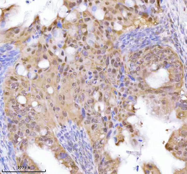
| Sample | Paraffin-embedded human endometrioid adenocarcinoma tissue; fixative not specified (datasheet A02076-2) |
| Fixation | Image fixative and duration unreported (datasheet A02076-2); verify before use. |
| Sectioning | 4–5 µm sections on charged slides (standard) |
| Deparaffinisation | Xylene, graded ethanol series to water (standard) |
| Antigen retrieval | Heat retrieval: EDTA pH 8.0 (datasheet A02076-2); 20 min, 95–100 °C (standard) |
| Peroxidase block | 3% H2O2, 10 min, room temperature (standard) |
| Blocking | 10% goat serum (datasheet A02076-2) |
| Primary antibody | Rabbit anti-RBBP8, 2-5 μg/ml (datasheet A02076-2) |
| Primary incubation | Overnight at 4 °C (datasheet A02076-2) |
| Detection | HRP-conjugated secondary, DAB chromogen (datasheet A02076-2) |
| Counterstain | Hematoxylin, blue, dehydrate and mount (standard) |
| Expected result | RBBP8-positive staining in glandular cells of appendix (HPA tissue IHC: Medium). HPA tissue profile: Nuclear and cytoplasmic expression in most tissues. No signal in the no-primary control. |
RBBP8 is a nuclear and chromosome-associated protein without a transmembrane segment (UniProt Q99708: subcellular location and topology). In paraffin-section IHC, expect nuclear staining, with possible cytoplasmic staining, in many tissues; HPA reports medium staining in several glandular epithelia and kidney tubular cells (HPA tissue IHC). The HPA IHC profile is Approved, with medium consistency against RNA data and a splice or transcript discrepancy, so interpret staining with controls (HPA tissue IHC).
| Glandular-cell nuclei stain in breast, colon, or appendix; some cytoplasmic color may accompany them (HPA tissue IHC). | This fits HPA's medium staining in those glandular cells and its broader nuclear and cytoplasmic tissue profile (HPA tissue IHC). Judge the result by the identified cells and compartments, rather than expecting identical color across every cell in the section (HPA tissue IHC: low tissue specificity; general IHC practice). |
| Color is confined to cell membranes or extracellular material, with no convincing nuclear staining. | A membrane-only or extracellular pattern does not fit RBBP8's nuclear and chromosome annotation or its lack of a transmembrane segment (UniProt Q99708). Check section morphology, detection background, and a known-positive control before assigning that color to RBBP8 (general IHC practice). Cytoplasmic color alone needs a more cautious judgment because HPA reports cytoplasmic tissue staining (HPA tissue IHC). |
| The strongest color appears in a cell population recorded as not detected, such as adipocytes or cardiomyocytes (HPA tissue IHC). | That distribution conflicts with the cited HPA observations for those cells, though HPA's Approved profile has medium RNA concordance and a splice or transcript discrepancy (HPA tissue IHC). Consider cross-reactivity or endogenous detection activity, then compare the suspect cells with an appropriate control section and a no-primary control (general IHC practice). |
| A uniform brown haze covers nuclei, cytoplasm, and tissue spaces, obscuring cell boundaries. | A haze without a readable cellular pattern cannot establish the reported RBBP8 distribution (HPA tissue IHC: nuclear and cytoplasmic profile; general IHC practice). Inspect the no-primary control and detection reagents; assess washing and blocking as general chromogenic IHC checks (general IHC practice). Avoid scoring diffuse color as positive cells until compartment boundaries are visible (general IHC practice). |
| No cellular signal is visible in glandular cells of a breast, colon, or appendix control section (HPA tissue IHC). | HPA records medium staining in those cell populations, so a blank control calls for review of the IHC workflow and antibody suitability (HPA tissue IHC; general IHC practice). Confirm the intended cells are present, inspect counterstain and detection controls, and compare sections processed in the same run (general IHC practice). A single blank field does not establish biological absence (general IHC practice). |
| IHC evidence and its limit | HPA rates tissue IHC Approved, with medium staining–RNA consistency and a splice or transcript discrepancy (HPA tissue IHC). Treat its cell-level observations as reference patterns, while keeping the discrepancy in mind when a sample differs (HPA tissue IHC). |
| Cell population and tissue | Medium staining is reported in glandular cells of appendix, breast, cervix, colon, duodenum, and gallbladder, plus esophageal squamous cells and kidney tubular cells (HPA tissue IHC). HPA records no detection in adipocytes, cardiomyocytes, and skeletal myocytes, among other listed cells (HPA tissue IHC). |
| Localization and damage context | UniProt places RBBP8 in the nucleus and on chromosomes and describes recruitment to DNA-damage sites in S/G2 (UniProt Q99708). HPA tissue IHC also reports cytoplasmic staining (HPA tissue IHC). Do not infer DNA damage, cell-cycle phase, or specificity from an ordinary chromogenic pattern alone (general IHC interpretation). |
| Isoforms and antibody evidence | UniProt lists three RBBP8 isoforms (UniProt Q99708). The payload supplies no epitope map or isoform-specific IHC result, so this section cannot assign a staining difference to one isoform. HPA039890 has Approved IHC status; HPA052946 has Supported ICC status, which is a separate application (HPA antibodies). |
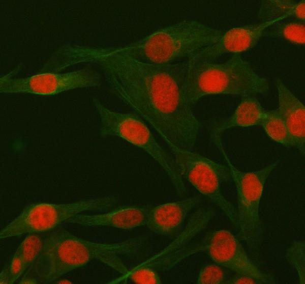
| IF/ICC Q: Where should fluorescence appear? | A: Mainly in the nucleoplasm, based on HPA's supported subcellular location; HPA lists ICC-IF images for MCF-7, U-251MG, and U2OS (HPA subcellular). That ICC-IF observation helps interpret localization but does not establish a paraffin-section IHC protocol or guarantee identical appearance in tissue (HPA subcellular; general IHC/IF practice). |
| Situation | Likely cause | Next action |
|---|---|---|
| Positive tissue is blank or unusually faint. | The observed result differs from HPA's medium glandular-cell or kidney tubular-cell staining; the cause cannot be assigned from that difference alone (HPA tissue IHC). | Verify that the cited cell population is present and review the IHC-validated antibody, antigen retrieval, primary incubation, and detection controls as general paraffin IHC checks (HPA antibodies; general IHC practice). No RBBP8-specific retrieval condition is supplied. |
| Signal is predominantly at membranes or in tissue spaces. | That location conflicts with UniProt's nuclear and chromosome annotation and lack of a transmembrane segment (UniProt Q99708). | Inspect a no-primary control and tissue morphology, then reassess detection background before scoring (general IHC practice). Retain a mixed nuclear and cytoplasmic result as a possibility because HPA reports both compartments in tissue (HPA tissue IHC). |
| Cells listed as not detected appear strongly positive. | The result differs from HPA's observations for adipocytes, cardiomyocytes, or skeletal myocytes; cross-reactivity or endogenous detection activity is possible (HPA tissue IHC; general IHC practice). | Check cell identity and compare a no-primary control with known-positive cells in the same run (general IHC practice). Record the mismatch rather than treating a single HPA entry as proof of biological absence (HPA tissue IHC: Approved, medium consistency). |
| Diffuse brown background obscures nuclei. | Nonspecific detection or incomplete washing can obscure compartment-level interpretation (general chromogenic IHC practice). | Inspect no-primary and detection controls, then review blocking, wash steps, and chromogen development using the assay's established workflow (general IHC practice). Score only cells whose boundaries and nuclear signal remain interpretable (general IHC practice). |
| Sections show patchy staining across otherwise comparable fields. | HPA reports low tissue specificity but varied cell-level staining, including medium, low, and not-detected entries; a patchy field alone has no single established cause (HPA tissue IHC). | Compare like cell populations across fields, confirm section quality, and check that control sections were processed together (general IHC practice). Do not attribute the variation to RBBP8 fixation sensitivity; no target-specific fixation evidence is supplied. |
| IHC and ICC-IF images seem to disagree about cytoplasmic signal. | HPA describes nuclear and cytoplasmic tissue IHC but mainly nucleoplasmic ICC-IF; the observations come from different applications (HPA tissue IHC; HPA subcellular). | Interpret each result against its own HPA application evidence and controls (HPA tissue IHC; HPA subcellular; general IHC/IF practice). For the paraffin-section decision, identify stained tissue cells and require an interpretable nuclear component. |



Comprehensive Human Protein Atlas IHC scoring per tissue (reliability: Approved — Medium consistency between antibody staining and RNA expression data. Caution, Splice and/or transcript discrepancy exists.). Rows are taken directly from the HPA tissue chart — click any row's HPA link to view the source.
| Tissue | Cell type | Level | Evidence | Source |
|---|---|---|---|---|
| Appendix | Glandular cells | Medium | Protein (IHC) | HPA → |
| Breast | Glandular cells | Medium | Protein (IHC) | HPA → |
| Cervix | Glandular cells | Medium | Protein (IHC) | HPA → |
| Colon | Glandular cells | Medium | Protein (IHC) | HPA → |
| Duodenum | Glandular cells | Medium | Protein (IHC) | HPA → |
| Tissue | Cell type | Level | Evidence | Source |
|---|---|---|---|---|
| Adipose tissue | Adipocytes | Not detected | Protein (IHC) | HPA → |
| Caudate | Glial cells | Not detected | Protein (IHC) | HPA → |
| Heart muscle | Cardiomyocytes | Not detected | Protein (IHC) | HPA → |
| Hippocampus | Glial cells | Not detected | Protein (IHC) | HPA → |
| Liver | Cholangiocytes | Not detected | Protein (IHC) | HPA → |
Troubleshoot RBBP8 staining in paraffin sections by checking retrieval, compartment, controls and scoring before interpreting differences between samples.
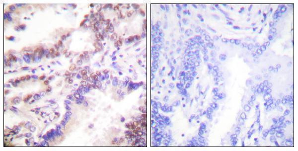
Catalog images show anti-RBBP8 IHC in human paraffin tissue sections and IF/ICC in HeLa cells; both antibodies list human and mouse reactivity (catalog image captions and reactivity lists).
A02076-2 has IHC images of human endometrioid adenocarcinoma, ovarian mucinous adenoma, and spleen paraffin sections, plus an IF/ICC image of HeLa cells (A02076-2 image captions). A02076 has an IHC image of a human lung carcinoma paraffin section with peptide blocking shown; IF/ICC is listed as an application without an IF image (A02076 image caption and application list).
Which to pick: For tissue IHC, choose A02076-2 when you want a documented starting condition: its paraffin-section caption specifies EDTA retrieval at pH 8.0 and 2 μg/ml primary antibody; A02076 offers a paraffin-section image with peptide blocking and a listed IHC dilution of 1:100–1:300 (A02076-2 and A02076 IHC captions; A02076 datasheet). For IF/ICC, A02076-2 has a HeLa image at 5 μg/ml, while A02076 lists IF/ICC without an IF image (A02076-2 IF caption; A02076 application list). Both list human and mouse reactivity, though the supplied images show human samples only; neither IHC caption reports the fixative (catalog reactivity lists and image captions).