This website uses cookies to ensure you get the best experience on our website.


- Table of Contents
Plan chromogenic RBFOX3 IHC in paraffin sections using nuclear staining in cerebellar granular cells as a positive reference (HPA tissue IHC). This guide covers the catalog antibody at 1:50 (datasheet M11954) and the risk of off-target staining (HPA tissue IHC).

Expected localisation, validated protocols, controls and antibodies — the at-a-glance facts below, then the full design guide.
| Expected localisation | Predominantly neuronal nuclei; cytoplasmic signal is possible (UniProt) | |
| Staining pattern | Strong nuclear staining in cerebellar granular cells (HPA tissue IHC) | |
| Antigen retrieval | EDTA pH 8.0 HIER, heat-mediated (datasheet M11954) | |
| Positive control | Cerebellum+4 more · see all | |
| Negative control | Adipose tissue+4 more · see all |
| Fixation | Keep paraffin-section fixation consistent (standard IHC practice; not target-specific) | |
| Caveat | Presumed off-target binding may confound staining (HPA tissue IHC) | |
| Regulation | Expression regulation unreported (UniProt) | |
| Isoform / epitope | 2 isoforms; epitope coverage is unknown (UniProt) |
The catalog antibody has a datasheet IHC-P protocol (datasheet: M11954). The published IHC protocols below cover human brain, piglet neocortex, and caudate putamen and substantia nigra (PMC4665621; PMC10605428; PMC9826750).
| Sample | Paraffin-embedded mouse brain tissue; fixative not specified (datasheet M11954) |
| Fixation | Image fixative and duration unreported (datasheet M11954); verify before use. |
| Sectioning | 4–5 µm sections on charged slides (standard) |
| Deparaffinisation | Xylene, graded ethanol series to water (standard) |
| Antigen retrieval | Heat retrieval: EDTA pH 8.0 (datasheet M11954); 20 min, 95–100 °C (standard) |
| Peroxidase block | 3% H2O2, 10 min, room temperature (standard) |
| Blocking | 10% goat serum (datasheet M11954) |
| Primary antibody | Rabbit monoclonal (clone AO-18) anti-RBFOX3, 1:50 (datasheet M11954) |
| Primary incubation | Overnight at 4 °C (datasheet M11954) |
| Detection | HRP-conjugated secondary, DAB chromogen (datasheet M11954) |
| Counterstain | Hematoxylin, blue, dehydrate and mount (standard) |
| Expected result | RBFOX3-positive staining in granular cells - nucleus of cerebellum (HPA tissue IHC: High). HPA tissue profile: Distinct expression in CNS. No signal in the no-primary control. |
RBFOX3 staining should concentrate in neuronal nuclei: UniProt describes it as largely restricted to neuronal nuclei, and HPA reports high staining in cerebellar granular cell nuclei (UniProt A6NFN3; HPA tissue IHC). HPA rates the tissue pattern “Enhanced” but notes medium agreement with RNA and presumed off-target binding; interpret unexpected staining cautiously (HPA tissue IHC). RBFOX3 has no transmembrane segment (UniProt A6NFN3 topology).
| Distinct nuclear staining in cerebellar granular cells; neuronal staining in cerebral cortex. | This matches HPA's high cerebellar granular cell nuclear and cerebral cortical neuronal staining. Score the nuclear pattern in its cell context; the cortex entry does not independently specify a compartment (HPA tissue IHC; UniProt A6NFN3 localization). |
| Predominantly cytoplasmic staining in neurons, without a clear nuclear pattern. | Review compartment assignment and controls: RBFOX3 is largely nuclear, although UniProt reports significant neuronal cytoplasmic localization in brains from HIV-infected individuals with cognitive impairment. Cytoplasmic staining alone is therefore context dependent, not automatic proof of an artefact (UniProt A6NFN3 localization). |
| Strong staining in cells outside the expected neuronal population. | Check cell identity and detection controls before calling it RBFOX3. HPA notes presumed off-target binding and cautions that staining can reflect protein from more than one gene. A positive signal alone cannot establish specificity in an unexpected cell type (HPA tissue IHC). |
| Diffuse color across cells, stroma, or a broad tissue area. | Assess the section alongside a no-primary control; widespread color without cell-specific nuclear enrichment can arise from nonspecific binding or endogenous detection activity (general IHC practice). HPA's tissue profile alone cannot assign the cause of background on this slide (HPA tissue IHC). |
| No signal in cerebellar granular cells or cerebral cortical neurons. | A blank result conflicts with HPA's high staining in these populations. First confirm the relevant cells are present, then review the IHC-validated antibody, detection run, and controls; absence on one section does not establish absent RBFOX3 expression (HPA tissue IHC; general IHC practice). |
| Anatomic and cellular context | HPA reports high cerebellar granular cell nuclear and cerebral cortical neuronal staining, with medium staining in caudate and hippocampal neurons. Compare like cell populations when judging signal strength (HPA tissue IHC). |
| Specificity of the tissue pattern | HPA calls the profile “Enhanced” while reporting medium staining–RNA consistency and presumed off-target binding. Its colon enterocyte result is medium; do not treat every non-neuronal positive as independently confirmed RBFOX3 (HPA tissue IHC). |
| Antibody-specific validation | The HPA030790 IHC result is “Enhanced”; HPA075862 and CAB078782 are “Supported.” These labels describe HPA validation for those antibodies and cannot be transferred to the catalog antibody without its own evidence (HPA antibody validation). |
| Compartment and protein forms | UniProt lists nuclear and cytoplasmic localization, two isoforms, and a full-length 1–312 chain with no signal peptide, propeptide, or transmembrane segment. No epitope position is supplied, so isoform-dependent IHC detection cannot be predicted here (UniProt A6NFN3). |
| IF/ICC Q: What localization is reported? | A: HPA reports supported nucleoplasmic localization in ICC-IF, with images from A-549, U-251MG, and U2OS. That observation is a compartment reference; it does not replace tissue IHC controls or establish an IF protocol (HPA subcellular ICC-IF). |
| Situation | Likely cause | Next action |
|---|---|---|
| Known-positive neuronal nuclei are blank. | The expected population may be absent from the section, or the staining run may have failed; HPA reports high signal in cerebellar granular cells and cortical neurons (HPA tissue IHC; general IHC practice). | Locate the relevant cells on the counterstain, inspect the positive control, then review the IHC-validated antibody and detection steps (general IHC practice). |
| Neurons appear cytoplasmic with little nuclear signal. | The pattern differs from the usual neuronal nuclear localization; UniProt also records a specific HIV-associated brain context with significant cytoplasmic localization (UniProt A6NFN3 localization). | Verify nuclear boundaries and compare a neuronal positive control. Document the specimen context before treating cytoplasmic staining as either genuine relocalization or artefact (general IHC practice; UniProt A6NFN3). |
| Unexpected non-neuronal cells stain strongly. | The signal may be off-target or detection-related; HPA explicitly notes presumed off-target binding. HPA also records medium colon enterocyte staining, so location alone cannot settle specificity (HPA tissue IHC). | Compare tissue morphology and no-primary controls; seek antibody-specific corroboration before assigning an unexpected positive cell population to RBFOX3 (HPA tissue IHC; general IHC practice). |
| Diffuse brown staining obscures nuclei. | Nonspecific binding or endogenous enzyme activity can contribute to chromogenic background (general IHC practice). HPA tissue levels do not identify the mechanism in an individual section (HPA tissue IHC). | Inspect a no-primary control and the detection reagents; adjust blocking, washes, or endogenous enzyme quenching according to the control result (general IHC practice). |
| Weak signal in caudate or hippocampus seems inconsistent with stronger cerebellum. | HPA reports medium neuronal staining in caudate and hippocampus versus high cerebellar granular cell nuclear staining; different recorded levels need not indicate a failed run (HPA tissue IHC). | Score each region against its own expected cell population and include a strong neuronal reference section in the run (HPA tissue IHC; general IHC practice). |
| Low non-neuronal staining is mistaken for a definitive positive. | HPA lists low staining in several non-neuronal populations and warns of presumed off-target binding; low signal is especially hard to interpret without controls (HPA tissue IHC). | Record cell type, compartment, and control behavior; reserve a positive RBFOX3 interpretation for a supported pattern or further antibody-specific corroboration (HPA tissue IHC; general IHC practice). |
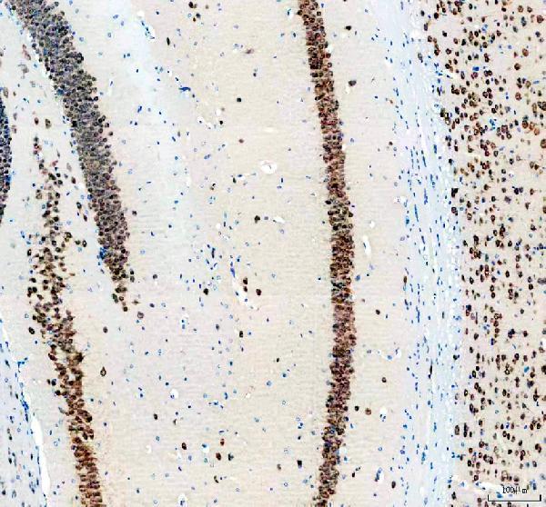
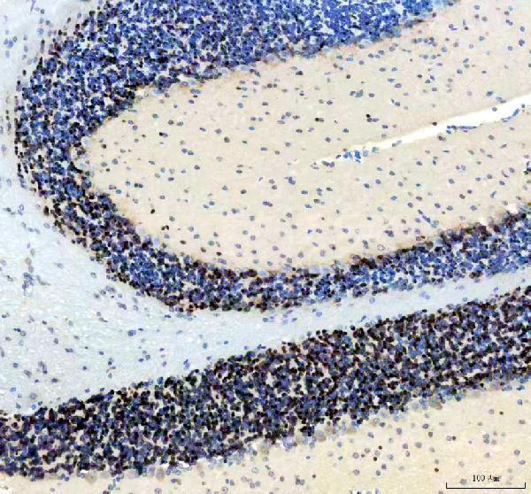
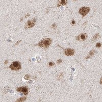


Comprehensive Human Protein Atlas IHC scoring per tissue (reliability: Enhanced — Medium consistency between antibody staining and RNA expression data. Caution, targets protein from more than one gene. Presumed off target binding observed and disregarded.). Rows are taken directly from the HPA tissue chart — click any row's HPA link to view the source.
| Tissue | Cell type | Level | Evidence | Source |
|---|---|---|---|---|
| Cerebellum | Granular cells - nucleus | High | Protein (IHC) | HPA → |
| Cerebral cortex | Neuronal cells | High | Protein (IHC) | HPA → |
| Caudate | Neuronal cells | Medium | Protein (IHC) | HPA → |
| Colon | Enterocytes | Medium | Protein (IHC) | HPA → |
| Hippocampus | Neuronal cells | Medium | Protein (IHC) | HPA → |
| Tissue | Cell type | Level | Evidence | Source |
|---|---|---|---|---|
| Adipose tissue | Adipocytes | Not detected | Protein (IHC) | HPA → |
| Adrenal gland | Glandular cells | Not detected | Protein (IHC) | HPA → |
| Appendix | Endocrine cells | Not detected | Protein (IHC) | HPA → |
| Bone marrow | Hematopoietic cells | Not detected | Protein (IHC) | HPA → |
| Breast | Adipocytes | Not detected | Protein (IHC) | HPA → |
Troubleshoot RBFOX3 staining in paraffin sections by checking retrieval, nuclear localisation and neuronal cell identity alongside appropriate controls.
The IHC-validated anti-RBFOX3 antibody has paraffin-section images from mouse and rat brain and cerebellum (M11954 IHC captions); human reactivity and IF/ICC applications are listed (M11954 catalog).
M11954 has IHC images from paraffin-embedded mouse and rat brain and cerebellum sections (M11954 IHC captions). Human, mouse and rat reactivity and IF/ICC applications are listed, but no IF image is supplied (M11954 catalog).
Which to pick: Choose M11954 for tissue IHC: its rabbit monoclonal antibody has paraffin-section images using EDTA retrieval at pH 8.0, 1:50 primary dilution and DAB detection; the fixative is unreported (M11954 catalog; M11954 IHC captions). For IF/ICC, M11954 lists those applications and a 1:50 IF dilution, but the payload supplies no IF image (M11954 catalog). For cross-species work, M11954 lists human, mouse and rat reactivity; its IHC images document mouse and rat sections only (M11954 catalog; M11954 IHC captions).