Click image to see more details
-

-
-
-
-
+12
Product Info Summary
| SKU: | M11954 |
|---|---|
| Size: | 100 μl |
| Reactive Species: | Human, Mouse, Rat |
| Host: | Rabbit |
| Application: | IF, IHC, ICC, WB |
Customers Who Bought This Also Bought

Product info
Product Name
Anti-NeuN RBFOX3 Rabbit Monoclonal Antibody
SKU/Catalog Number
M11954
BM4354 is an alternative SKU for this antibody, used in previous lots.
Size
100 μl
Form
Liquid
Description
Boster Bio Anti-NeuN RBFOX3 Rabbit Monoclonal Antibody catalog # M11954. Tested in WB, IHC, ICC/IF, IF applications. This antibody reacts with Human, Mouse, Rat.
Storage & Handling
Store at -20°C for one year. For short term storage and frequent use, store at 4°C for up to one month. Avoid repeated freeze-thaw cycles.
Cite This Product
Anti-NeuN RBFOX3 Rabbit Monoclonal Antibody (Boster Biological Technology, Pleasanton CA, USA, Catalog # M11954)
Host
Rabbit
Contents
Rabbit IgG in stabilizing components, phosphate buffered saline, pH 7.4, 150mM NaCl, 0.02% sodium azide and 50% glycerol.
*This antibody is supplied in a stabilized formulation.
Compatibility with conjugation reactions depends on the chemistry of the conjugation method used.
For conjugation methods that are not compatible with the stabilizing components present in this formulation, a carrier-free antibody format is required.
Clonality
Monoclonal
Clone Number
AO-18
Isotype
Rabbit IgG
Immunogen
A synthesized peptide derived from human NeuN
Reactive Species
M11954 is reactive to RBFOX3 in Human, Mouse, Rat
Observed Molecular Weight
46, 50 kDa
Calculated molecular weight
33.9 kDa
Antibody Validation
Boster validates all antibodies on WB, IHC, ICC, Immunofluorescence, and ELISA with known positive control and negative samples to ensure specificity and high affinity, including thorough antibody incubations.
Application & Images
Applications
M11954 is guaranteed for IF, IHC, ICC, WB Boster Guarantee
Recommend Dilution
WB 1:500-2000
IHC 1:50-200
IF 1:50-200
ICC/IF 1:50-200
Tested application
Suggested blocking solution with 5% non-fat milk or BSA; (*)Recommended protein loading: 20-40 µg per lane
Use TE buffer pH 9.0 for antigen retrieval; (*) citrate buffer pH 6.0 is an alternative.
Validation Images & Assay Conditions
Click image to see more details
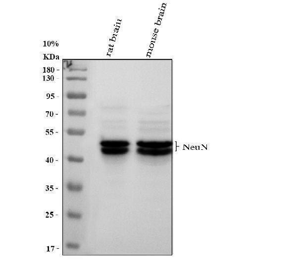
Western blot analysis of NeuN using anti-NeuN antibody (M11954).
Electrophoresis was performed on a 10% SDS-PAGE gel at 80V (Stacking gel) / 120V (Resolving gel) for 2 hours. The sample well of each lane was loaded with 30 ug of sample under reducing conditions.
Lane 1: rat brain tissue lysates,
Lane 2: mouse brain tissue lysates.
After electrophoresis, proteins were transferred to a nitrocellulose membrane at 150 mA for 50-90 minutes. Blocked the membrane with 5% non-fat milk/TBS for 1.5 hour at RT. The membrane was incubated with rabbit anti-NeuN antigen affinity purified monoclonal antibody (M11954) at 1:1000 overnight at 4°C, then washed with TBS-0.1%Tween 3 times with 5 minutes each and probed with a goat anti-rabbit IgG-HRP secondary antibody at a dilution of 1:5000 for 1.5 hour at RT. The signal is developed using an ECL Plus Western Blotting Substrate (Catalog # AR1196-200) with Tanon 5200 system. A specific band was detected for NeuN at approximately 46, 50 kDa. The expected band size for NeuN is at 34 kDa.

Click image to see more details
IHC analysis of NeuN using anti-NeuN antibody (M11954).
NeuN was detected in a paraffin-embedded section of mouse brain tissue. Heat mediated antigen retrieval was performed in EDTA buffer (pH 8.0, epitope retrieval solution). The tissue section was blocked with 10% goat serum. The tissue section was then incubated with 1:50 rabbit anti-NeuN Antibody (M11954) overnight at 4°C. Peroxidase Conjugated Goat Anti-rabbit IgG was used as secondary antibody and incubated for 30 minutes at 37°C. The tissue section was developed using HRP Conjugated Rabbit IgG Super Vision Assay Kit (Catalog # SV0002) with DAB as the chromogen.

Click image to see more details
IHC analysis of NeuN using anti-NeuN antibody (M11954).
NeuN was detected in a paraffin-embedded section of mouse cerebellum tissue. Heat mediated antigen retrieval was performed in EDTA buffer (pH 8.0, epitope retrieval solution). The tissue section was blocked with 10% goat serum. The tissue section was then incubated with 1:50 rabbit anti-NeuN Antibody (M11954) overnight at 4°C. Peroxidase Conjugated Goat Anti-rabbit IgG was used as secondary antibody and incubated for 30 minutes at 37°C. The tissue section was developed using HRP Conjugated Rabbit IgG Super Vision Assay Kit (Catalog # SV0002) with DAB as the chromogen.

Click image to see more details
IHC analysis of NeuN using anti-NeuN antibody (M11954).
NeuN was detected in a paraffin-embedded section of rat brain tissue. Heat mediated antigen retrieval was performed in EDTA buffer (pH 8.0, epitope retrieval solution). The tissue section was blocked with 10% goat serum. The tissue section was then incubated with 1:50 rabbit anti-NeuN Antibody (M11954) overnight at 4°C. Peroxidase Conjugated Goat Anti-rabbit IgG was used as secondary antibody and incubated for 30 minutes at 37°C. The tissue section was developed using HRP Conjugated Rabbit IgG Super Vision Assay Kit (Catalog # SV0002) with DAB as the chromogen.

Click image to see more details
IHC analysis of NeuN using anti-NeuN antibody (M11954).
NeuN was detected in a paraffin-embedded section of rat cerebellum tissue. Heat mediated antigen retrieval was performed in EDTA buffer (pH 8.0, epitope retrieval solution). The tissue section was blocked with 10% goat serum. The tissue section was then incubated with 1:50 rabbit anti-NeuN Antibody (M11954) overnight at 4°C. Peroxidase Conjugated Goat Anti-rabbit IgG was used as secondary antibody and incubated for 30 minutes at 37°C. The tissue section was developed using HRP Conjugated Rabbit IgG Super Vision Assay Kit (Catalog # SV0002) with DAB as the chromogen.

Click image to see more details
NeuN (Rbfox3) was expressed in different organs at protein level. (A) Western blot analysis; (B) semi-quantitative analysis of the results of Western blotting (Mean ± SD, n = 3); (C) β -actin expressed in different organs at protein level, * P < 0.05 vs. brain, ANOVA; (D) β -actin and Gapdh expressed in different organs at mRNA level. * P < 0.05 vs. brain, ANOVA; (E) Lambs expressed in different organs at mRNA level. FPKM = value of fragments per kilobase of transcript sequence per million base pairs sequenced, *, P < 0.05, Mann Whiteney test (two-tailed). Download full-size image DOI:
Index in PubMed under a CC BY license. PMID: 31938576

Click image to see more details
Knockdown FCGR2B improved hippocampal neuronal excitability. A Representative images of Golgi staining of the hippocampal neuronal spines from the mice. B IHC assay was performed to examine the expression of NeuN in hippocampus of mice. C IHC assay was used to examine the expression of c-fos and GABAA in hippocampus of mice. D The expressions of c-fos, CaMKII, GABAA, and GABAARAP in hippocampus of mice were detected by Western blot. E TUNEL staining was conducted to assess cell apoptosis in hippocampus. F IF was used to detect BrdU and Ki67 positive cells in hippocampaltissue to analyzecell proliferation * P < 0.05, ** P < 0.01, *** P < 0.001 Full size image
Index in PubMed under a CC BY license. PMID: 40537751

Click image to see more details
The hippocampal neuron in DM mice showed the morphological changes. A The work flow chart of how to construct a DM mouse model. B - C The levels of blood glucose and body weight of mice were assessed. D The level of insulin of mice was assessed. E - F Morris water maze test was evaluated the learning and memory ability of mice by the escape latency time, number of crossing platform and swimming time in quadrant. G The recognition index among 4 groups in the novel object recognition test. H H&E staining evaluated the pathological changes of hippocampus. I Neuronal damage of the hippocampal region was assessed by Nissl staining. J IHC assay was used to examine the expression of NeuN in hippocampus of mice. * P < 0.05, ** P < 0.01, *** P < 0.001 Full size image
Index in PubMed under a CC BY license. PMID: 40537751

Click image to see more details
FCGR2B were up-regulated in hippocampus of DM mice. A qRT-PCR was performed to detect the expression of ALB, AREG and FCGR2B mRNA expression in hippocampus of mice. B Western blot was conducted to detect the ALB, AREG and FCGR2B protein expression in hippocampus of mice. C IHC assay was employed to examine the ALB, AREG and FCGR2B protein expression in hippocampus of mice. D IF staining was utilized to detect the expression of FCGR2B and NeuN in hippocampus of mice. E Western blot was performed to detect the SHC1 protein expression in hippocampus of mice. F IF staining was performed to detect the expression of SHC1 and NeuN in hippocampus of mice. G Western blot was used to detect the p-PI3K and p-AKT protein expression in hippocampus of mice. *** P < 0.001 Full size image
Index in PubMed under a CC BY license. PMID: 40537751

Click image to see more details
Quantitative analysis of NeuN-positive cells in rat organs and cultured cells (mean ± SD, n = 3). Photos can be seen in (rat organs) and (cultured cells). (A) NeuN-positive cell rate in rat organs; (B) NeuN-positive cell rate in cultured cells. Download full-size image DOI:
Index in PubMed under a CC BY license. PMID: 31938576

Click image to see more details
Immunofluorescence (IF) analysis of different cell lines (bar = 20 µm). (A–C) 293T cell; (D–F) A549 cell; (G–I) BRL3A cell; (J–L) Caco2 cell; (M–O) HL7702 cell; (P–R) PC12Adh cell; (S–U) atrial muscle cell. Green fluorescence was stained to locate NeuN by FITC-linked secondary antibody, and blue fluorescence was stained to locate nucleus by Hoechst 33342. Download full-size image DOI:
Index in PubMed under a CC BY license. PMID: 31938576

Click image to see more details
Double immunofluorescence (IF) analysis of rat heart (bar = 20 µm). (A–D) Cardiac muscle fibers in transection; (E–H) cardiac muscle fibers in longitudinal section. Red fluorescence was stained to locate Myl3 by Alexa Fluor ® 647-linked secondary antibody, green fluorescence was stained to locate NeuN by FITC-linked secondary antibody, and blue fluorescence was stained to locate nucleus by Hoechst 33342. Download full-size image DOI:
Index in PubMed under a CC BY license. PMID: 31938576

Click image to see more details
Immunofluorescence (IF) assays verified in four organs (bar = 20 µm). (A–C) Heart; (D–F) lung; (G–I) liver; (J–L) Kidney; (M–O) kidney (Negative control). Red fluorescence was stained to locate NeuN by another rabbit anti-NeuN monoclonal antibody (ab177487, Abcam), and blue fluorescence was stained to locate nucleus by Hoechst 33342. Negative control was only stained with Alexa Fluor ® 647-linked secondary antibody and Hoechst 33342. Download full-size image DOI:
Index in PubMed under a CC BY license. PMID: 31938576

Click image to see more details
Immunofluorescence (IF) and immunohistochemistry (IHC) assays of different organs (bar = 20 µm). (A–D) Brain; (E–H) heart; (I–L), liver; (M–P) lung; (Q–T) kidney; (U–X) stomach; (Y–BB) duodenum; (CC–FF) ileum. As for IF, green fluorescence was stained to locate NeuN (BM4354, Boster) by FITC-linked secondary antibody, and blue fluorescence was stained to locate nucleus by Hoechst 33342. As for IHC, NeuN was stained in brown. Download full-size image DOI:
Index in PubMed under a CC BY license. PMID: 31938576

Click image to see more details
Knockdown FCGR2B could promote the PI3K/AKT signaling pathway in vivo. A The work flow chart of how to construct a DM mouse model. B The mRNA levels of FCGR2B and SHC1 were assessed by qRT-PCR. C The protein levels of FCGR2B and SHC1 were evaluated by Western blot. D The protein level of SHC1 was determined by Western blot. E IF staining was used to detect the expression of FCGR2B and NeuN in hippocampus of mice. F IF staining was performed to detect the expression of SHC1 and NeuN in hippocampus of mice. G The expressions of p-PI3K and p-AKT in hippocampus of mice were evaluated by Western blot. ** P < 0.01, *** P < 0.001 Full size image
Index in PubMed under a CC BY license. PMID: 40537751

Click image to see more details
Heterochromatin changes are associated with Tau pathology in AD temporal lobes at intermediate stages. (A) WB of heterochromatin markers HP1α and H3K9me3 in human brain samples at BS1/2 (N = 7), BS3/4 (N = 6) and BS5/6 (N = 8) and relative quantification. Histone H3 and Actin are the housekeeping genes. HP1α and H3K9me3 fold change is reported in the graphs. H3K9me3 has been normalized on total H3 histone. (B) Immunofluorescence and relative quantification of H3K9me3 in human brain samples at BS1/2 (N = 6), BS3/4 (N = 5) and BS5/6 (N = 5). Red: NeuN; yellow: H3K9me3; green: pAT8; blue: DAPI. Scale bar: 20 µm * p < 0.05; ** p < 0.01.
Index in PubMed under a CC BY license. PMID: 39830212
Specific Publications For Anti-NeuN RBFOX3 Rabbit Monoclonal Antibody (M11954)
Loading publications
Recommended Resources
Here are featured tools and databases that you might find useful.
- Boster's Pathways Library
- Protein Databases
- Bioscience Research Protocol Resources
- Data Processing & Analysis Software
- Photo Editing Software
- Scientific Literature Resources
- Research Paper Management Tools
- Molecular Biology Software
- Primer Design Tools
- Bioinformatics Tools
- Phylogenetic Tree Analysis
Customer Reviews
Have you used Anti-NeuN RBFOX3 Rabbit Monoclonal Antibody?
Share your experimental results or join a short interview to earn up to $1,000 in product credits or other rewards.
0 Reviews For Anti-NeuN RBFOX3 Rabbit Monoclonal Antibody
Customer Q&As
Have a question?
Find answers in Q&As, reviews.
Can't find your answer?
Submit your question
5 Customer Q&As for Anti-NeuN RBFOX3 Rabbit Monoclonal Antibody
Question
I see that the anti-NeuN Rabbit Monoclonal antibody M11954 works with IF, what is the protocol used to produce the result images on the product page?
Verified Customer
Verified customer
Asked: 2019-11-29
Answer
You can find protocols for IF on the "support/technical resources" section of our navigation menu. If you have any further questions, please send an email to support@bosterbio.com
Boster Scientific Support
Answered: 2019-11-29
Question
Please see the WB image, lot number and protocol we used for cerebellum using anti-NeuN Rabbit Monoclonal antibody M11954. Please let me know if you require anything else.
Verified Customer
Verified customer
Asked: 2019-05-03
Answer
Thank you very much for the data. Our lab team are working to resolve this as quickly as possible, and we appreciate your patience and understanding! You have provided everything we needed. Please let me know if there is anything you need in the meantime.
Boster Scientific Support
Answered: 2019-05-03
Question
Is a blocking peptide available for product anti-NeuN Rabbit Monoclonal antibody (M11954)?
Verified Customer
Verified customer
Asked: 2018-07-30
Answer
We do provide the blocking peptide for product anti-NeuN Rabbit Monoclonal antibody (M11954). If you would like to place an order for it please contact support@bosterbio.com and make a special request.
Boster Scientific Support
Answered: 2018-07-30
Question
My lab would like to test anti-NeuN Rabbit Monoclonal antibody M11954 on mouse cerebellum for research purposes, then I may be interested in using anti-NeuN Rabbit Monoclonal antibody M11954 for diagnostic purposes as well. Is the antibody suitable for diagnostic purposes?
A. Parker
Verified customer
Asked: 2016-08-16
Answer
The products we sell, including anti-NeuN Rabbit Monoclonal antibody M11954, are only intended for research use. They would not be suitable for use in diagnostic work. If you have the means to develop a product into diagnostic use, and are interested in collaborating with us and develop our product into an IVD product, please contact us for more discussions.
Boster Scientific Support
Answered: 2016-08-16
Question
Will anti-NeuN Rabbit Monoclonal antibody M11954 work for IF with cerebellum?
P. Roberts
Verified customer
Asked: 2016-07-01
Answer
According to the expression profile of cerebellum, RBFOX3 is highly expressed in cerebellum. So, it is likely that anti-NeuN Rabbit Monoclonal antibody M11954 will work for IF with cerebellum.
Boster Scientific Support
Answered: 2016-07-01
