This website uses cookies to ensure you get the best experience on our website.


- Table of Contents
Plan RBP5 staining in paraffin sections using hepatocytes and glomerular cells as high-staining reference populations (HPA tissue IHC). The catalog antibody lists an IHC dilution of 1:100–1:300 (datasheet).

Expected localisation, validated protocols, controls and antibodies — the at-a-glance facts below, then the full design guide.
| Expected localisation | Cytoplasmic tissue staining (HPA tissue IHC) | |
| Staining pattern | Variable cytoplasmic staining across tissues (HPA tissue IHC) | |
| Antigen retrieval | Citrate pH 6.0 HIER, 95–98 °C, 20 min (rule: cytoplasmic / membrane antigen) | |
| Positive control | Kidney+4 more · see all | |
| Negative control | Smooth muscle |
| Fixation | Matched tissue-IHC evidence does not establish the fixation claim. Validate the specimen-specific method before use. (selected-SKU IHC image A30467) | |
| Caveat | Antibody staining and RNA show medium consistency (HPA tissue IHC) | |
| Regulation | Reduced in hepatocellular carcinoma (UniProt) | |
| Isoform / epitope | No annotated isoforms; one chain, residues 1–135 (UniProt) |
Compare the catalog antibody’s IHC-P protocol (datasheet) with the published chromogenic RBP5 IHC procedure for paraffin-embedded keloid and skin tissue (PMC11251391).
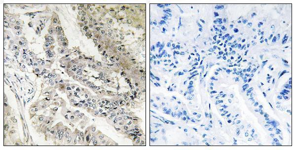
| Sample | Paraffin-embedded human lung carcinoma tissue; fixative not specified (datasheet A30467) |
| Fixation | Image fixative and duration unreported (datasheet A30467); verify before use. |
| Sectioning | 4–5 µm sections on charged slides (standard) |
| Deparaffinisation | Xylene, graded ethanol series to water (standard) |
| Antigen retrieval | Heat-induced epitope retrieval in citrate buffer, pH 6.0, 20 min at 95–98 °C (standard rule: cytoplasmic / membrane antigen) |
| Peroxidase block | 3% H2O2, 10 min, room temperature (standard) |
| Blocking | 10% normal serum of the secondary host, 30 min, room temperature (standard) |
| Primary antibody | Rabbit anti-RBP5, 1:100 - 1:300 (datasheet A30467) |
| Primary incubation | Overnight at 4 °C (standard) |
| Detection | HRP-polymer secondary, DAB chromogen 5–10 min (standard) |
| Counterstain | Hematoxylin, blue, dehydrate and mount (standard) |
| Expected result | RBP5-positive staining in cells in glomeruli of kidney (HPA tissue IHC: High). HPA tissue profile: Cytoplasmic expression at variable levels in all tissues. No signal in the no-primary control. |
RBP5 is a cytoplasmic protein with no transmembrane segment (UniProt P82980). In paraffin-section IHC, expect cytoplasmic staining in glomerular cells and hepatocytes, with high staining also reported in selected lymphoid and epithelial cells (HPA tissue IHC). HPA describes variable cytoplasmic expression across tissues; its tissue IHC reliability is Enhanced, with medium consistency between staining and RNA data and external verification pending (HPA tissue IHC).
| Cytoplasmic staining in glomerular cells or hepatocytes, with recognizable tissue structure. | This matches HPA's High staining in kidney glomerular cells and liver hepatocytes (HPA tissue IHC). Score cell type, compartment and intensity together; the presence of brown signal alone does not establish a correct result (general IHC practice). |
| Predominantly nuclear or sharply cell-surface staining, without a convincing cytoplasmic component. | That distribution conflicts with the cytoplasmic UniProt annotation and HPA tissue IHC profile (UniProt P82980; HPA tissue IHC). Check the counterstain and morphology, then compare a known-positive section and detection controls before interpreting it as RBP5 (general IHC practice). |
| Strong staining in smooth muscle cells, especially when expected positive cells lack staining. | HPA reports smooth muscle cells as Not detected (HPA tissue IHC). An unexpected pattern raises concern for nonspecific binding or endogenous detection activity; verify it with a reagent-omission control and the expected positive-cell pattern (general IHC practice). |
| Diffuse color across cells and surrounding tissue, with little distinction between compartments. | An indiscriminate deposit cannot be scored confidently as cytoplasmic RBP5 (UniProt P82980; general IHC practice). Review blocking, washing and chromogen development, and compare the reagent-omission control to assess background from the detection system (general IHC practice). |
| No convincing cytoplasmic staining in glomerular cells or hepatocytes. | These are HPA High cell populations, so a blank result merits a technical check before a biological interpretation (HPA tissue IHC; general IHC practice). Review the catalog antibody's IHC-P instructions and run a documented positive tissue alongside the specimen (general IHC practice). |
| Target location and topology | RBP5 is annotated in the cytoplasm and has no transmembrane segment (UniProt P82980). Evaluate staining within cells rather than treating a cell-surface outline as the expected target distribution (general IHC practice). |
| Cell-specific tissue pattern | HPA reports High staining in glomerular cells, hepatocytes, germinal center cells, splenic white-pulp cells, keratinocytes and thyroid glandular cells; adipocytes are Medium and smooth muscle cells are Not detected (HPA tissue IHC). Compare like cell types when judging intensity. |
| Tissue IHC evidence strength | The HPA tissue IHC reliability label is Enhanced, while staining and RNA show medium consistency and external verification is pending (HPA tissue IHC). Use the reported pattern as an interpretation reference, and investigate discordant specimens before assigning a biological explanation. |
| Annotated processing and variants | UniProt lists one chain spanning residues 1–135, with no signal peptide, propeptide, annotated isoforms, glycosylation sites or modified residues (UniProt P82980). These annotations give no basis for predicting a secreted or processed staining pattern. |
| IHC versus IF localization | HPA tissue IHC describes variable cytoplasmic expression, whereas HPA ICC-IF places RBP5 mainly at the Golgi apparatus and additionally in vesicles (HPA tissue IHC; HPA subcellular). Interpret each image in its assay context; the ICC-IF observation does not define a paraffin-section protocol. |
| Situation | Likely cause | Next action |
|---|---|---|
| Expected positive tissue shows little or no signal. | The result disagrees with HPA's High staining in glomerular cells and hepatocytes; the image alone does not identify the technical cause (HPA tissue IHC; general IHC practice). | Confirm those cell types are present, check the IHC-validated antibody's documented IHC-P conditions, and process a positive section with the specimen (general IHC practice). |
| The slide is uniformly brown or has diffuse haze. | Detection background can obscure a cell-specific cytoplasmic pattern (general IHC practice; HPA tissue IHC). | Inspect a reagent-omission control; review blocking, washes and chromogen development using the detection system's instructions (general IHC practice). |
| Strong nuclear or cell-border signal dominates. | The dominant compartment disagrees with cytoplasmic RBP5 annotation and the HPA tissue IHC pattern (UniProt P82980; HPA tissue IHC). | Check morphology and counterstain, compare a known-positive section, and investigate nonspecific or detection-related staining before scoring (general IHC practice). |
| Smooth muscle stains strongly while the expected positive cells are weak. | HPA reports smooth muscle cells as Not detected and glomerular cells and hepatocytes as High (HPA tissue IHC). | Verify cell identity and examine reagent-omission and positive-tissue controls; treat the discordant pattern as unresolved until the controls support interpretation (general IHC practice). |
| Kidney and liver show different apparent staining strengths. | HPA reports cytoplasmic expression at variable levels across tissues, even though glomerular cells and hepatocytes are both listed as High (HPA tissue IHC). | Compare the specified cell populations and assess staining against background within each section; avoid scoring an entire organ from one field (general IHC practice). |
| Why does an ICC-IF image look more Golgi-centered than the tissue IHC image? | HPA reports mainly Golgi and additional vesicular localization in ICC-IF, while its tissue IHC profile is cytoplasmic (HPA subcellular; HPA tissue IHC). | Record the assay and compartment with each observation, then interpret the paraffin-section result against the tissue IHC pattern (general IHC practice; HPA tissue IHC). |
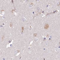


Comprehensive Human Protein Atlas IHC scoring per tissue (reliability: Enhanced — Medium consistency between antibody staining and RNA expression data. Pending external verification.). Rows are taken directly from the HPA tissue chart — click any row's HPA link to view the source.
| Tissue | Cell type | Level | Evidence | Source |
|---|---|---|---|---|
| Kidney | Cells in glomeruli | High | Protein (IHC) | HPA → |
| Liver | Hepatocytes | High | Protein (IHC) | HPA → |
| Lymph node | Germinal center cells | High | Protein (IHC) | HPA → |
| Skin | Keratinocytes | High | Protein (IHC) | HPA → |
| Spleen | Cells in white pulp | High | Protein (IHC) | HPA → |
| Tissue | Cell type | Level | Evidence | Source |
|---|---|---|---|---|
| Smooth muscle | Smooth muscle cells | Not detected | Protein (IHC) | HPA → |
Troubleshoot RBP5 staining in paraffin sections using the selected tissue image, expression references, and compartment controls (catalog caption A30467; HPA; UniProt P82980).
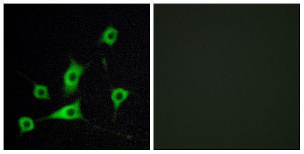
A30467 has IHC data from human lung carcinoma paraffin sections (IHC image caption) and IF data from LOVO cells (IF image caption); listed reactivity is human, mouse and rat (catalog: reactivity).
A30467 will render with an IHC figure from paraffin-embedded human lung carcinoma tissue, including a peptide-blocked comparison (IHC image caption). Its IF figure shows LOVO cells with a peptide-blocked comparison (IF image caption); the catalog lists IHC, IF and ICC applications and human, mouse and rat reactivity (catalog: applications and reactivity).
Which to pick: Choose A30467 for tissue IHC: its figure shows paraffin-embedded human lung carcinoma tissue (IHC image caption); the fixative is unreported (IHC image caption). Choose A30467 for IF/ICC because both applications are listed, although its pictured IF example is limited to LOVO cells (catalog: applications; IF image caption). For cross-species work, A30467 lists human, mouse and rat reactivity and is polyclonal (catalog: reactivity; dilution_raw: Polyclonal).