This website uses cookies to ensure you get the best experience on our website.


- Table of Contents
Plan RHOG chromogenic IHC around cytoplasmic staining in a subset of immune cells (HPA tissue IHC). Bone marrow hematopoietic cells show high staining (HPA tissue IHC); the catalog antibody starts at 5 μg/mL, with IHC validation reported in mouse tissue (datasheet).

Expected localisation, validated protocols, controls and antibodies — the at-a-glance facts below, then the full design guide.
| Expected localisation | Cytoplasmic staining in immune cells (HPA tissue IHC); membrane localization at the molecular level (UniProt) | |
| Staining pattern | A subset of immune cells shows cytoplasmic staining (HPA tissue IHC) | |
| Antigen retrieval | Citrate pH 6.0 HIER, 95–98 °C, 20 min (rule: cytoplasmic / membrane antigen) | |
| Positive control | Bone marrow+4 more · see all | |
| Negative control | Adipose tissue+4 more · see all |
| Fixation | Keep fixation consistent across sections (standard IHC practice; not target-specific) | |
| Caveat | Staining varies across immune-cell subsets (HPA tissue IHC) | |
| Regulation | No expression regulator annotated (UniProt) | |
| Isoform / epitope | No isoforms; mature chain spans residues 1–188 (UniProt) |
The catalog antibody protocol is followed by one published RHOG IHC protocol for paraffin sections (PMC4232683: Methods).
| Sample | Tissue sections; selected-image fixative not specified (standard IHC workflow) |
| Fixation | Image fixative and duration unreported (datasheet A03485); verify before use. |
| Sectioning | 4–5 µm sections on charged slides (standard) |
| Deparaffinisation | Xylene, graded ethanol series to water (standard) |
| Antigen retrieval | Heat-induced epitope retrieval in citrate buffer, pH 6.0, 20 min at 95–98 °C (standard rule: cytoplasmic / membrane antigen) |
| Peroxidase block | 3% H2O2, 10 min, room temperature (standard) |
| Blocking | 10% normal serum of the secondary host, 30 min, room temperature (standard) |
| Primary antibody | Rabbit anti-RHOG, 5 μg/mL (datasheet A03485) |
| Primary incubation | Overnight at 4 °C (standard) |
| Detection | HRP-polymer secondary, DAB chromogen 5–10 min (standard) |
| Counterstain | Hematoxylin, blue, dehydrate and mount (standard) |
| Expected result | RHOG-positive staining in hematopoietic cells of bone marrow (HPA tissue IHC: High). HPA tissue profile: Cytoplasmic expression in a subset of immune cells. No signal in the no-primary control. |
RHOG staining in paraffin sections should be cytoplasmic in a subset of immune cells, with possible peripheral accentuation because UniProt places RHOG at the cell membrane without a transmembrane segment (HPA: tissue IHC profile; UniProt P84095: subcellular location and topology). HPA rates its tissue IHC evidence Enhanced, while describing only medium consistency between antibody staining and RNA expression (HPA: tissue IHC reliability).
| Strong cytoplasmic staining in bone marrow hematopoietic cells or tonsil germinal center cells. | This fits two high staining compartments; score the named cells rather than treating every cell in the section as positive (HPA: High in both compartments). |
| Predominantly nuclear staining in cells otherwise expected to be positive. | This conflicts with the reported cytoplasmic IHC pattern and cell membrane annotation; check whether the signal follows nuclei in a no-primary control (HPA: tissue IHC profile; UniProt P84095: cell membrane; standard IHC practice). |
| Strong staining in bronchial respiratory epithelium or esophageal squamous epithelium. | HPA reports RHOG as undetected in those cell types; investigate cross-reactivity or endogenous detection activity before calling the signal RHOG (HPA: Not detected in both compartments; standard IHC practice). |
| Color spreads across tissue and empty areas without clear cell boundaries. | This is inconsistent with a selected-cell cytoplasmic pattern; assess nonspecific staining and detection background with appropriate controls (HPA: tissue IHC profile; standard IHC practice). |
| No staining in bone marrow hematopoietic cells on the test section. | A negative result there conflicts with HPA's high staining report; verify tissue preservation, retrieval, primary antibody and detection performance before interpreting another section as RHOG-negative (HPA: High in bone marrow hematopoietic cells; standard IHC practice). |
| Choice of tissue compartment | Bone marrow hematopoietic cells, splenic red pulp cells and tonsil germinal center cells are high staining examples; lymph node non-germinal center cells are medium (HPA: tissue IHC levels). |
| Antibody evidence | HPA039871 is a rabbit polyclonal antibody with Enhanced IHC validation, meaning its pattern was supported by independent antibodies or orthogonal data; this does not establish every new run's specificity (HPA: antibody validation; standard IHC practice). |
| Localization and topology | UniProt assigns RHOG to the cell membrane and lists no transmembrane segment; HPA describes cytoplasmic staining in a subset of immune cells, so a crisp membrane rim is not required for a positive IHC call (UniProt P84095: location and topology; HPA: tissue IHC profile). |
| Processing and epitope position | UniProt lists a chain at residues 1–188 and a propeptide at 189–191; without an antibody epitope map, those annotations cannot predict which compartment will stain or whether processing affects detection (UniProt P84095: processing). |
| Endogenous detection activity | Background from endogenous detection activity can mimic specific chromogenic signal; a no-primary control helps assess it, especially when unexpected cells stain (standard IHC practice). |
| IF/ICC: is the IHC pattern an IF localization reference? | Use the membrane annotation as biological context, but HPA supplies no main ICC-IF location or cell-line images here; it does not provide an IF pattern to validate against (UniProt P84095: cell membrane; HPA: subcellular record). |
| Situation | Likely cause | Next action |
|---|---|---|
| Bone marrow hematopoietic cells are blank. | The expected high staining compartment is absent, or the IHC detection workflow has failed (HPA: High in bone marrow hematopoietic cells; standard IHC practice). | Confirm the annotated cell population, then review the run's retrieval, primary antibody and detection controls before scoring a negative result (standard IHC practice). |
| Tonsil germinal center staining is weak. | The observed intensity falls below HPA's high category, though intensity can vary between IHC runs (HPA: High in tonsil germinal center cells; standard IHC practice). | Check staining of a known positive compartment in the same run and review the antibody's documented IHC conditions before changing interpretation (HPA: high staining compartments; standard IHC practice). |
| Bronchial respiratory epithelium stains strongly. | That cell type is reported as undetected, raising concern for nonspecific primary staining or endogenous detection activity (HPA: Not detected in bronchial respiratory epithelial cells; standard IHC practice). | Compare a no-primary control and the tissue's expected immune-cell staining; investigate persistent epithelial signal before assigning it to RHOG (HPA: cytoplasmic immune-cell profile; standard IHC practice). |
| Nuclei dominate the signal. | Nuclear dominance conflicts with the reported cytoplasmic IHC profile and membrane annotation (HPA: tissue IHC profile; UniProt P84095: cell membrane). | Inspect the counterstain and no-primary control, then review antibody specificity and detection background (standard IHC practice). |
| Diffuse color obscures cell boundaries. | The section cannot be assessed against HPA's subset-specific cytoplasmic pattern while background dominates (HPA: tissue IHC profile; standard IHC practice). | Check no-primary and detection controls, then optimize routine blocking or wash conditions as indicated by those controls (standard IHC practice). |
| Staining varies sharply among immune-cell groups. | HPA reports high staining in some named compartments and medium staining in lymph node non-germinal center cells; mixed-cell averages can hide that difference (HPA: tissue IHC levels). | Identify and score the annotated cell populations separately, keeping the HPA compartment labels with each comparison (HPA: tissue IHC levels; standard IHC practice). |


Comprehensive Human Protein Atlas IHC scoring per tissue (reliability: Enhanced — Medium consistency between antibody staining and RNA expression data.). Rows are taken directly from the HPA tissue chart — click any row's HPA link to view the source.
| Tissue | Cell type | Level | Evidence | Source |
|---|---|---|---|---|
| Bone marrow | Hematopoietic cells | High | Protein (IHC) | HPA → |
| Skin | Fibrohistiocytic cells | High | Protein (IHC) | HPA → |
| Spleen | Cells in red pulp | High | Protein (IHC) | HPA → |
| Tonsil | Germinal center cells | High | Protein (IHC) | HPA → |
| Lymph node | Non-germinal center cells | Medium | Protein (IHC) | HPA → |
| Tissue | Cell type | Level | Evidence | Source |
|---|---|---|---|---|
| Adipose tissue | Adipocytes | Not detected | Protein (IHC) | HPA → |
| Breast | Adipocytes | Not detected | Protein (IHC) | HPA → |
| Bronchus | Respiratory epithelial cells | Not detected | Protein (IHC) | HPA → |
| Cerebellum | Cells in granular layer | Not detected | Protein (IHC) | HPA → |
| Colon | Endothelial cells | Not detected | Protein (IHC) | HPA → |
Troubleshoot RHOG staining in paraffin sections by checking retrieval, cell identity, staining compartment, and controls before comparing signal between samples.
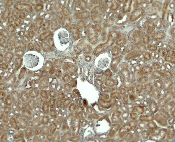
A03485 has real RHOG IHC data from mouse kidney tissue (image caption: mouse kidney). Its listed reactivity covers human, mouse and rat; the pictured IHC result is from mouse tissue (catalog: reactivity; image caption).
A03485 will render with an IHC image of mouse kidney tissue stained at 5 μg/mL (image caption). It lists IHC-P and human, mouse and rat reactivity, while the catalog specifies IHC validation in mouse samples (catalog: applications, reactivity and validation).
Which to pick: Choose A03485 for paraffin-section IHC: IHC-P is listed, and its own image shows mouse kidney staining at 5 μg/mL (catalog: IHC-P; image caption: mouse kidney, 5 μg/mL). No IF/ICC application or image is supplied for A03485, so there is no IF/ICC pick here; the fixative used for the pictured tissue is unreported (catalog: applications and images; image caption). For work across species, A03485 is the only listed choice and is described as reactive with human, mouse and rat, but its stated IHC validation is in mouse samples (catalog: reactivity and validation).