This website uses cookies to ensure you get the best experience on our website.
- Table of Contents
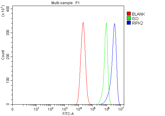
2 Citations 4 Q&As


1 Citations

Facts about Receptor-interacting serine/threonine-protein kinase 2.

Contributes to the tyrosine phosphorylation of the guanine exchange factor ARHGEF2 via Src tyrosine kinase resulting in NF-kappaB activation by NOD2. The polyubiquitinated protein mediates the recruitment of MAP3K7/TAK1 into IKBKG/NEMO and induces'Lys-63'-linked polyubiquitination of IKBKG/NEMO and subsequent activation of IKBKB/IKKB.
| Human | |
|---|---|
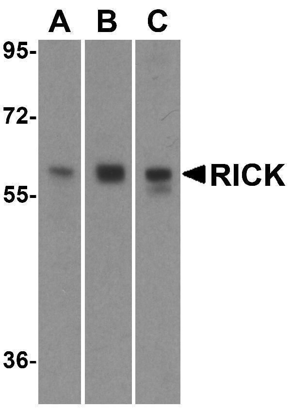
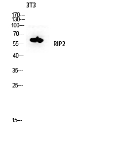
| Gene Name: | RIPK2 |
| Uniprot: | O43353 |
| Entrez: | 8767 |

| Belongs to: |
|---|
| protein kinase superfamily |

CARD3; CARD-containing IL-1 beta ICE-kinase; CARD-containing interleukin-1 beta-converting enzyme (ICE)-associated kinase; CARD-containing interleukin-1 beta-converting enzyme-associated kinase; CARDIAKCCK; EC 2.7.11; EC 2.7.11.1; GIG30; growth-inhibiting gene 30; receptor interacting protein 2; receptor-interacting protein (RIP)-like interacting caspase-like apoptosisregulatory protein (CLARP) kinase; Receptor-interacting protein 2; receptor-interacting serine/threonine-protein kinase 2; receptor-interacting serine-threonine kinase 2; RICK; RICKGIG30; RIP2; RIP-2; RIP2CARD-carrying kinase; RI
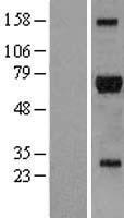
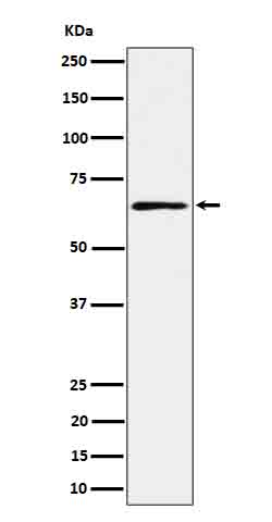
Mass (kDA):
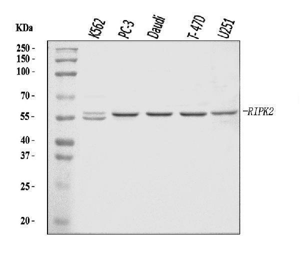
61.195 kDA

| Human | |
|---|---|
| Location: | 8q21.3 |
| Sequence: | 8; NC_000008.11 (89757816..89791064) |
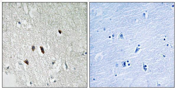
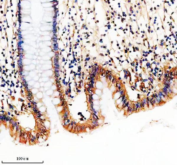
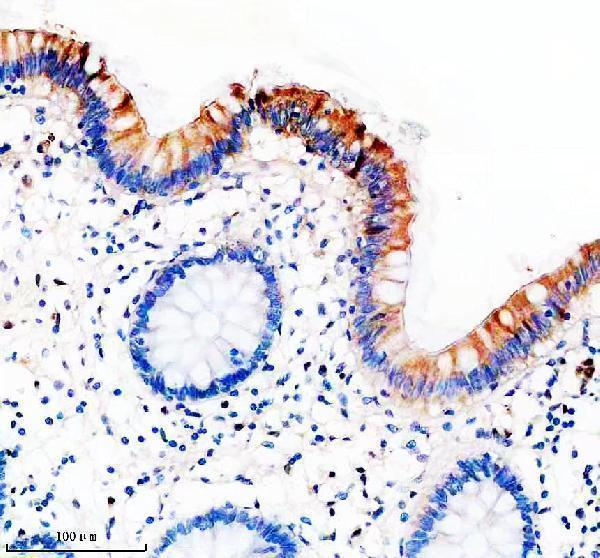
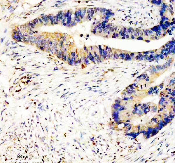
Detected in heart, brain, placenta, lung, peripheral blood leukocytes, spleen, kidney, testis, prostate, pancreas and lymph node.
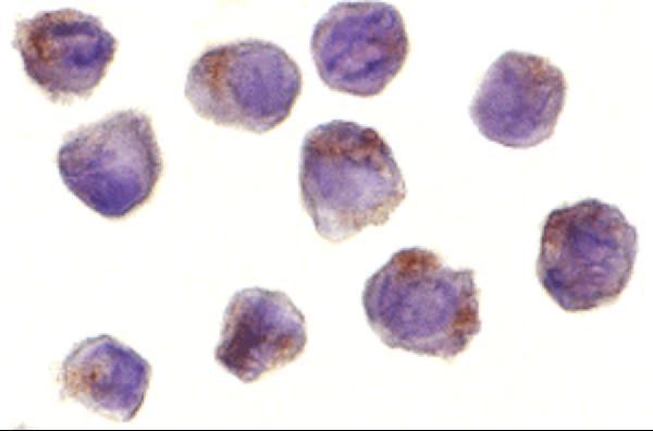
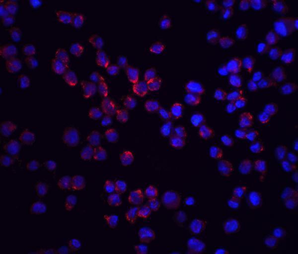
Cytoplasm.


PMID: 9575181 by Inohara N., et al. RICK, a novel protein kinase containing a caspase recruitment domain, interacts with CLARP and regulates CD95-mediated apoptosis.
PMID: 9642260 by McCarthy J.V., et al. RIP2 is a novel NF-kappaB-activating and cell death-inducing kinase.