This website uses cookies to ensure you get the best experience on our website.


- Table of Contents
Plan chromogenic RNF7 IHC in paraffin sections using the IHC-validated antibody M05620 at 1:25 (datasheet M05620). Compare staining in kidney tubule cells, bone marrow hematopoietic cells and splenic red pulp cells, while accounting for the reported low consistency between antibody staining and RNA expression (HPA tissue IHC).

Expected localisation, validated protocols, controls and antibodies — the at-a-glance facts below, then the full design guide.
| Expected localisation | Cytoplasmic staining in several tissues (HPA tissue IHC) | |
| Staining pattern | Cytoplasmic staining across several tissue cell types (HPA tissue IHC) | |
| Antigen retrieval | Citrate pH 6 HIER, heat-mediated (datasheet M05620) | |
| Positive control | Bone marrow+4 more · see all | |
| Negative control | Bronchus+4 more · see all |
| Fixation | Use formaldehyde-fixed paraffin sections (selected-SKU IHC image M05620); keep fixation consistent (standard IHC practice; not target-specific) | |
| Caveat | Antibody staining has low consistency with RNA expression (HPA tissue IHC) | |
| Regulation | Expression varies by tissue (UniProt) | |
| Isoform / epitope | Four isoforms; epitope coverage is unspecified (UniProt; datasheet M05620) |
The catalog antibody’s IHC-P protocol is paired with one published RNF7 protocol for paraffin sections (PMC9107170).
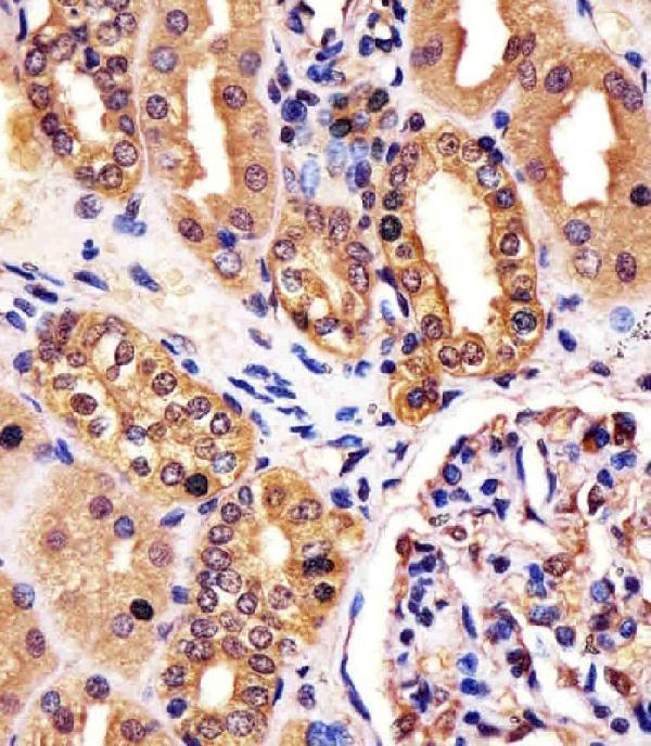
| Sample | Formaldehyde-fixed, paraffin-embedded human kidney tissue (datasheet M05620) |
| Fixation | Image formalin-fixed; duration unreported (datasheet M05620); verify before use. |
| Sectioning | 4–5 µm sections on charged slides (standard) |
| Deparaffinisation | Xylene, graded ethanol series to water (standard) |
| Antigen retrieval | Heat retrieval: Citrate pH 6 (datasheet M05620); 20 min, 95–100 °C (standard) |
| Peroxidase block | 3% H2O2, 10 min, room temperature (standard) |
| Blocking | 10% normal serum of the secondary host, 30 min, room temperature (standard) |
| Primary antibody | Rabbit anti-RNF7, 1:25 (datasheet M05620) |
| Primary incubation | 1 hours at 37°C (datasheet M05620) |
| Detection | HRP-polymer secondary, DAB chromogen 5–10 min (standard) |
| Counterstain | Hematoxylin, blue, dehydrate and mount (standard) |
| Expected result | RNF7-positive staining in hematopoietic cells of bone marrow (HPA tissue IHC: High). HPA tissue profile: Cytoplasmic expression in several tissues. No signal in the no-primary control. |
In paraffin-section IHC, expect RNF7 staining mainly in the cytoplasm of cells within several tissues, particularly bone-marrow hematopoietic cells, kidney tubular cells and splenic red-pulp cells (HPA tissue IHC: High). RNF7 has no transmembrane segment and is annotated in both cytoplasm and nucleus (UniProt Q9UBF6). Treat the tissue pattern as a guide: the HPA IHC antibody is Approved, but its staining has low consistency with RNA expression (HPA tissue IHC).
| Clear cytoplasmic staining in hematopoietic cells, kidney tubular cells or splenic red-pulp cells. | This matches the reported high-staining cell populations and cytoplasmic tissue profile (HPA tissue IHC: High; cytoplasmic expression). Judge the named cells rather than the overall darkness of a mixed tissue section (general IHC practice). |
| Staining appears predominantly at cell borders or outside cells, with little identifiable cytoplasmic signal. | A membrane-only or extracellular pattern does not match the reported tissue-IHC profile (HPA tissue IHC: cytoplasmic expression). Review morphology and detection controls before calling it RNF7-specific (general IHC practice). |
| Strong staining occurs mainly in a cell population reported as not detected, such as lung alveolar cells. | This conflicts with that HPA tissue observation (HPA tissue IHC: alveolar cells Not detected). Consider antibody cross-reactivity or endogenous detection activity, then check cell identity and controls (general IHC practice). |
| Color spreads across the section or obscures cell boundaries and the counterstain. | Diffuse background prevents a reliable compartment or cell-type call (general IHC practice). It is not the discrete cytoplasmic pattern reported across several tissues (HPA tissue IHC: cytoplasmic expression). |
| No signal appears in hematopoietic cells of bone marrow or tubular cells of kidney. | An assay failure is possible because both are reported high-staining populations (HPA tissue IHC: High). Check tissue preservation, staining controls and detection performance before interpreting absence as biological loss (general IHC practice). |
| IHC tissue pattern | The strongest listed staining is in bone-marrow hematopoietic cells, kidney tubular cells and splenic red-pulp cells (HPA tissue IHC: High). Several listed glandular and neuronal populations stain at medium levels (HPA tissue IHC: Medium), so intensity depends on the cells examined. |
| Antibody evidence | The listed antibody, HPA036995, is Approved for IHC; the tissue record also reports low consistency between antibody staining and RNA expression (HPA antibodies: IHC Approved; HPA tissue IHC: reliability description). Treat an unexpected result as requiring controls. |
| Subcellular interpretation | UniProt places RNF7 in cytoplasm and nucleus, while HPA tissue IHC describes cytoplasmic expression (UniProt Q9UBF6: subcellular location; HPA tissue IHC: profile). Nuclear staining alone therefore needs assessment against the IHC tissue pattern; it is not settled by the location annotation. |
| Protein form and epitope | RNF7 has 4 listed isoforms and a chain spanning residues 2–113, with no signal peptide, propeptide or transmembrane segment (UniProt Q9UBF6: isoforms, processing, topology). Without an antibody epitope map, these facts cannot predict isoform coverage or staining strength. |
| Modified residues | UniProt lists N-acetylalanine at residue 2 and CK2-dependent phosphothreonine at residue 10 (UniProt Q9UBF6: modified residues). Their effects on this antibody's IHC signal are unreported; do not infer a retrieval requirement or fixation sensitivity from them. |
| IF/ICC Q: Should the IHC cytoplasmic pattern also be expected in IF/ICC? | A: HPA ICC-IF reports mainly nucleoplasmic localization with additional cytosolic signal (HPA subcellular: supported locations). Interpret IF/ICC using its own localization evidence; the paraffin-section IHC profile describes cytoplasmic staining in tissues (HPA tissue IHC). |
| Situation | Likely cause | Next action |
|---|---|---|
| A high-staining reference population is blank. | The run may have failed, or the selected section may lack the expected cells (general IHC practice); bone-marrow hematopoietic and kidney tubular cells are reported High (HPA tissue IHC). | Confirm cell identity and section quality, then review the run's positive control, primary-antibody step and detection reagents (general IHC practice). Do not score biological absence until the run works. |
| A reported negative cell population stains strongly. | Cross-reactivity or endogenous detection activity may contribute (general IHC practice); lung alveolar cells and heart cardiomyocytes are reported Not detected (HPA tissue IHC). | Check a no-primary control and the cells actually stained; assess endogenous enzyme blocking for chromogenic detection (general IHC practice). Compare the result with HPA at the cell-type level. |
| The whole section has diffuse brown background. | Excess detection signal or inadequate blocking can obscure cell-specific staining (general IHC practice). Diffuse signal cannot establish the cytoplasmic profile reported for RNF7 (HPA tissue IHC). | Review no-primary controls, blocking, reagent concentration, washes and chromogen development time; adjust one general IHC workflow variable at a time (general IHC practice). |
| Signal is confined to cell borders or extracellular material. | This location conflicts with the reported cytoplasmic tissue pattern and RNF7's lack of a transmembrane segment (HPA tissue IHC: profile; UniProt Q9UBF6: topology). | Recheck morphology and background controls, then seek independent antibody evidence before assigning RNF7 specificity (general IHC practice). Do not score border-only color as cytoplasmic positivity. |
| Nuclear staining dominates a paraffin-section IHC result. | RNF7 has a nuclear annotation and nucleoplasmic ICC-IF signal (UniProt Q9UBF6; HPA subcellular), but the supplied tissue-IHC profile describes cytoplasmic expression (HPA tissue IHC). | Record nuclear and cytoplasmic scores separately, check controls, and compare like-for-like IHC tissue images before deciding whether the pattern is credible (general IHC practice). |
| Cardiomyocytes stain despite the HPA IHC negative listing. | UniProt reports RNF7 expression in heart, while HPA reports cardiomyocytes Not detected by tissue IHC (UniProt Q9UBF6: tissue specificity; HPA tissue IHC: heart muscle). These observations use different evidence types. | Confirm the stained cells and run controls, then report the discrepancy as assay-specific evidence rather than treating either source as a guaranteed IHC result (general IHC practice). |


Comprehensive Human Protein Atlas IHC scoring per tissue (reliability: Approved — Low consistency between antibody staining and RNA expression data.). Rows are taken directly from the HPA tissue chart — click any row's HPA link to view the source.
| Tissue | Cell type | Level | Evidence | Source |
|---|---|---|---|---|
| Bone marrow | Hematopoietic cells | High | Protein (IHC) | HPA → |
| Kidney | Cells in tubules | High | Protein (IHC) | HPA → |
| Spleen | Cells in red pulp | High | Protein (IHC) | HPA → |
| Adrenal gland | Glandular cells | Medium | Protein (IHC) | HPA → |
| Appendix | Glandular cells | Medium | Protein (IHC) | HPA → |
| Tissue | Cell type | Level | Evidence | Source |
|---|---|---|---|---|
| Bronchus | Respiratory epithelial cells | Not detected | Protein (IHC) | HPA → |
| Esophagus | Squamous epithelial cells | Not detected | Protein (IHC) | HPA → |
| Heart muscle | Cardiomyocytes | Not detected | Protein (IHC) | HPA → |
| Hippocampus | Glial cells | Not detected | Protein (IHC) | HPA → |
| Lung | Alveolar cells | Not detected | Protein (IHC) | HPA → |
Use matched controls and cell-level scoring to troubleshoot RNF7 staining in paraffin sections; interpret patterns alongside documented tissue and subcellular evidence.
RNF7 IHC has a human kidney paraffin-section image (M05620 image caption); human and mouse reactivity is listed (M05620 catalog). IF/ICC is listed for a second antibody without an image (M05620-1 catalog).
M05620 is listed for IHC-P in human and mouse, with an IHC image from formaldehyde-fixed, paraffin-embedded human kidney sections (M05620 catalog; M05620 image caption). M05620-1 is listed for IHC and IF/ICC in human, mouse and rat, but has no IHC or IF image in the payload (M05620-1 catalog).
Which to pick: For paraffin-section tissue IHC, choose M05620 because its own image shows staining in formaldehyde-fixed human kidney sections (M05620 image caption); it is a rabbit polyclonal antibody (M05620 catalog). For IF/ICC, consider the rabbit monoclonal M05620-1, which lists those applications but provides no IF image; its IHC fixative is unreported (M05620-1 catalog). For rat samples, M05620-1 is the listed option; both SKUs list human and mouse reactivity (M05620-1 catalog; M05620 catalog).