RPL23A / Large ribosomal subunit protein uL23 · IHC design guide

Design Immunohistochemistry for RPL23A

Plan RPL23A paraffin IHC using the cytoplasmic tissue pattern and high staining in ciliated-cell rootlets of the fallopian tube and nasopharynx (HPA tissue IHC). Interpret intensity cautiously because antibody staining has low consistency with RNA expression (HPA tissue IHC).

Evidence assembled Oct 2026 · For research use; verify linked source records and product datasheet before use
Immunohistochemistry protocol sheet for RPL23A (IHC for RPL23A): expected localisation Cytoplasmic tissue staining (HPA tissue IHC); nuclear location annotated (UniProt), antibody A06803, validated IHC image, and IHC protocol steps
Printable RPL23A IHC protocol sheet — expected localisation Cytoplasmic tissue staining (HPA tissue IHC); nuclear location annotated (UniProt), antibody A06803, controls and protocol steps. Open the full RPL23A IHC guide →

RPL23A Immunohistochemistry Experimental Design Guide

Expected localisation, validated protocols, controls and antibodies — the at-a-glance facts below, then the full design guide.

Must know before staining
Expected localisation Cytoplasmic tissue staining (HPA tissue IHC); nuclear location annotated (UniProt)
Staining pattern Cytoplasmic in most tissues; high in ciliated-cell rootlets (HPA tissue IHC)
Antigen retrieval Citrate pH 6.0 HIER, heat-mediated (datasheet A06803)
Positive control ⓘ Fallopian tube+4 more · see all
Negative control ⓘ Adipose tissue+4 more · see all
Important caveats
Reasons your staining may differ from the expected pattern.
Fixation Keep fixation consistent across sections. (standard IHC practice; not target-specific)
Caveat Antibody staining has low consistency with RNA expression (HPA tissue IHC)
Regulation No staining regulator annotated (UniProt)
Isoform / epitope No isoforms annotated; mature chain spans residues 2–156 (UniProt)
Section 1

Recommended RPL23A IHC & IF Protocols

The catalog antibody protocol uses citrate pH 6.0 retrieval (datasheet A06803). The published option below reports RPL23A IHC staining in tissue sections studied for NEC-related intestinal injury (PMC13578613).

Recommended immunohistochemistry (IHC-P) protocol parameters
SampleParaffin-embedded rat brain tissue; fixative not specified (datasheet A06803)
FixationImage fixative and duration unreported (datasheet A06803); verify before use.
Sectioning4–5 µm sections on charged slides (standard)
DeparaffinisationXylene, graded ethanol series to water (standard)
Antigen retrievalHeat retrieval: Citrate pH 6.0 (datasheet A06803); 20 min, 95–100 °C (standard)
Peroxidase block3% H2O2, 10 min, room temperature (standard)
Blocking10% normal serum of the secondary host, 30 min, room temperature (standard)
Primary antibodyRabbit anti-RPL23A, 1:50-1:200 (datasheet A06803)
Primary incubationOvernight at 4 °C (standard)
DetectionHRP-polymer secondary, DAB chromogen 5–10 min (standard)
CounterstainHematoxylin, blue, dehydrate and mount (standard)
Expected resultRPL23A-positive staining in ciliated cells (ciliary rootlets) of fallopian tube (HPA tissue IHC: High). HPA tissue profile: Cytoplasmic expression in most tissues. No signal in the no-primary control.
💡Decision noteStart with heat-mediated citrate pH 6.0 retrieval (datasheet A06803); the published excerpt does not specify retrieval conditions (PMC13578613 methods).
Section 2

What Is the Expected RPL23A Staining Pattern?

RPL23A is a cytoplasmic and nuclear ribosomal protein with no transmembrane segment (UniProt P62750: location and topology). In tissue IHC, expect cytoplasmic staining in many cell types, with high staining at ciliary rootlets in fallopian tube and nasopharynx ciliated cells (HPA: tissue IHC). HPA rates the tissue pattern Approved but reports low consistency between antibody staining and RNA expression; interpret unexpected patterns cautiously (HPA: tissue IHC reliability).

What am I looking at on my slide?
Cytoplasmic staining across a tissue, with strong ciliary-rootlet staining in ciliated cells.This fits the reported broad cytoplasmic pattern and high signal in fallopian tube or nasopharynx ciliated cells (HPA: tissue IHC). Compare identifiable cells and compartments rather than assigning one intensity to every cell.
Signal is confined to cell borders, luminal material or extracellular space.That distribution conflicts with the reported intracellular locations and absence of a transmembrane segment (UniProt P62750: location and topology). Check morphology, detection deposits and controls before calling it RPL23A; nuclear signal alone is not disqualifying (UniProt P62750: nucleus).
Strong staining appears in an unexpected cell population while expected cells remain unstained.Compare cell identity with HPA's cell-specific observations: adipocytes and bone marrow hematopoietic cells were not detected (HPA: tissue IHC). Antibody cross-reactivity or endogenous detection activity is possible; verify with appropriate controls (general IHC practice).
Diffuse color covers tissue and spaces between cells, obscuring cell boundaries.This is difficult to score as intracellular RPL23A (UniProt P62750: location). Nonspecific binding or chromogen background can produce such a pattern; review blocking, washing and a control without primary antibody (general IHC practice).
No signal appears in ciliated cells of fallopian tube or nasopharynx.Those ciliary rootlets are reported High and provide useful positive reference cells (HPA: tissue IHC). First confirm that ciliated cells are present, then review antibody application and detection controls; one negative section does not establish protein absence (general IHC practice).
💡Expected RPL23A appearanceCall a section positive when identifiable cells show intracellular, mainly cytoplasmic staining, especially strong ciliary-rootlet signal in fallopian tube or nasopharynx ciliated cells (HPA: tissue IHC); isolated membrane or extracellular color is suspect (UniProt P62750: location and topology).
How each factor affects the staining
Cell-specific reference patternsHigh ciliary-rootlet staining is reported in fallopian tube and nasopharynx, whereas adrenal glandular cells and colon endothelial cells are Medium (HPA: tissue IHC). An unstained adipocyte is a cell-specific observation, not evidence that every cell in adipose tissue is negative (HPA: tissue IHC).
Intracellular locationRPL23A functions in cytoplasmic ribosomes and enters the nucleus during ribosomal-subunit assembly (UniProt P62750: function and location). Judge an IHC pattern against cell morphology: cytoplasmic signal is broadly expected, and nuclear signal may be biologically plausible (HPA: tissue IHC; UniProt P62750: nucleus).
IHC evidence and antibody choiceHPA lists HPA051754 as Approved for IHC and HPA077474 as Approved for ICC, with no IHC status listed for HPA077474 (HPA: antibody validation). The tissue profile is Approved with low staining–RNA consistency, so use morphology and controls when interpreting discrepancies (HPA: tissue IHC reliability).
IF/ICC Q: where should fluorescence appear?HPA reports approved localization to nucleoli fibrillar centers, endoplasmic reticulum and cytosol in ICC-IF (HPA: subcellular). That cellular map can inform interpretation, but it does not prescribe an IF protocol or imply that every compartment will resolve in chromogenic tissue IHC.
Why is my staining missing, weak or wrong?
SituationLikely causeNext action
A section containing identifiable ciliated cells has no ciliary-rootlet signal.High signal is reported at these rootlets in fallopian tube and nasopharynx (HPA: tissue IHC); a failed staining or detection step is possible (general IHC practice).Confirm the rootlets are present, then check the IHC-validated antibody's stated application and the run's positive and detection controls (general IHC practice).
Color is diffuse across tissue and empty spaces.This does not match intracellular RPL23A localization (UniProt P62750: location); nonspecific binding or detection background may obscure the pattern (general IHC practice).Compare a control without primary antibody, inspect washing and blocking, and score only localized cellular signal that remains distinguishable from background (general IHC practice).
Staining outlines membranes or fills lumens without intracellular signal.RPL23A has no transmembrane segment and is reported in cytoplasm and nucleus (UniProt P62750: topology and location).Inspect for deposited chromogen or tissue artifacts and repeat with appropriate detection controls before attributing the outline to RPL23A (general IHC practice).
Unexpected cells stain strongly while reported positive cells do not.Cell identity may have been misread, or cross-reactivity or endogenous detection activity may contribute (general IHC practice); HPA reports cell-specific, uneven staining (HPA: tissue IHC).Recheck morphology against a reported positive cell population and use a control without primary antibody to assess detection-related color (HPA: tissue IHC; general IHC practice).
A low-signal tissue looks negative.HPA reports Low staining in hepatocytes, lung alveolar cells and several other populations (HPA: tissue IHC); faint signal can be hard to distinguish from background (general IHC practice).Evaluate the section alongside a reported High ciliary-rootlet reference and matched background controls before interpreting the low-signal population (HPA: tissue IHC; general IHC practice).
Nuclear staining dominates with little cytoplasmic staining.Nuclear residence is biologically plausible (UniProt P62750: location), although HPA describes cytoplasmic expression in most tissues (HPA: tissue IHC).Check whether signal follows recognizable nuclei, compare cytoplasmic reference cells, and review background controls before accepting the distribution (HPA: tissue IHC; general IHC practice).

Sample controls for RPL23A IHC & IF

🧪Run fallopian tube first: ciliary rootlets in ciliated cells should stain (HPA: High). Use adipose tissue as a negative comparator, focusing on adipocytes (HPA: Not detected); on the fallopian tube slide, adjacent cells should lack the distinct ciliary-rootlet pattern, though their RPL23A status is not established by these rows (HPA: High in ciliated-cell ciliary rootlets).
Positive control tissue: Fallopian tube (Ciliated cells (ciliary rootlets), HPA High)
Negative control tissue: Adipose tissue (HPA Not detected)
ICC-IF cell lines (HPA subcellular resource): HPA ICC-IF images show RPL23A in A-549, RT-4, U2OS, with annotated localisation: Nucleoli fibrillar center (approved), Endoplasmic reticulum (approved), Cytosol (approved) (HPA subcellular).
Technical controls: Include no-primary (secondary-only) and matched rabbit polyclonal isotype controls (caption: rabbit pAb), plus RPL23A knockout material or cognate-peptide competition if available. Quench endogenous peroxidase for chromogenic IHC and check background around the ciliary rootlets (HPA: High in fallopian-tube ciliated-cell ciliary rootlets).
⚠️Feasibility: No target-specific fixation window or fixation effect is reported in the supplied evidence; the selected A06803 paraffin-section caption does not state a fixative (caption: fixative unreported). The caption uses high-pressure retrieval in 10 mM citrate, pH 6.0, before IHC, but does not establish that retrieval is required; the evidence also does not establish whether frozen sections or IF are easier (caption: paraffin-section IHC; HPA: ICC-IF images). Ciliary rootlets are small, so section plane and diffuse chromogen can make their pattern difficult to judge (HPA: High in ciliated-cell ciliary rootlets).

HPA tissue IHC evidence for RPL23A

Comprehensive Human Protein Atlas IHC scoring per tissue (reliability: Approved — Low consistency between antibody staining and RNA expression data.). Rows are taken directly from the HPA tissue chart — click any row's HPA link to view the source.

Positive expression · recommended positive controls

TissueCell typeLevelEvidenceSource
Fallopian tube Ciliated cells (ciliary rootlets) High Protein (IHC) HPA →
Nasopharynx Ciliated cells (ciliary rootlets) High Protein (IHC) HPA →
Adrenal gland Glandular cells Medium Protein (IHC) HPA →
Appendix Glandular cells Medium Protein (IHC) HPA →
Colon Endothelial cells Medium Protein (IHC) HPA →

Undetected expression · recommended negative controls

TissueCell typeLevelEvidenceSource
Adipose tissue Adipocytes Not detected Protein (IHC) HPA →
Bone marrow Hematopoietic cells Not detected Protein (IHC) HPA →
Bronchus Basal cells Not detected Protein (IHC) HPA →
Cerebellum Cells in granular layer Not detected Protein (IHC) HPA →
Cerebral cortex Endothelial cells Not detected Protein (IHC) HPA →
Section 3

Advanced RPL23A IHC Tips

Troubleshoot RPL23A chromogenic IHC in paraffin sections by checking retrieval, cell type, compartment and controls before comparing staining intensity (UniProt P62750; standard IHC practice).

How should I adjust retrieval when RPL23A staining is weak in paraffin sections?
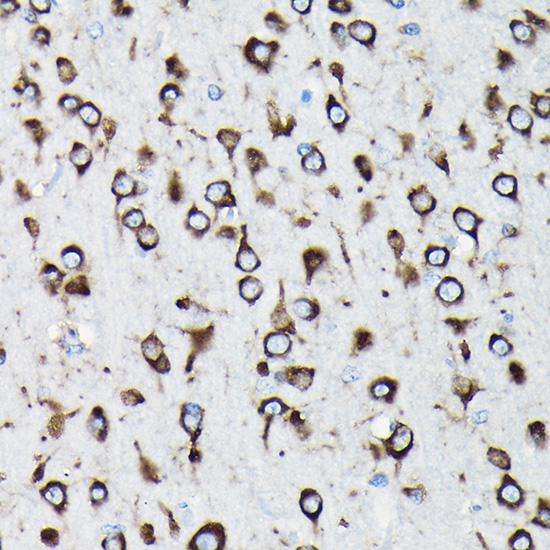
Begin with heat-mediated citrate antigen retrieval at pH 6.0 for paraffin sections (datasheet A06803). The selected rat brain image used high-pressure retrieval in 10 mM citrate at pH 6.0 and primary antibody at 1:100; its caption does not state the fixative (caption A06803). If staining remains weak, compare heating for 10 and 20 min on adjacent sections while keeping detection conditions constant (standard IHC optimization). Evaluate morphology and background alongside signal, and check whether staining remains predominantly cytoplasmic, as expected in most tissues (standard IHC practice; HPA tissue IHC profile).
Could fixation explain inconsistent RPL23A staining between paraffin blocks?
The selected paraffin-section caption does not identify its fixative, and target-specific fixation sensitivity for RPL23A is unknown (caption A06803). For new specimens, record fixative, time to fixation and fixation duration; compare matched samples fixed for 6 h and 24 h only as a controlled optimization (standard IHC practice). For existing blocks, stain adjacent sections together with the same retrieval and 1:100 starting dilution used in the selected image (caption A06803; standard IHC practice). Judge any difference against morphology and staining controls before attributing it to fixation, since processing and detection also affect chromogenic signal (standard IHC practice).
Should an RPL23A IHC stain appear cytoplasmic, nuclear or both?
Expect predominantly cytoplasmic staining across most tissues in chromogenic IHC (HPA tissue IHC profile). Nuclear staining can also be plausible because RPL23A enters the nucleus for ribosomal-subunit assembly, although a nuclear chromogen deposit alone cannot establish that process (UniProt P62750 localisation; standard IHC interpretation). Higher-resolution IF places RPL23A in the nucleoli fibrillar center, endoplasmic reticulum and cytosol (HPA subcellular, approved locations). In IHC, use a hematoxylin counterstain and inspect individual cell boundaries before scoring nuclear or cytoplasmic signal; compare the same compartment across sections stained in one run (standard IHC practice).
How do isoforms or modified residues affect interpretation of lost RPL23A staining?
UniProt lists 0 RPL23A isoforms and a mature chain spanning residues 2–156, so an isoform switch is unsupported as an explanation for a changed IHC pattern (UniProt P62750 processing and isoforms). Several modified residues are recorded, including residue 2 methylation and modifications at residues 41, 43, 45 and 70 (UniProt P62750 modified residues). Their effect on this antibody is unknown because the supplied evidence does not map its epitope (supplied antibody evidence). Compare retrieval conditions and, if available, an independently validated antibody with a distinct epitope before assigning epitope masking or modification as the cause (standard IHC validation practice).
How can IF help verify an ambiguous RPL23A IHC pattern?
Use IF/ICC as a separate check of the IHC pattern, since HPA reports RPL23A in the nucleoli fibrillar center, endoplasmic reticulum and cytosol by subcellular imaging (HPA subcellular). For tissue multiplexing, pair RPL23A with a cilia marker such as acetylated tubulin when examining ciliated cells, whose rootlets show high IHC staining (HPA tissue IHC; standard IF marker practice). Select a far-red fluorophore when shorter wavelengths show tissue autofluorescence, and include single-color controls (standard IF practice). Because RPL23A has no transmembrane segment, pilot mild permeabilization, such as 0.1% Triton X-100 for 5 min, to access intracellular epitopes without assuming an antibody epitope position (UniProt P62750 topology; standard IF optimization).
What should I check when RPL23A DAB staining is diffuse or widespread?
First compare a no-primary control with the stained section to identify signal from secondary reagents or the chromogenic workflow (standard IHC practice). Include an endogenous peroxidase block, for example 3% hydrogen peroxide for 10 min, before peroxidase-based DAB detection (standard IHC practice). If the control is clean but staining remains diffuse, titrate the primary around the caption's 1:100 starting point and assess blocking, washes and retrieval intensity one variable at a time (caption A06803; standard IHC optimization). Broad cytoplasmic staining is biologically plausible for RPL23A, so judge background against cell boundaries and tissue morphology rather than distribution alone (HPA tissue IHC profile; standard IHC interpretation).
How should I score RPL23A chromogenic staining across tissue samples? ⚠ ANSWER MARKED FOR VERIFICATION
Define the cell population and cytoplasmic compartment before scoring, because HPA reports cytoplasmic expression in most tissues and different levels across listed cell types (HPA tissue IHC). An H-score can combine the percentage of viable cells at intensity grades 0–3 into a 0–300 score; report percent positive cells separately (standard IHC scoring practice). For spatial comparisons, report positive-cell density per mm² of viable annotated tissue and normalize intensity comparisons to cell number or measured compartment area (standard image-analysis practice). Keep exposure, chromogen development and thresholds consistent, and interpret RNA concordance cautiously because HPA rates staining-to-RNA consistency as low (standard IHC practice; HPA reliability description).
How can I distinguish convincing RPL23A signal from staining artefact?
A convincing IHC pattern has cellular boundaries and fits the predominantly cytoplasmic tissue profile; strong ciliary-rootlet staining in ciliated cells is another supplied reference pattern (HPA tissue IHC). Nuclear signal is possible given RPL23A nuclear import, but isolated extracellular or edge-heavy deposits do not follow its recorded cellular localisation (UniProt P62750 localisation; standard IHC artefact assessment). Exclude torn edges and necrotic regions from scoring, and compare suspect deposits with a no-primary control and the endogenous peroxidase block (standard IHC practice). Treat a missing signal in one cell population cautiously, because HPA lists some cell types as undetected and reports low consistency between staining and RNA data (HPA tissue IHC; HPA reliability description).
Boster reagents

Best RPL23A / Large ribosomal subunit protein uL23 IHC Antibodies

Two anti-RPL23A antibodies have IHC images of paraffin sections from rat brain and human tissues (catalog IHC captions). Both list human, mouse and rat reactivity; one also lists IF/ICC (catalog applications).

Real IHC data Immunohistochemistry of paraffin-embedded rat brain using RPL23A Rabbit pAb at dilution of 1:100 .Perform high pressure antigen retrieval with 10 mM citrate buffer pH 6.0 before commencing with IHC staining protocol.
Anti-60S ribosomal protein L23a RPL23A Antibody
Cat # A06803
Real IHC data Immunohistochemical analysis of RPL23A Antibody (C-term) on paraffin-embedded Human breast carcinoma tissue. Tissue was fixed with formaldehyde at room temperature. Heat induced epitope retrieval was performed by EDTA buffer (pH9. 0). Samples were incubated with primary antibody(1:100) for 1 hour at room temperature. Undiluted CRF Anti-Polyvalent HRP Polymer antibody was used as the secondary antibody.
Anti-RPL23A Antibody (C-term)
Cat # A06803-1

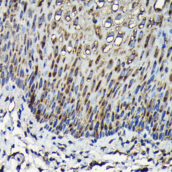
A06803 has IHC images from paraffin-embedded rat brain, human esophagus and human placenta; IF/ICC is listed, but no IF image is supplied (catalog captions and applications). A06803-1 has IHC images from paraffin-embedded human breast carcinoma and kidney (catalog captions).

Which to pick: For tissue IHC, choose A06803 for its 1:50–1:200 range and citrate pH 6.0 retrieval shown on paraffin-embedded rat brain; its fixative is unreported (catalog dilution and rat brain caption). Choose A06803-1 when the documented paraffin-section, paraffin-embedded human breast carcinoma workflow with EDTA pH 9.0 retrieval and 1:100 primary dilution fits your sample (catalog breast carcinoma caption). For IF/ICC, choose A06803 because it lists those applications and 1:50–1:100; both SKUs list human, mouse and rat reactivity, but the supplied IHC images show rat and human samples only (catalog applications, dilutions, reactivity and captions). The selected A06803 tissue-IHC caption documents paraffin sections, but does not specify the fixative (selected-SKU IHC image A06803).

Each figure is that product's own IHC / IF validation image from its datasheet.

References

  1. UniProt Consortium. UniProt entry P62750 (RL23A_HUMAN, Large ribosomal subunit protein uL23).
  2. Human Protein Atlas. RPL23A tissue IHC expression (reliability: Approved).
  3. Human Protein Atlas. RPL23A subcellular location (ICC-IF): Localized to the nucleoli fibrillar center, endoplasmic reticulum and cytosol..
  4. Human Protein Atlas. RPL23A antibody validation summary (2 antibodies).
  5. HERC3 directly targets RPL23A for ubiquitination degradation and further regulates Colorectal Cancer proliferation and the cell cycle. International journal of biological sciences 2022 — PMC9134906.
  6. FBXO5 regulates RPL23A to promote MDM2-mediated p53 degradation and facilitate malignant progression of breast cancer. Breast cancer research : BCR 2026 — PMC13555960.
  7. Multiomics analyses revealed the roles of the epithelial-mesenchymal transition and novel early candidate diagnostic biomarkers in neonatal necrotising enterocolitis. Clinical and translational medicine 2026 — PMC13578613.
  8. PubMed PMID:8662070 — UniProt-cited evidence.
  9. PubMed PMID:9417910 — UniProt-cited evidence.
  10. PubMed PMID:14702039 — UniProt-cited evidence.