RPL4 / Large ribosomal subunit protein uL4 · IHC design guide

Design Immunohistochemistry for RPL4

Plan paraffin section IHC for RPL4 using the reported cytoplasmic tissue pattern (HPA tissue IHC). Compare staining with appropriate controls, since antibody staining has low concordance with RNA expression (HPA tissue IHC).

Evidence assembled Oct 2026 · For research use; verify linked source records and product datasheet before use
Immunohistochemistry protocol sheet for RPL4 (IHC for RPL4): expected localisation Cytoplasm in most tissues (HPA tissue IHC), antibody A05175-1, validated IHC image, and IHC protocol steps
Printable RPL4 IHC protocol sheet — expected localisation Cytoplasm in most tissues (HPA tissue IHC), antibody A05175-1, controls and protocol steps. Open the full RPL4 IHC guide →

RPL4 Immunohistochemistry Experimental Design Guide

Expected localisation, validated protocols, controls and antibodies — the at-a-glance facts below, then the full design guide.

Must know before staining
Expected localisation Cytoplasm in most tissues (HPA tissue IHC)
Staining pattern Cytoplasmic staining across most tissues (HPA tissue IHC)
Antigen retrieval EDTA pH 8.0 HIER, heat-mediated (datasheet A05175-1)
Positive control ⓘ Adrenal gland+4 more · see all
Negative control ⓘ Adipose tissue+4 more · see all
Important caveats
Reasons your staining may differ from the expected pattern.
Fixation Keep fixation consistent across sections. (standard IHC practice; not target-specific)
Caveat Staining reliability is uncertain owing to low RNA concordance (HPA tissue IHC)
Regulation No expression regulation annotated (UniProt)
Isoform / epitope No isoforms annotated; mature chain spans residues 2–427 (UniProt)
Section 1

Recommended RPL4 IHC & IF Protocols

The catalog antibody’s IHC-P protocol (datasheet: A05175-1) is accompanied by published protocols for colorectal cancer tissue (PMC13474844) and rat lung tissue (PMC9761115).

Recommended immunohistochemistry (IHC-P) protocol parameters
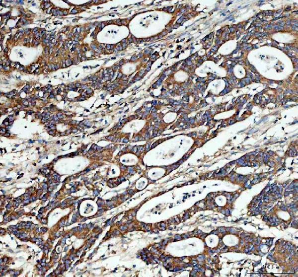
SampleParaffin-embedded human colon cancer tissue; fixative not specified (datasheet A05175-1)
FixationImage fixative and duration unreported (datasheet A05175-1); verify before use.
Sectioning4–5 µm sections on charged slides (standard)
DeparaffinisationXylene, graded ethanol series to water (standard)
Antigen retrievalHeat retrieval: EDTA pH 8.0 (datasheet A05175-1); 20 min, 95–100 °C (standard)
Peroxidase block3% H2O2, 10 min, room temperature (standard)
Blocking10% goat serum (datasheet A05175-1)
Primary antibodyRabbit anti-RPL4, 1:50 recommended; image 1:100 (datasheet A05175-1)
Primary incubationOvernight at 4 °C (datasheet A05175-1)
DetectionHRP-conjugated secondary, DAB chromogen (datasheet A05175-1)
CounterstainHematoxylin, blue, dehydrate and mount (standard)
Expected resultRPL4-positive staining in glandular cells of adrenal gland (HPA tissue IHC: Medium). HPA tissue profile: Cytoplasmic expression in most tissues. No signal in the no-primary control.
💡Decision noteStart with heat-mediated EDTA at pH 8.0 for the catalog antibody (datasheet: A05175-1); the rat lung protocol used 0.01 M sodium citrate (PMC9761115).
Section 2

What Is the Expected RPL4 Staining Pattern?

RPL4 is a cytoplasmic ribosomal protein with no transmembrane segment (UniProt P36578: function, location, topology). In paraffin sections, expect cytoplasmic staining in many cell types, including colon glandular cells, bronchial respiratory epithelial cells and cerebral cortex neurons (HPA tissue IHC: Medium). Treat the pattern as provisional: HPA rates the tissue IHC profile Uncertain because antibody staining and RNA expression show low consistency (HPA tissue IHC: reliability).

What am I looking at on my slide?
Cytoplasmic staining in colon glandular cells, bronchial respiratory epithelial cells or cerebral cortex neurons.This matches the reported compartment and examples of Medium tissue staining (HPA tissue IHC: profile and levels; UniProt P36578: cytoplasm). Assess the named cells within each section; a positive cell population does not imply that every cell in that tissue must stain (HPA tissue IHC: cell-specific entries).
Staining is confined to nuclei, cell borders or an apparent membrane rim, with little cytoplasmic signal.A dominant noncytoplasmic IHC pattern conflicts with the reported tissue profile and cytoplasmic location; check specificity and staining controls (HPA tissue IHC: cytoplasmic profile; UniProt P36578: cytoplasm, no transmembrane segment). Focal nucleolar signal alone is less decisive because that additional location is Uncertain in ICC-IF (HPA subcellular).
A purported positive result is strongest in adipocytes, cardiomyocytes or endometrial stromal cells.These named cell populations are reported Not detected by tissue IHC (HPA tissue IHC: negative entries). Check cell identification, primary antibody controls and detection controls for possible cross-reactivity or endogenous activity (general IHC practice). HPA's Uncertain tissue reliability makes this a flag for verification, not proof of an artefact (HPA tissue IHC: reliability).
Color spreads evenly across tissue, empty spaces or unrelated cell populations.Diffuse staining that obscures cellular boundaries is difficult to score as cytoplasmic RPL4 (HPA tissue IHC: cytoplasmic profile). Review blocking, wash steps, chromogen development and detection controls for nonspecific or endogenous signal (general IHC practice); do not interpret a uniform haze as broad expression.
A section expected to contain colon glandular cells shows no detectable staining.HPA reports Medium staining in that cell population, so first confirm the cells are present and compare a documented positive section processed in the same run (HPA tissue IHC: colon glandular cells; general IHC practice). An absent result may reflect an assay failure, but the Uncertain HPA rating prevents treating one negative slide as biological absence (HPA tissue IHC: reliability).
💡Expected RPL4 appearanceCall a section positive when identifiable colon glandular cells, bronchial respiratory epithelial cells or cerebral cortex neurons show predominantly cytoplasmic, approximately Medium staining (HPA tissue IHC: positive entries; UniProt P36578: cytoplasm); isolated nuclear, membrane-rim or uniform background color requires verification (HPA tissue IHC: cytoplasmic profile; general IHC practice).
How each factor affects the staining
Reported tissue pattern and confidenceHPA describes cytoplasmic expression in most tissues but rates tissue IHC Uncertain because staining and RNA data have low consistency (HPA tissue IHC: profile, reliability). Use the named cell populations as comparison points, not as an absolute expression map.
Cell population within a tissueHPA reports Medium staining in colon glandular cells but Not detected in adipocytes and cardiomyocytes (HPA tissue IHC: positive and negative entries). Score the specified cells rather than assigning one intensity to an entire organ section.
Antibody validationThe listed antibody HPA034600 is rated Uncertain for IHC and Supported for ICC (HPA antibodies: validation status). The ICC rating does not upgrade confidence in paraffin-section IHC; corroborate unexpected IHC patterns with appropriate controls (general IHC practice).
Compartment and topologyUniProt places RPL4 in the cytoplasm and lists no transmembrane segment (UniProt P36578: location, topology). HPA ICC-IF supports endoplasmic reticulum and cytosol localization; nucleoli and nuclear bodies are additional Uncertain locations (HPA subcellular).
Target-specific fixation sensitivityNo target-specific fixation effect is supplied by UniProt or HPA here (source scope). Optimize section preparation using routine IHC controls if needed (general IHC practice); do not infer an RPL4 retrieval requirement or predict a changed tissue result from these records.
Why is my staining missing, weak or wrong?
SituationLikely causeNext action
No signal in colon glandular cells.The expected Medium cell-population result is missing, though the tissue IHC record is Uncertain (HPA tissue IHC: colon, reliability).Confirm glandular cells are present; check a documented positive section, primary antibody inclusion and detection reagents from the same run (general IHC practice).
Strong staining in adipocytes or cardiomyocytes.Those cells are reported Not detected, so cell misidentification or assay background is possible (HPA tissue IHC: negative entries; general IHC practice).Recheck morphology and compare primary-omission and detection controls before calling expression; treat disagreement as unresolved if controls are clean (general IHC practice; HPA tissue IHC: Uncertain).
Mostly nuclear or membrane-rim staining.This differs from the cytoplasmic tissue pattern and lacks support from the no-transmembrane topology (HPA tissue IHC: profile; UniProt P36578: topology).Check cellular boundaries and control sections; review antibody specificity before scoring. Interpret small nucleolar foci cautiously because their ICC-IF assignment is Uncertain (general IHC practice; HPA subcellular).
Uniform brown haze obscures cells.Background or overdeveloped chromogen can prevent compartment scoring (general IHC practice).Compare a primary-omission control, inspect blocking and washes, and adjust detection development within the assay workflow (general IHC practice).
Staining varies sharply between adjacent sections.Cell composition, section handling or detection variation can change the apparent pattern (general IHC practice).Compare the same named cell population in matched sections and include a positive control in each run; avoid whole-tissue intensity comparisons (general IHC practice; HPA tissue IHC: cell-specific entries).
ICC-IF appears positive but paraffin IHC does not.HPA rates the listed antibody Supported for ICC and Uncertain for IHC, so the applications have different validation evidence (HPA antibodies: validation status).Assess each assay with its own controls and report the IHC result as unresolved; do not transfer the ICC confidence rating to paraffin IHC (general IHC practice; HPA antibodies: validation status).

Sample controls for RPL4 IHC & IF

🧪Run adrenal gland first: its glandular cells should show medium RPL4 staining (HPA: adrenal gland, glandular cells Medium). Run adipose tissue as the negative comparator because adipocytes are listed as not detected (HPA: adipose tissue, adipocytes Not detected); non-glandular cells on the adrenal slide can provide a background reference, but their negative status is unreported (HPA: adrenal gland row specifies glandular cells only).
Positive control tissue: Adrenal gland (Glandular cells, HPA Medium)
Negative control tissue: Adipose tissue (HPA Not detected)
ICC-IF cell lines (HPA subcellular resource): HPA ICC-IF images show RPL4 in A-431, U-251MG, U2OS, ASC52telo, with annotated localisation: Endoplasmic reticulum (approved), Cytosol (supported) (HPA subcellular).
Technical controls: Include no-primary (secondary-only) and concentration-matched rabbit IgG isotype controls, plus an RPL4 knockout specimen or a validated peptide-block control (selected tissue-IHC caption: rabbit primary; standard IHC control practice). Block endogenous peroxidase and inspect the adrenal slide for background before interpreting DAB staining (selected tissue-IHC caption: peroxidase secondary and DAB; standard chromogenic IHC practice).
⚠️Feasibility: A target-specific fixation window or fixation effect is unreported, and the exact A05175-1 tissue-IHC caption does not state a fixative (selected tissue-IHC caption: fixative not stated). The demonstrated paraffin-section procedure uses heat-mediated EDTA retrieval at pH 8.0 and primary antibody at 1:100, but retrieval dependence has not been established (selected tissue-IHC caption). Neither frozen sections nor IF are established as easier here; adrenal-specific artefacts are unreported, so assess background with the controls above (HPA: adrenal gland Medium; selected tissue-IHC caption: paraffin-section IHC).

HPA tissue IHC evidence for RPL4

Comprehensive Human Protein Atlas IHC scoring per tissue (reliability: Uncertain — Low consistency between antibody staining and RNA expression data. Pending external verification.). Rows are taken directly from the HPA tissue chart — click any row's HPA link to view the source.

Positive expression · recommended positive controls

TissueCell typeLevelEvidenceSource
Adrenal gland Glandular cells Medium Protein (IHC) HPA →
Appendix Glandular cells Medium Protein (IHC) HPA →
Bronchus Respiratory epithelial cells Medium Protein (IHC) HPA →
Caudate Neuronal cells Medium Protein (IHC) HPA →
Cerebellum Purkinje cells Medium Protein (IHC) HPA →

Undetected expression · recommended negative controls

TissueCell typeLevelEvidenceSource
Adipose tissue Adipocytes Not detected Protein (IHC) HPA →
Endometrium Cells in endometrial stroma Not detected Protein (IHC) HPA →
Epididymis Glandular cells Not detected Protein (IHC) HPA →
Heart muscle Cardiomyocytes Not detected Protein (IHC) HPA →
Kidney Cells in glomeruli Not detected Protein (IHC) HPA →
Section 3

Advanced RPL4 IHC Tips

Troubleshoot RPL4 staining in paraffin sections by checking retrieval, cytoplasmic localisation, controls and scoring before interpreting differences between samples.

What should I change if RPL4 staining is weak after antigen retrieval?
Start with heat-mediated retrieval in EDTA at pH 8.0 (datasheet A05175-1). The selected paraffin-section example used this retrieval before 10% goat-serum blocking and primary-antibody incubation at 1:100 overnight at 4°C (datasheet A05175-1). If staining remains weak, compare a modestly longer heating step or an alternative retrieval buffer on matched sections, keeping antibody concentration and detection constant (standard IHC practice). Include a section processed with the documented EDTA condition in each comparison so any gain can be judged against tissue damage or background (standard IHC practice). Score cytoplasmic staining rather than treating every nuclear signal as recovered antigen (UniProt P36578 localisation; HPA subcellular).
Could fixation explain inconsistent RPL4 staining between paraffin blocks?
Target-specific fixation sensitivity is unknown: the selected image identifies a paraffin-embedded section but does not state its fixative (datasheet A05175-1). Record the fixative and processing history for each block, then compare sections of similar thickness using identical EDTA pH 8.0 retrieval and detection (standard IHC practice; datasheet A05175-1). If blocks differ in fixation, run them together with a shared reference section and assess morphology alongside cytoplasmic signal (standard IHC practice; UniProt P36578 localisation). Do not assign a weak result to overfixation solely because another block stains more strongly; antibody accessibility, tissue condition and detection background also need checking (standard IHC practice).
How should I assess cytoplasmic, perinuclear and nuclear RPL4 staining?
Assess the cytoplasm first: RPL4 is a large-ribosomal-subunit component with cytoplasmic localisation (UniProt P36578 function and localisation). A perinuclear pattern can be considered alongside the reported endoplasmic-reticulum and cytosolic locations, while nucleolar and nuclear-body locations carry uncertain support (HPA subcellular). Compare staining within intact cells against a no-primary control and the expected morphology after counterstaining; diffuse precipitate across cell boundaries is suspect (standard IHC practice). Record nuclear or nucleolar staining separately from cytoplasmic staining rather than merging them into one score (standard IHC practice; HPA subcellular). Recheck section edges and damaged areas before assigning any unusual compartment as a biological finding (standard IHC practice).
Could an isoform or modified epitope explain discordant RPL4 staining?
No isoforms are annotated for this record, and no antibody epitope is supplied, so isoform-specific staining cannot be inferred (UniProt P36578 isoforms; datasheet A05175-1). RPL4 has no annotated transmembrane segment and has multiple modified residues, including acetylated lysines and methylated arginine (UniProt P36578 topology and modified residues). Those annotations do not establish that a modification blocks this antibody; test that possibility only if epitope-mapping information becomes available (UniProt P36578 modified residues; standard IHC practice). Meanwhile, compare matched sections using EDTA pH 8.0 retrieval and the documented 1:100 primary dilution, and investigate discordance with an independent antibody whose epitope is known (datasheet A05175-1; standard IHC practice).
How can I investigate an IHC pattern with multiplex immunofluorescence?
Treat fluorescence as a separate assay: the selected antibody example documents chromogenic IHC in a paraffin section, not IF validation (datasheet A05175-1). Multiplex RPL4 with a marker for the expected cell type, such as an epithelial marker when examining glandular cells, and check that signals occupy the same cells (HPA tissue IHC; standard IF practice). Choose spectrally separated fluorophores and consider a far-red RPL4 channel if tissue autofluorescence obscures shorter wavelengths; inspect unstained and no-primary controls (standard IF practice). Because RPL4 is cytoplasmic and lacks a transmembrane segment, optimise permeabilisation for intracellular access rather than assuming a cell-surface epitope (UniProt P36578 localisation and topology; standard IF practice). Interpret apparent nucleolar overlap cautiously because that location has uncertain support (HPA subcellular).
What causes widespread brown signal without clear cellular boundaries?
Check a no-primary section first: staining there points to the detection system or tissue-derived signal rather than demonstrated RPL4 binding (standard IHC practice). In a peroxidase/DAB workflow, block endogenous peroxidase and inspect pigment or necrotic material before increasing antibody dilution (standard IHC practice). The selected example used 10% goat serum, a peroxidase-conjugated secondary for 30 minutes at 37°C, and DAB development (datasheet A05175-1). Compare shorter DAB development and stronger washing on adjacent sections while preserving the documented EDTA pH 8.0 retrieval as a reference (datasheet A05175-1; standard IHC practice). Accept signal only when it follows intact cellular cytoplasm rather than coating spaces and section edges (UniProt P36578 localisation; standard IHC practice).
How should I score RPL4 staining across tissue samples? ⚠ ANSWER MARKED FOR VERIFICATION
Define the cell population and cytoplasmic compartment before scoring, because the reported tissue profile is cytoplasmic but its antibody-staining reliability is uncertain (HPA tissue IHC; UniProt P36578 localisation). For comparable regions, report the percentage of positive intact cells and an intensity-based H-score from 0–300, or count positive cells per mm² when cell density matters (standard IHC practice). Normalise counts to the annotated tissue area or eligible cell population, and apply the same threshold, retrieval and imaging settings to every section (standard IHC practice). Keep edge, necrotic and strongly pigmented regions out of the scored area, and report them separately if they affect sampling (standard IHC practice).
When is an unexpected RPL4-positive region likely to be artefact?
A convincing positive pattern lies within intact cell cytoplasm, consistent with RPL4's ribosomal role and reported cytoplasmic distribution (UniProt P36578 function and localisation; HPA tissue IHC). Check the cell identity: HPA reports medium glandular-cell staining in colon but no detected signal in several other listed cell populations, with overall tissue-IHC reliability marked uncertain (HPA tissue IHC). Signal concentrated at section edges, in necrotic areas, or outside cell boundaries warrants review against morphology and a no-primary control (standard IHC practice). Nuclear-only staining needs separate scrutiny because HPA calls nucleolar and nuclear-body localisation uncertain (HPA subcellular). Persistent brown signal in a no-primary section should prompt an endogenous-peroxidase and detection-control check before biological interpretation (standard IHC practice).
Boster reagents

Best RPL4 / Large ribosomal subunit protein uL4 IHC Antibodies

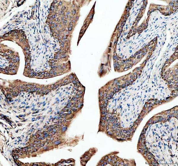
Anti-RPL4 antibodies have IHC images from human, mouse and rat paraffin sections (A05175-1 and A05175 captions; A05175-1 reactivity) and IF images from human U-2 OS cells (M05175 caption).

Real IHC data IHC analysis of Ribosomal protein L4/RPL4 using anti-Ribosomal protein L4/RPL4 antibody (A05175-1). Ribosomal protein L4/RPL4 was detected in a paraffin-embedded section of human colon cancer tissue. Heat mediated antigen retrieval was performed in EDTA buffer (pH 8.0, epitope retrieval solution). The tissue section was blocked with 10% goat serum. The tissue section was then incubated with 1:100 rabbit anti-Ribosomal protein L4/RPL4 Antibody (A05175-1) overnight at 4°C. Peroxidase Conjugated Goat Anti-rabbit IgG was used as secondary antibody and incubated for 30 minutes at 37°C. The tissue section was developed using HRP Conjugated Rabbit IgG Super Vision Assay Kit (Catalog # SV0002) with DAB as the chromogen.
Anti-Ribosomal protein L4/RPL4 Antibody
Cat # A05175-1
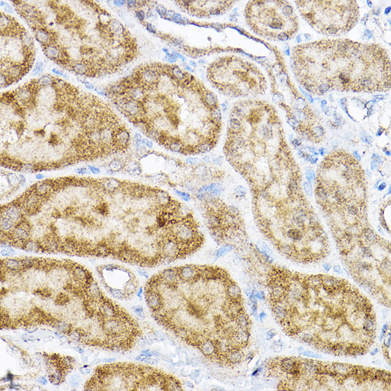
Real IHC data Immunohistochemistry of paraffin-embedded rat kidney using RPL4 Rabbit pAb at dilution of 1:100 .Perform high pressure antigen retrieval with 10 mM citrate buffer pH 6.0 before commencing with IHC staining protocol.
Anti-60S ribosomal protein L4 RPL4 Antibody
Cat # A05175
Real IF data Immunofluorescent analysis of 4% paraformaldehyde-fixed, 0.1% Triton X-100 permeabilized U-2 OS (human osteosarcoma cell line) cells labeling RPL4 with M05175 at 1/25 dilution, followed by Dylight® 488-conjugated goat anti-rabbit IgG secondary antibody at 1/200 dilution (green). Immunofluorescence image showing cytoplasm and nucleus staining on U-2 OS cell line. Cytoplasmic actin is detected with Dylight® 554 Phalloidin at 1/100 dilution (red).The nuclear counter stain is DAPI (blue).
Anti-RPL4 Antibody (N-Term)
Cat # M05175

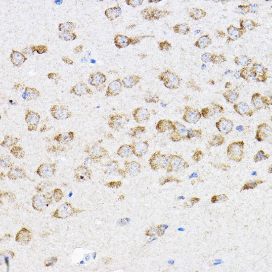
A05175-1 has IHC images from human colon cancer, mouse bladder and rat bladder paraffin sections (A05175-1 captions). A05175 has IHC images from rat kidney and mouse brain paraffin sections (A05175 captions); M05175 has IF data from human U-2 OS cells (M05175 caption).

Which to pick: For tissue IHC, choose A05175-1 for its human, mouse and rat reactivity and IHC application listing (A05175-1 catalog), or A05175 for its illustrated rat kidney and mouse brain staining (A05175 captions). For IF/ICC, choose polyclonal A05175-1 when both applications or human, mouse and rat reactivity matter (A05175-1 catalog), or polyclonal M05175 for IF in human cells (M05175 catalog and caption). The IHC captions describe paraffin sections with EDTA pH 8.0 retrieval for A05175-1 and citrate pH 6.0 retrieval for A05175; neither reports the tissue fixative (A05175-1 and A05175 captions).

Each figure is that product's own IHC / IF validation image from its datasheet.

References

  1. UniProt Consortium. UniProt entry P36578 (RL4_HUMAN, Large ribosomal subunit protein uL4).
  2. Human Protein Atlas. RPL4 tissue IHC expression (reliability: Uncertain).
  3. Human Protein Atlas. RPL4 subcellular location (ICC-IF): Mainly localized to the endoplasmic reticulum and cytosol. In addition localized to the nucleoli and nuclear bodies..
  4. Human Protein Atlas. RPL4 antibody validation summary (1 antibodies).
  5. An epithelial cell fate-driven predictive model for liver metastasis risk in primary colorectal cancer through single-cell and multi-omics integration. Journal of translational medicine 2026 — PMC13474844.
  6. CCT8 drives colorectal cancer progression via the RPL4-MDM2-p53 axis and immune modulation. BMC medical genomics 2025 — PMC12008926.
  7. Effects of short-term waterfall forest aerosol air exposure on rat lung proteomics. Annals of translational medicine 2022 — PMC9761115.
  8. Pan-cancer analysis of the prognostic and immunological role of RPL4. Heliyon 2024 — PMC11305218.
  9. PubMed PMID:8268230 — UniProt-cited evidence.
  10. PubMed PMID:11875025 — UniProt-cited evidence.
  11. PubMed PMID:14702039 — UniProt-cited evidence.